


Cicatrization of Oncological Wounds by Autologous Fibrin Biopolymer Dressing Treatment: A Case Series
Abstract
Introduction. Traditional therapies used to treat chronic wounds are often expensive and, in general, are not adequate to support healing. A promising alternative to conventional dressings is the autologous biopolymer FM, full of cytokines and growth factors that accelerate the healing process of wounds of various etiologies. Materials and Methods. The authors report 3 cases in which FM was used to treat chronic oncological wounds that had been conventionally treated for more than 6 months with no sign of healing. Results. Among the 3 reported cases, there was complete healing of 2 wounds. The other lesion did not heal, mainly due to the location (at the base of the skull). However, it significantly reduced its area, extension, and depth. No adverse effects or hypertrophic scar formation were recorded, and the patients reported an absence of pain from the second week of FM application. Conclusions. The proposed FM dressing approach was effective in healing and speeding up tissue regeneration. It can also be considered one of the most versatile delivery systems to the wound bed, as it is an excellent carrier of growth factors and leukocytes.
Introduction
Chronic wounds are characterized by a persistent inflammatory phase, prolonged infection, and failure of the immune system cells to respond to environmental stimuli. These lesions represent a substantial challenge for conventional dressings since the wounds remain open for months or even years,1,2 negatively affecting a patient’s quality of life and leading to considerably high medical costs.3
Traditional therapies are often expensive, time-consuming, and have limited effectiveness4 since they cannot provide the growth factors essential for healing.5-7 Generally, these conventional wound dressings repair the wound by forming connective tissues, but they do not restore the tissue’s functional integrity.8
One natural biomaterial candidate for treating acute or chronic wounds is the FM formed from fibrin-rich plasma. This autologous 3D biopolymer is easily prepared and is full of cytokines and growth factors paramount to wound healing.9
In the current work, the authors studied the use of autologous FM as a dressing in treating chronic oncological wounds, evaluating the reduction of the wound area, healing, and adverse effects.
Materials and Methods
The study, which was approved by the Human Research Ethics Committee (Protocol # 51787321.9.0000.0098), included 3 patients from the Erasto Gaertner Hospital in Curitiba, Brazil, who presented with oncological wounds that had been treated with conventional therapy for more than 6 months with no sign of healing. All the lesions were from surgical resection of tumors: Patient A had neurofibromatosis with melanoma in the cervical region, Patient B had fibrosarcoma at the base of the skull, and Patient C had breast carcinoma.
The patients went through a peripheral venous puncture in the upper limb with a vacutainer-needle system coupled to the closed system tube free of anticoagulants, and 30 mL to 80 mL of blood was extracted from each patient using 10 mL tubes. The tubes were centrifugated (Centrifuge 80-2b 12 tube; Centribio) at 770 g for 12 minutes at 20°C. After centrifugation, the fibrin-rich plasma clot was isolated with a sterile clamp and scissor, discarding the red cells that adhered to its end. The exudate (growth factor rich) was reserved to irrigate the wound.
The FM was applied weekly on the wound bed according to the following protocol: cleaning the wound with 0.9% saline solution; drying the surface area; irrigating with exudate (which remained from the blood centrifugation); applying the FM; applying nonadherent gauze; and covering with a secondary coverage (sterile gauze and bandages). Patients were instructed to change only the secondary coverage daily.
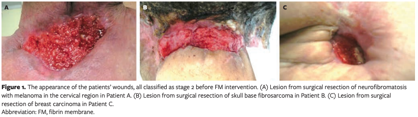
The wounds were documented with a digital camera, measured, and evaluated in their appearance and odor every week until healed. The oncological wounds were staged as follows: stage 1 was intact skin, reddish or violet-colored tissue; stage 2 was an open wound reaching the dermis and epidermis with superficial ulcerations, no or small amounts of exudate, occasional pain, and odor; stage 3 was a wound contemplating the subcutaneous tissue with necrosis and presence of exudate and odor; and stage 4 was a tumor wound with the invasion of deep tissues and presence of exudate in considerable quantity, odor, and pain.10 The 3 wounds studied were initially classified as stage 2. The wounds’ appearance before the FM intervention can be seen in Figure 1.
Results
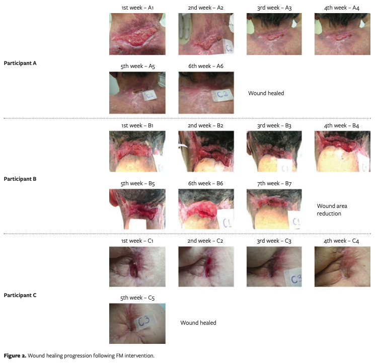
On the first day, the patients reported pain and a bad smell. The authors noted the presence of biofilm, exudate (as seen in Figure 1), and a bad odor. Figure 2 presents the images of the wounds as treatment progressed.
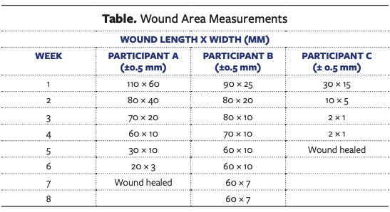
In Figure 2, one can see that wounds from Patients A and C were wholly healed after 6 and 5 applications of FM, respectively. In Patient B, the healing process was slower (7 weeks), presenting a 44% area reduction. The measurements taken each week (Table) demonstrate the fast-healing process of the lesions week by week and are confirmed by the images from Figure 2. As of the time of writing, Patient B continues with the treatment.
The 3 patients also reported pain relief after the second week of treatment with the FM, and the odor initially classified as grade I for the 3 wounds decreased to grade 0 after the second week. Reports of discomfort felt by patients were also recorded weekly by the principal researcher nurse. None of the cases presented phlogistic signs and/or symptoms indicative of wound infection or adverse reactions.
Discussion
Autologous FM, either alone or with other materials, has been used as a biological scaffold for stem cells or primary cells to regenerate various tissues11 because of its ability to induce angiogenesis and to release growth factors, leukocytes, and cytokines.12-14
Although Patients A and C had comorbidities and were older than Patient B, their wounds were healed fully. Even for Patient A, a former smoker, the wound was completely healed in 6 weeks. With no comorbidities, Patient B had the wound area reduced with no complete healing in 7 weeks, which may be explained by the large size of the wound and its location, where frequent movement can slow up the wound healing.
In each patient, the volume of exudate decreased week by week, which was evidence of the healing process. Together with cytokines and platelet cells, the exudate components contain growth factors responsible for tissue regeneration.15
Limitations
The results suggest the great benefits of autologous FM for the healing of chronic oncological wounds. However, the weakness of this study is the limited number of cases studied. The next step is to apply this protocol to a larger population of participants in randomized clinical trials. Additional research is needed to gather evidence of the effectiveness of the technology as a viable therapeutic alternative for the treatment of chronic oncological wounds.
Conclusion
The autologous FM dressing demonstrated efficacy in healing recalcitrant oncologic wounds with no adverse effects nor the formation of hypertrophic scars. In Patient B, even without complete healing, the lesion showed scar tissue and edge closure. It is worth noting that before the FM treatment, all 3 wounds had remained open for more than 6 months even though the patients had been treated weekly with conventional therapy. Therefore, it is possible to say that, compared with conventional dressings, the FM is a lower-cost alternative that is easy to prepare and apply. These features justify the effort for continuing its clinical evaluation in chronic wounds, especially in immunosuppressed patients.
Acknowledgments
Authors: Chayane K.L. de Carvalho, MSc; Beatriz Luci Fernandes, PhD; Michel M. Dalmedico, PhD; and Mauren Abreu de Souza, PhD
Acknowledgments: The authors thank Adriana Stelzner Brozoski for the clinical care of the patients, Emerson Czachorowski, and the Erasto Gaertner Hospital for their support in the feasibility of the clinical trial.
Affiliations: Pontifícia Universidade Católica do Paraná, Paraná, Brazil
Disclosure: The authors declare no financial or other conflicts of interest.
Correspondence: Beatriz Luci Fernandes, PhD, Pontificia Universidade Católica do Paraná, Programa de Pós-graduação em Tecnologia em Saúde, Rua Imaculada Conceição, 1155, Prado Velho, Curitiba, PR, Brazil 80320-030; beatriz.fernandes@pucpr.br
References
1. Shao M, Hussain, Z, Thu HE, et al. Emerging trends in therapeutic algorithm of chronic wound healers: recent advances in drug delivery systems, concepts-to-clinical application and future prospects. Crit Rev Ther Drug Carrier Syst. 2017;34(5):387-452. doi:10.1615/CritRevTherDrugCarrierSyst.2017016957
2. Pinto NR, Ubilla M, Zamora Y, Del Rio V, Ehrenfest DMD, Quirynen M. Leucocyte- and platelet-rich fibrin (L-PRF) as a regenerative medicine strategy for the treatment of refractory leg ulcers: a prospective cohort study. Platelets. 2018;29(5):468-475. doi:10.1080/09537104.2017.1327654
3. Dehkordi AN, Babaheydari FM, Chehelgerdi M, Dehkordi SR. Skin tissue engineering: wound healing based on stem-cell-based therapeutic strategies. Stem Cell Res Ther. 2019;10(1):111. doi:10.1186/s13287-019-1212-2
4. Pang C, Ibrahim A, Bulstrode NW, Ferretti P. An overview of the therapeutic potential of regenerative medicine in cutaneous wound healing. Int Wound J. 2017;14(3):450-459. doi:10.1111/iwj.12735
5. Prabhu R, Vijayakumar C, Chandra AAB, et al. Efficacy of homologous, platelet-rich plasma dressing in chronic non-healing ulcers: an observational study. Cureus. 2018;10(2):e2145. doi:10.7759/cureus.2145
6. Dong R, Guo B. Smart wound dressings for wound healing. Nanotoday. 2021;41:101290. doi:10.1016/j.nantod.2021.101290
7. Weller CD, Team V, Sussman G. First-line interactive wound dressing update: a comprehensive review of the evidence. Front Pharmacol. 2020;11:155. doi:10.3389/fphar.2020.00155
8. Borena BM, Martens A, Broeckx SY, et al. Regenerative skin wound healing in mammals: state-of-the-art on growth factor and stem cell based treatments. Cell Physiol Biochem. 2015;36(1):1-23. doi:10.1159/000374049
9. De Carvalho CKL, Fernandes BL, De Souza MA. Autologous matrix of platelet-rich fibrin in wound care settings: a systematic review of randomized clinical trials. J Funct Biomater. 2020;11(2):31. doi:10.3390/jfb11020031
10. De Carvalho MMC, Macêdo WTP, Carneiro RB, de Cássia Lima Xavier E, Peixoto IVP. Skin lesions in oncology palliative care: observational study. Res Society Develop. 2021;10(6):e7510615350. doi:10.33448/rsd-v10i6.15350
11. Ahmed TA, Dare EV, Hincke M. Fibrin: a
versatile scaffold for tissue engineering applications. Tissue Eng Part B Rev. 2008;14(2):199-215. doi:10.1089/ten.teb.2007.0435
12. Mirhaj M, Tavakoli M, Varshosaz J, et al. Platelet rich fibrin containing nanofibrous dressing for wound healing application: fabrication, characterization and biological evaluations. Biomater Adv. 2021;17:112541. doi:10.1016/j.msec.2021.112541
13. Sari R, Larasati GS, Kuncorowati NG, Syaify A. Platelet-rich fibrin (PRF) membranes accelerate open wound healing better than amniotic membranes: a histological study on the proliferation phase. Wound Med. 2020;31:100190. doi:10.1016/j.wndm.2020.100190
14. Dohan DM, Choukroun J, Diss A, et al. Platelet-
rich fibrin (PRF): a second-generation platelet concentrate. Part I: technological concepts and evolution. Oral Surg Oral Med Oral Pathol Oral Radiol Endod. 2006;101(3):e37-e44. doi:10.1016/j.tripleo.2005.07.008
15. Spear M. Wound exudate--the good, the bad, and the ugly. Plast Surg Nurs. 2012;32(2):77-79. doi:10.1097/PSN.0b013e318256d638


