Venous Leg Ulcers: Potential Algorithms of Care
Abstract
Management of VLUs can be challenging, depending on wound complexity, and may require the use of several treatment modalities to achieve complete wound closure or significant wound area reduction. This review presents a systematic approach to management of VLUs based on previous literature and the authors’ clinical experience, with consideration given to wound size, etiology, and responses to prior treatment. Techniques described include debridement (autolytic, enzymatic, sharp/surgical), compression therapy, physical therapy, medical adjuncts, and cellular- and tissue-based therapy. The algorithm of care for VLUs is multimodal. Appropriate diagnostic studies must be performed, including venous duplex and appropriate pathophysiology to confirm the diagnosis of VLU. After the correct diagnosis is confirmed, appropriate treatment may commence. All patients should undergo appropriate wound debridement; the exact modality used is dependent on wound characteristics. Patients must also adhere to consistent compression therapy. Any underlying venous disease that is amenable to surgical intervention should be addressed. Treatment with a medical adjunct and physical therapy are recommended. For patients who do not achieve significant wound area reduction, the addition of CTP is recommended. Use of these methods should result in substantial wound area reduction and/or wound closure.
Abbreviations
ABI, ankle-brachial index; CI, confidence interval; CTP, cellular and tissue-based therapy; CVD, chronic venous disease; CVI, chronic venous insufficiency; ESCHAR, Effect of Surgery and Compression on Healing and Recurrence; EVRA, Early Venous Reflux Ablation; FBD, fetal bovine dermis; MPFF, micronized purified flavonoid fraction; NPWT, negative pressure wound therapy; RCT, randomized clinical trial; SIS, small intestinal submucosa; STSG, split-thickness skin graft; VLU, venous leg ulcer.
Introduction
The first priority when managing a VLU is making the appropriate diagnosis. A clinical history and physical examination are paramount. Appropriate noninvasive imaging is also necessary, along with measurement of ABI. VLUs are open lesions between the knee and ankle joint that occur in the presence of venous disease.1 Such wounds are not necessarily the result of venous hypertension. Thus, there is somewhat of a disconnect between the diagnosis of an ulcer that occurs in the leg with underlying venous disease and an ulcer that occurs secondary to underlying venous disease. A large number of studies do not make a distinction between these 2 very distinct wound types.2
The development of venous ulcers is a debilitating consequence of CVI. Tissue breakdown and ulceration results from chronic dermal inflammation, leading to dermatitis. The inflammatory response stimulated by venous hypertension causes extravasation of macromolecules and red blood cell products into the dermal interstitium and alterations in the extracellular matrix that deposits disorganized collagen, which allows for persistent and sustained dermal injury.3 The characteristic skin changes seen are brawny induration, thickening, edema, and eventual tissue breakdown. The Clinical-Etiology-Anatomy-Pathophysiology classification system is often used to aid in identifying CVI clinically. This descriptive classification is composed of 7 stages, from C0 (no venous disease) to C6 (open and active ulcer).4 This system takes into account clinical signs including the presence of varicose veins, reticular veins, telangiectasia, edema, pigmentation, skin changes, and presence of ulcers. Although the diagnosis of CVI is primarily clinical, it is important to confirm with imaging studies. However, there is a role for other dermatologic evaluation. A comprehensive history is also necessary, including evaluation for other skin manifestations of inflammatory conditions, such as rheumatoid arthritis, systemic lupus, and scleroderma. Other atypical diseases and hematologic disorders such as sickle cell anemia, thalassemia, hemochromatosis, and protein S and protein C deficiency may also manifest as chronic skin conditions. Although these diseases can occur in the setting of venous disease, they are not caused by venous disease. Therefore, treatment in these cases is aimed at the primary underlying pathophysiology.
A study published in 2018 in France followed the French health authorities’ guidelines that if there is no clinical improvement after 6 months and/or atypical clinical signs persist, biopsy should be performed.5 In that study, biopsy findings were positive in 5% of the lower extremity ulcers that fit these criteria. In patients without a clear etiology of leg ulceration, however, early biopsy is indicated because waiting 6 months could potentially delay diagnosis.
Although the majority of lower extremity ulcers are attributed to CVI, a significant number of lower extremity ulcers are the result of both arterial and venous disease. These so-called mixed ulcers account for up to 26% of lower extremity ulcers, and they are defined by the absence of pedal pulses, reduced ABI (<0.90), inflow stenosis greater than 50%, superficial or deep venous insufficiency, and/or deep venous thrombosis at ultrasound.6 As previously mentioned, physical examination and noninvasive imaging studies are important in determining the etiology of the wound. Without a correct diagnosis, a patient may undergo weeks or months of treatment without obtaining the desired results.
Another potential contributor to nonhealing lower extremity ulceration is the relationship between the venous system and the lymphatic system. In patients with CVI, the lymphatic systems can be overloaded, leading to phlebolymphedema—that is, combined venous and lymphatic insufficiency.7 When lymphatic channels become overloaded, increased fibrosis of interstitial tissue is observed with large protein concentration in the tissue, which ultimately results in skin infections and ulceration.
Diagnostic Imaging Modalities Used to Evaluate Venous Disease
While there are invasive and complex imaging studies that can be performed to help diagnose CVD—such as venography, venous pressure measurements, computed tomography venography, and magnetic resonance venography—by far the most commonly used imaging test to evaluate for CVD is noninvasive duplex ultrasound. In 2020, Bhatt et al8 published a systematic review of 43 studies evaluating the accuracy of diagnostic tests for an episode of deep venous thrombosis of the lower extremities. The pooled estimated sensitivity and specificity of whole-leg ultrasonography were 94.0% (95% CI, 91.3–95.9) and 97.3% (95% CI, 94.8–98.6), respectively. In the setting of CVI and VLU, however, the goal of duplex ultrasound is to map out normal and abnormal venous pathways and identify the sources of level of obstruction and incompetence. Visual cues are noted, such as vein size and reflex dilation due to chronic pooling with incompetent great saphenous veins greater than 15 mm in diameter.9 In addition to using duplex ultrasound for direct evaluation of obstruction, there has been increased emphasis on Doppler evaluation of flow patterns. The venous valves may appear structurally damaged at ultrasound, but more importantly, the function of the valves can be determined by observing the direction of flow. Certain maneuvers are used to elicit venous reflux; the preferred method is a standardized technique with calibrated inflation and rapid deflation with a pneumatic cuff applied distally to the suspect segment; use of this maneuver ensures sufficient retrograde flow to close normal valves.10 The most commonly used cutoff value in the literature according to multiple prospective studies is 1 second for identifying deep vein reflux in the femoral or popliteal veins and greater than 0.5 seconds in the superficial veins and deep veins in the calf.11 Clinical guidelines of the Society for Vascular Surgery and the American Venous Forum recommend the use of duplex ultrasound-guided treatment of veins located near active VLU when the veins have a diameter greater than 3.5 mm and reflux time greater than or equal to 0.5 seconds.
Importance and Effect of Compression Therapy
Compression, elevation, and local wound care have long been accepted as the first-line approaches in the management of chronic venous ulcers. Rigid bandages have been documented for the treatment of extremity edema from as early as the 17th century.13 While advances in technology have broadly expanded the range of materials and devices available for use to provide compression, the general principle of managing CVD remains the same.
Compression therapy is known to improve venous and lymphatic outflow, reduce venous hypertension, and prevent reflux.14 Furthermore, multiple studies have demonstrated that compression therapy reduces local inflammation, improves cutaneous oxygenation, and allows for improved healing.12,15,16 Traditionally, compression therapy has been used with caution in patients with peripheral vascular disease and diabetes given the underlying perfusion deficits in these patients. However, a study by Rother et al17 in 2020 demonstrated that low-level, graded compression can be safe for use in this patient population.
The 3 main types of compression are compression stockings, layered bandaging, and programmed pneumatic compression devices. It is generally accepted that compression stockings are appropriate for patients with uncomplicated varicose veins; however, such treatment may not be suitable for patients with more severe disease. Individual RCTs have reported inconsistent data regarding the superiority of multilayer vs single-layer compression. This may be owing to the generally small sample sizes of these studies and the possible risk of bias. When comparing the data collectively, however, multicomponent compression therapy was found to be the superior treatment for venous ulcer disease. A 2012 Cochrane Review of 48 RCTs (4321 patients) showed multicomponent compression to be the most widely used compression system.18 Multilayered compression has been demonstrated to be more efficacious than single-layer compression in regard to healing rate and time to healing, with higher compression stockings being superior to those with lower compression when trying to maintain lower extremity edema reduction.19,20 No difference between 2-layer and 4-layer compression therapy was noted at 3 months.18
Medical Management of the Symptoms of Venous Disease
Pentoxifylline is a rheologic agent known to improve VLU healing, especially in ulcers with a duration of more than 1 year.21 Pentoxifylline is a methylxanthine derivative with good oral absorption; it is typically administered 3 times per day at a dose of 400 mg, with lower doses recommended in patients with significant renal failure. Benefits may be observed after 2 to 4 months of therapy. Pentoxifylline has several mechanisms of action, including increasing the deformability of erythrocytes and inhibiting neutrophil adhesion and activation. It is also a known inhibitor of tumor necrosis factor α, which has been hypothesized to impair healing of VLUs.21,22
The other medications/supplements that receive significant attention for the treatment of VLU are flavonoids. Available in the United States, flavonoids are a diverse group of naturally occurring phlebotropic compounds that are commonly used as food supplements.23 Horse chestnut seed extract, derived from Aesculus hippocastanum, contains flavonoids.24 In several randomized controlled trials, this extract was found to be safe and effective in the management of edema associated with venous disease. MPFF consists of 90% diosmin and 10% hesperidin and is reported to improve venous tone, support lymphatic drainage, and protect the microcirculation owing to the inhibition of activation, migration, and adhesion of leukocytes, thus effectively reducing release of inflammatory mediators.25 In the treatment of CVI, MPFF 500 mg twice daily is prescribed; when used for 6 months, MPFF has been shown to decrease the inflammatory response and the clinical symptoms of CVI.23 In a Cochrane Review of 9 studies (1075 participants) examining the role of flavonoids in the management of VLUs, MPFF was shown to improve the healing rate in the treatment of VLUs; however, the quality of the studies was poor.26
Nutrition is another factor to consider in the medical management of VLUs. A patient’s nutritional status is of the utmost importance in healing a chronic wound. Many patients with chronic wounds are malnourished at baseline; malnourishment is a risk factor for the development of venous stasis ulcers. Additionally, patients with VLUs tend to leak fluid and thus, lose nutrients from their ulcers, thereby worsening their nutritional status. Because of the nutrient deficiencies that may be prevalent in patients with VLU, it has been suggested that oral nutritional supplements be prescribed to aid in wound healing.27 Unfortunately, no well-powered prospective randomized controlled trials have been done to study the efficacy of oral supplementation in patients with VLU.
Indications for Procedural Intervention in Venous Disease
Modern technologies such as duplex ultrasound have significantly advanced the ability to identify patients for whom surgical intervention may be of some benefit. For example, Shami et al28 found that 53% of patients with CVI and resultant ulceration showed reflux solely in the surgically treatable superficial veins, whereas in 32% the combined superficial and deep veins were affected and 15% had reflux in only the deep veins. Generally, indications for surgical treatment of CVI are based on the patient’s symptoms and lack of improvement or disease progression after a trial of compression therapy and lifestyle modification. This approach was initially supported in the oft-cited ESCHAR study, a randomized controlled study in which patients with venous ulceration were randomized to either compression therapy and leg elevation only, or to superficial venous (open) surgery in addition to compression therapy and leg elevation. While overall wound healing was not significantly different between the 2 treatment arms, ulcer recurrence was significantly reduced with the addition of surgery (P <.0001).29
However, in the past decade, open surgery has almost completely been replaced by endovenous treatment modalities. Although the open techniques are often quite successful, they are associated with significant postprocedural morbidity. Thus, endovenous treatment modalities have become the standard of care owing to decreased postprocedural morbidity and pain compared with open surgery, as well as faster return to normal activity. In a large meta-analysis comparing the results of radiofrequency ablation, endovenous laser ablation, sclerotherapy, and surgery, van den Bos et al30 reported success rates of 84% and 94%, respectively, for radiofrequency ablation and endovenous laser ablation, compared with 78% for surgery and 77% for sclerotherapy. Those authors proposed that minimally invasive techniques were at least as effective as traditional open techniques.
The findings of the ESCHAR study were accepted until 2018, when the results of the EVRA trial were reported. The EVRA trial comprised 450 patients with VLU treated at 20 centers in the United Kingdom. Patients were randomized to receive either endovenous ablation of superficial venous reflux or compression therapy alone. For patients randomized to compression therapy, consideration was given to endovenous ablation after 6 months if the wound remained open or after the wound was fully healed. The study found that patients who received early ablation experienced faster wound healing of VLU (hazard ratio for ulcer healing, 1.38; 95% CI, 1.13–1.68; P =.001).31
Anatomic considerations are also important when deciding on surgical interventions. The most frequent veins of interest are the great and small saphenous veins. Other veins that have less commonly been implicated in symptomatic venous insufficiency, but which can be treated if identified, are the intersaphenous vein in the popliteal fossa, perforating veins between the deep and superficial systems, and gastrocnemius veins.32 Care must be taken not to plan an intervention that incompletely addresses the offending vessels and thus may allow the reflux to continue or worsen. The primary concern is a patient with significant deep venous obstructive etiology with reflux in the superficial system. At times, the superficial system is dilated secondary to being the primary conduit of blood out of the leg, thus rendering the valves incompetent.
Debridement of VLU
VLUs with extensive necrotic or devitalized tissue warrant surgical intervention. Surgical debridement is required for patients with the sequelae of infected wounds, including sepsis, osteomyelitis, and severe cellulitis.33,34 These patients require prompt treatment to avoid wound progression, worsening infection, and, in some cases, limb loss. A 2015 Cochrane Review of debridement for the management of VLUs evaluated 10 studies with a total of 715 participants.33 Most of these studies were small. A range of debridement methods were used, including autolytic methods such as nonadherent dressings, very small beads, biocellulose dressings, honey, gels, gauze, and methods utilizing enzymes. No studies evaluated sharp debridement. The most common type of debridement performed was autolytic debridement. There was no significant difference in efficacy between methods; however, there was evidence to suggest that ulcers with large amounts of slough from which more than 50% of slough was removed after 4 weeks were more likely to heal by 12 weeks, and there was some evidence to suggest that ulcers debrided using honey were more likely to heal by 12 weeks than ulcers debrided with hydrogel.33 However, in a retrospective analysis of a prospective trial of 366 patients with VLU, the 68 patients debrided at initial presentation in addition to at least 1 subsequent visit exhibited a 47% closure rate at 12 weeks, whereas 298 patients who did not undergo initial debridement had a 30% closure rate.35
Few data exist to support the use of enzymatic debridement for VLU. The role of enzymatic debridement is to remove necrotic and/or devitalized tissue in patients with wounds that do not require or cannot otherwise tolerate surgical debridement. Often, these wounds show no signs of acute infection and are usually smaller in size. A subset of patients for whom surgical debridement would be optimal but who for various reasons are not agreeable to debridement in the operating room will opt for enzymatic debridement. Some VLUs are excruciatingly painful, making bedside mechanical and/or sharp debridement incredibly difficult, if not impossible; these patients would also benefit from enzymatic debridement. Application of topical debriding agents can save these patients a trip to the operating room, thus eliminating the additional morbidity associated with undergoing anesthesia. Enzymatic debridement agents consist of exogenous enzymes that act on a wound to remove necrotic tissue, debris, and eschar. Currently, the only commercially available product for the enzymatic debridement of VLUs is clostridium histolyticum collagenase (Santyl; Smith & Nephew).
Another enzymatic debridement agent that has been shown to be effective in wound care, specifically in the treatment of burn wounds, is bromelain. This extract is derived from pineapple; it can be harvested from either the fruit itself or the stem. Bromelain contains proteolytic enzymes and nonenzymatic substances. A phase 2 clinical trial of EscharEx (MediWound), a bioactive debriding agent comprising proteolytic enzymes enriched in bromelain, is underway to investigate its efficacy in treating patients with nonhealing VLU in the United States and Switzerland.36 This multicenter, double-blinded, randomized controlled trial is designed to evaluate the product versus placebo gel and versus the nonsurgical standard of care. After patients have undergone complete debridement, they may undergo additional procedures (if needed) to achieve complete wound closure.36 Should this bioactive debridement agent prove to be a viable treatment option, its use has the potential to affect the care of this population.
Role of NPWT
NPWT has been used to stimulate wound closure. It functions by the application of negative pressure (intermittent or continuous), resulting in removal of exudate, macrophage mobilization, and stimulation of senescent cells and angiogenesis.32 NPWT has been shown to increase blood flow and granulation tissue in preparation for delayed wound closure.37 Some NPWT delivery systems also have the capability to instill topical wound solutions, which can further increase their utility and efficacy. The purpose of instillation of topical wound solutions is to reduce bacterial burden, which has not been shown to occur with simple NPWT. Instillation introduces topical wound solution into the wound; this solution remains in the wound bed for a predetermined amount of time before being removed by negative pressure in order to remove contaminants and debris, thus effectively decreasing the wound bioburden.37
The use of NPWT with STSG has been investigated to improve the rate of wound closure in patients with VLUs. STSGs have been shown to be quite effective in achieving wound closure in burn wounds, with high rates of graft uptake in this patient population. However, this same success has not been replicated in chronic wounds, owing to the location and shape of chronic wounds, which can pose a challenge for STSG placement and lead to increased graft failure rates. Ross et al38 retrospectively reviewed the records of patients who underwent STSG followed by immediate fixation with NPWT at a single center from January 2004 to January 2011. Each patient underwent wound bed preparation, followed by STSG and immediate placement of NPWT, with NPWT maintained for 96 hours. Graft uptake was recorded after removal of the NPWT device (4 days postprocedure) and at 2 weeks and 4 weeks. Greater than 92% graft uptake was seen across all wound types with use of NPWT immediately after STSG.38
Yang et al39 performed a cost analysis of large chronic VLUs treated with NPWT with instillation prior to STSG. This single-center, retrospective review examined 10 massive (≥100 cm2) VLUs in 7 patients with venous insufficiency as documented at ultrasound. Each patient initially underwent surgical debridement of VLU in the operating room with immediate placement of NPWT with instillation using 0.125% sodium hypochlorite solution. On postoperative day 7, the patients returned to the operating room for STSG, after which NPWT was placed and remained for 96 hours before hospital discharge. Eight of 10 patients achieved complete wound closure by 6 months after treatment; in the 2 remaining wounds, the percentage of graft uptake was 70% and 80%. The overall cost of this protocol, including hospital stay and all follow-up visits, was $27 000, compared with an estimated cost of $28 000 for standard compression therapy.39
Indications for Physical Therapy in Patients with VLU
Numerous articles support the use of increased exercise and range of motion exercises in treating patients with VLU. Additionally, many studies note that physical inactivity and inability to ambulate independently are independent risk factors for failure of VLU closure.2 One meta-analysis that included 5 RCTs comprising 190 patients with VLU noted that increased physical exercise was associated with a 14% increase in leg ulcer closure.40 When the physical therapy included progressive resistance exercise plus prescribed physical activity, there was a 27% increase in wound closure. Patients apparently performed best if they were at an exercise facility. Patients who followed the most aggressive physical therapy regimen did so at an exercise facility; they performed multiple sets of calf raises and partial squats, with or without weights. In addition, they walked on a treadmill for 30 minutes or cycled 3 times weekly as well as performed ankle flexibility and range of motion exercises.41 However, the authors of the current review find such a regimen too aggressive for most of their patients. By extrapolating from the findings of 2 other studies,42,43 the authors prescribe 10 dorsiflexion movements per foot per waking hour and 2000 steps per day to their patients. For patients in the United States who are on Medicare, an attempt is made to procure 8 weeks of in-home physical therapy. Increased range of motion heel raises and strengthening 3 times per week with stair-step exercise are also prescribed.
Indication for CTP
Studies indicate that VLUs commonly recur and have been found to persist for more than 1 year in 25% to 50% of patients.44-46 CTPs have been engineered to help accelerate wound healing by providing a tissue graft to the chronic wound bed in order to stimulate cell migration, angiogenesis, and epithelialization. Wounds are considered chronic after 6 weeks of nonhealing persistent venous ulcer even with receiving standard compression therapy.44 Generally, the guideline for the closure of a VLU is that if 40% area reduction has not occurred by 4 weeks, the ulcer will likely have a very poor prognosis.47 Thus, local coverage for most CTPs requires failure to achieve a reduction in wound area by 40% to 50% after 4 weeks of appropriate compression therapy.
The only product approved by the US Food and Drug Administration for the closure of VLUs is Apligraf (Organogenesis), a bilayered living skin equivalent, which has received premarket approval.48 This product performs best in VLUs that have been open for longer than 1 year. However, owing to cost considerations and frequency of application required, other modalities have been reviewed. Many other less robust studies have shown high rates of healing with certain CTPs (ie, extracellular matrix graft, acellular fish skin graft, FBD) compared with standard compression therapy.49-51
Institutional Quality Analysis Studies
Based upon the fact that most patients do not close their VLU with simply 1 exogenous product, the authors undertook a variety of quality assurance outcomes reviews for patients who were diagnosed with VLUs. Patients presented for weekly office visits, adherence to multilayer compression was strongly emphasized, and appropriate venous interventions were performed. Although patients were generally adherent with taking pentoxifylline, this was not universal. The value analysis and quality assurance committees have a process for assessing possible augmentation of strategies to enhance wound healing and ensure the best patient outcomes at the lowest cost. The review of these patients’ outcomes was completely de-identified from all demographic material that could be used to identify the patients. The sole assessment was application of product and wound area reduction over time.
The first of the studies that current authors conducted at their institution involved placement of a matrix comprising a synthetic resorbable polyelectrolyte multilayer nanofilm composed of cationic poly(allylamine hydrochloride) and anionic polyacrylic acid (MicroLyte AG; Imbed Biosciences). This matrix is designed to act as a functional molecular template to facilitate granulation in the wound bed. The nanofilm matrix is coated with a 20-µm layer of polyvinyl alcohol to provide moisture management. The nanofilm matrix contains a low level of ionic and metallic silver (<25 µg/cm2) to prevent microbial contamination and colonization of the matrix.52 This metallic silver is thought to be antimicrobial, based on well-documented components of silver therapy and the ability of silver to kill bacteria.53 Therefore, the concept was to apply this very thin layer directly to a wound immediately under a porcine SIS decellularized product (Oasis; Cook Biotech, Inc). This evaluation was designed to include 8 applications of both products placed at the same time in 4 patients. Each patient was seen in clinic weekly and received applications of both products to their wounds. The wounds were measured each week and monitored for changes in size and evidence of improved wound healing. Additionally, wound culture data were obtained to investigate the potential alteration of bioburden of the wounds. Wound area reduction was observed in all 4 patients; the average percentage decrease in wound area at 8 weeks was 44%. One patient achieved complete wound closure before receiving the final application of the product. There was also a decrease in bacteria present in the wounds of 2 patients, as reflected by wound culture data. After reviewing the data at the conclusion of the study, however, it was thought that there would be a better response to potentially 2 to 3 weeks of wound bed preparation with the nanofilm alone prior to application of the SIS. If a future large study is designed to assess this combination, it is recommended that the therapy be metachronous rather than synchronous.
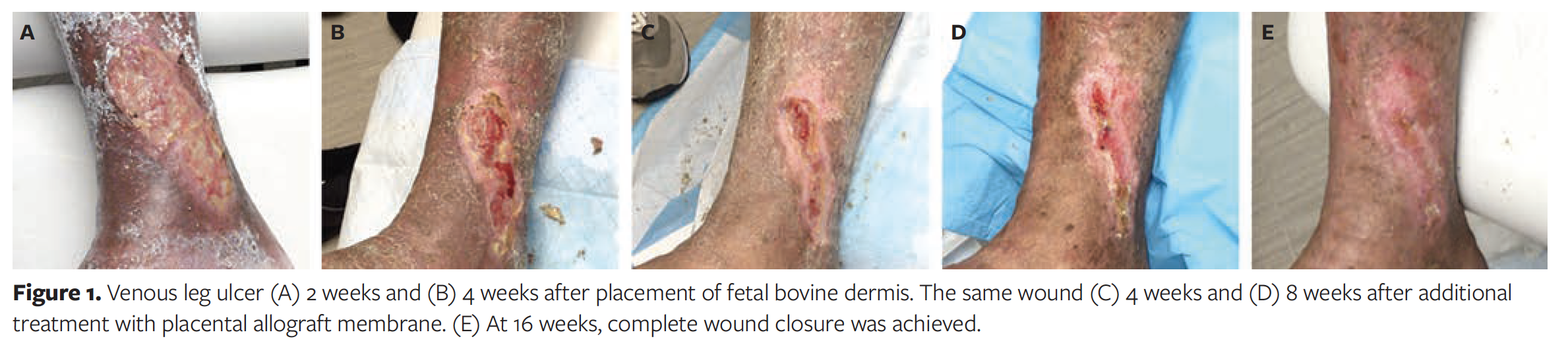
One of the most frequently used skin substitutes at the authors’ institution is FBD (PriMatrix; Integra LifeSciences). This acellular bovine dressing contains physiologic levels of collagen that work to accelerate wound healing, and it has been shown to be effective in wound bed preparation.54 The authors of the current review primarily use this as a single application product and later apply an autograft over it, especially for wounds greater than 40 cm2. Therefore, in patients with VLU who have received FBD, a metachronous product other than autogenous STSG is desired. To this end, the authors of the current review decided to investigate the use of a topical freeze-dried placental allograft membrane (AmnioExcel Plus; Integra LifeSciences) for potential synergistic effect. Placental allograft membrane is a tri-layer allograft consisting of amnion-chorion-amnion layers containing extracellular matrix, cytokines, and growth factors to promote wound healing.55
The authors of this review treated 5 patients with VLUs that were present for longer than 4 weeks but less than 2 years. Each patient was treated for 4 weeks with FBD followed by 8 weeks of the topical freeze-dried placental allograft membrane. Patients were seen in clinic each week to monitor progress, and they also wore multilayer compression for the duration of treatment as well as follow-up period. Wound progress was monitored subjectively by the clinician, and specialized software (inSight; ekare, Inc) was used to capture accurate and consistent objective wound measurements. Overall, treatment was well tolerated by all patients, and significant wound area reduction was demonstrated (Figure 1). Two patients achieved complete wound closure. The average wound area reduction was 6.1 cm2, and the average wound area reduction percentage was 43%. Which of the 2 products studied was the primary contributor to ultimate wound closure is unknown, however. In general, the faster wound healing trajectory occurred after the application of FBD. However, it is important to note that when small wounds get smaller, they close at a slower rate.
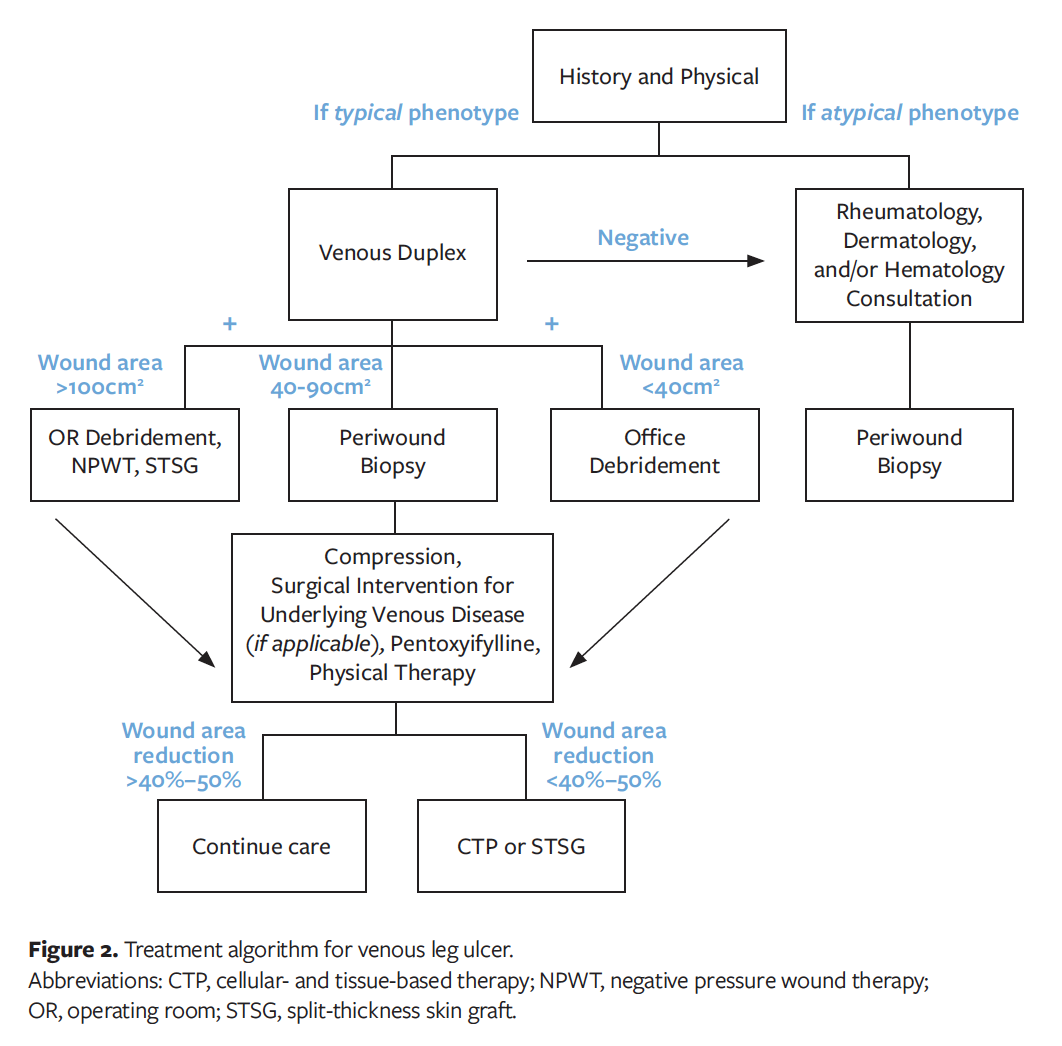
In the inpatient setting at the authors’ institution, 2 different algorithms of care are used in the management of ulcers greater than or equal to 40 cm2 (Figure 2). For wounds greater than or equal to 100 cm2, debridement with tangential hydrosurgery is performed in the operating room and NPWT with instillation is applied for 7 days; this is followed by STSG, bolstered with NPWT. Such treatment requires a total of 11 days in the hospital. Favorable outcomes data with this method have been previously published.38 For wounds (measuring 40–99 cm2) treated at the current authors’ institution, debridement is performed in the operating room with tangential hydrosurgery, and FBD is placed. The FBD is bolstered with NPWT for 96 hours, after which the patient is discharged home with multilayer compression with a plan to undergo STSG in 4 weeks. In this second category, the current authors undertook a prospective quality assurance trial to replace the FBD with a “synthetic dermal replacement scaffold that is composed of polyurethane open-cell foam” as described by Dearman et al56 and evaluated in a porcine study. This material is meant to act as a temporizing dressing prior to STSG after tangential debridement has been performed. The polyurethane open cell foam has been described as a product that allows for the integration of vascular tissue and ingrowth of surrounding collagen fibers that create a neodermis.57 On day 21, the polyurethane open cell foam is removed so that the patient can subsequently undergo STSG, as was done in a study by Wagstaff et al.58 The outcomes of this study were noted as follows: all 5 patients underwent wide surgical excision at the base of the wound prior to application of the foam, and the foam was then affixed with NPWT for a minimum of 4 days. In 4 patients, very good engraftment was achieved, with removal of the outer membrane and explantation of the material at approximately day 21. The 4 patients for whom the material was in place for 21 days underwent successful STSG, with close to 100% take. The sole patient for whom the polyurethane open cell foam was unsuccessful had a very dense infection, and the product sloughed off at day 7. The primary question that remains concerns the quality of the histology of the neodermis that is created. As of the time of this writing, no histologic analysis was available for review, and owing to the quality assurance nature of the authors’ project such assessment was not possible.
Limitations
The authors of the current review recognize that this algorithm of care may not apply to all patients and that most treatments will be left at the discretion of the surgeon. In addition, the institutional quality assurance projects using different CTPs that were carried out at the authors’ institution had very small sample sizes, and thus the same results may not be seen when conducted on larger groups of patients.
Conclusion
There is a very significant and appropriate algorithm of care for the treatment of VLUs (Figure 2). This algorithm is multimodal, and the early components are especially important. Appropriate diagnostic studies are necessary, including venous duplex ultrasound, as well as appropriate skin pathophysiologic analysis and patient history for a conclusive diagnosis of VLU. If the patient has other significant diseases as well, it is reasonable to perform a skin biopsy. Wounds that do not seem to be consistent with VLU should be evaluated by specialists in hematology, rheumatology, and/or potentially dermatology, and periwound biopsy should be considered. In addition, sufficient evidence exists to warrant early intervention and closure of the great saphenous and small saphenous venous reflux as well as of perforator reflux in the periwound area. The addition of pentoxifylline 400 mg 3 times daily—as well as a prescription for physical therapy with range of motion, gait training, and strengthening of the calf muscle pump 3 times weekly—is appropriate. All of this must be done in conjunction with appropriate compression therapy. Additionally, the authors of this review strongly suggest at least 1 sharp debridement to the VLU base, which should occur relatively early in the therapeutic algorithm (ie, within the first 2 weeks of care).
In the authors’ experience, both wound size and wound duration are important. Wounds that have been open for more than 1 year should be considered for CTP therapy, and it should be started sooner rather than later. For wounds that are 100 cm2 or larger, closure in the operating room with wide debridement and application of NPWT and STSG should be performed. For wounds between 40 cm2 and 99 cm2 in size, debridement in the operating room with application of a scaffold is recommended. Historically, the authors have had very good results with FBD; however, newer low-cost options may continue to come to market and should be considered. For wounds smaller than 40 cm2 and of less than 1-year duration, early debridement in the office with application of the other aforementioned therapies is adequate.
The small quality assurance projects discussed in this review demonstrate that many synergies may exist between products. In general, it should not be assumed that wound closure will occur with just 1 product. VLU is a full-thickness wound that closes from the inside out. Therefore, fixing the internal problem is mandatory. This can be done in conjunction with re-creating a functional dermis by performing debridement and applying appropriate and/or autogenous graft technologies.
Acknowledgments
Authors: Crystal V. James, MD1; Quinn Murray, MD1; So Youn Park, MD2; Nazanin Khajoueinejad, MD2; Jani Lee, MD2; Keval Ray, MD2; and John C. Lantis II, MD1
Affiliations: 1Mount Sinai West/Morningside Hospitals, Department of Surgery, New York, NY; 2The Mount Sinai Hospital, Department
of Surgery, New York, NY
Disclosure: Dr. James received honorarium from Imbed Biosciences for a presentation at a conference on the topic of wound care. Dr. Lantis serves as a consultant for Integra LifeSciences and is currently Principal Investigator in the EscharEx clinical trial.
Correspondence: Crystal V. James, MD; Mount Sinai West Medical Center, 1000 10th Ave, New York, NY 10019; Crystal.James2@mountsinai.org
References
1. Scottish Intercollegiate Guideline Network (SIGN) Edinburgh: SIGN; 1998. The Care of Patients with Chronic Leg Ulcer. Guideline 26.
2. Marston WA, Ennis WJ, Lantis JC II, et al. Baseline factors affecting closure of venous leg ulcers. J Vasc Surg Venous Lymphat Disord. 2017;5(6):829-835. doi:10.1016/j.jvsv.2017.06.017
3. Raffetto JD, Mannello F. Pathophysiology of chronic venous disease. Int Angiol. 2014;33(3):212-221.
4. Lurie F, Passman M, Meisner M, et al. The 2020 update of the CEAP classification system and reporting standards. J Vasc Surg Venous Lymphat Disord. 2020;8(3):342-352. doi:10.1016/j.jvsv.2019.12.075.
5. Stansal A, Khayat K, Duchatelle V, et al. Quand poser l’indication d’une biopsie cutanée chez un patient porteur d’ulcère de jambe? Étude rétrospective sur 143 biopsies consécutives [When to ask for a skin biopsy in a patient with leg ulcer? Retrospective study of 143 consecutive biopsies]. J Med Vasc. 2018;43(1):4-9. doi:10.1016/j.jdmv.2017.10.003.
6. Hedayati N, Carson JG, Chi YW, Link D. Management of mixed arterial venous lower extremity ulceration: a review. Vasc Med. 2015;20(5):479-486. doi:10.1177/1358863X15594683
7. Lee BB. Phlebolymphedema: neglected outcome of combined venous and lymphatic insufficiency. Vasc Specialist Int. 2020;36(1):1-3. doi:10.5758/vsi.2020.36.1.1
8. Bhatt M, Braun C, Patel P, et al. Diagnosis of deep vein thrombosis of the lower extremity: a systematic review and meta-analysis of test accuracy. Blood Adv. 2020;4(7):1250-1264. doi:10.1182/bloodadvances.2019000960
9. Min RJ, Khilnani NM, Golia P. Duplex ultrasound evaluation of lower extremity venous insufficiency. J Vasc Interv Radiol. 2003;14(10):1233-1241. doi:10.1097/01.rvi.0000092663.72261.37
10. van Bemmelen PS, Bedford G, Beach K, Strandness DE. Quantitative segmental evaluation of venous valvular reflux with duplex ultrasound scanning. J Vasc Surg. 1989;10(4):425-431. doi:10.1067/mva.1989.14123
11. Labropoulos N, Tiongson J, Pryor L, et al. Definition of venous reflux in lower-extremity veins. J Vasc Surg. 2003;38(4):793-798. doi:10.1016/s0741-5214(03)00424-5
12. O'Donnell TF Jr, Passman MA, Marston WA, et al. Management of venous leg ulcers: clinical practice guidelines of the Society for Vascular Surgery ® and the American Venous Forum. J Vasc Surg. 2014;60(2 Suppl):3S-59S. doi:10.1016/j.jvs.2014.04.049
13. O'Meara S, Cullum NA, Nelson EA. Compression for venous leg ulcers. Cochrane Database Syst Rev. 2009;(1):CD000265. doi:10.1002/14651858.CD000265.pub2
14. Partsch H, Mortimer P. Compression for leg wounds. Br J Dermatol. 2015;173(2):359-369. doi:10.1111/bjd.13851
15. Chiang N, Rodda O, Oldham S, Sleigh J, Vasudevan T. Effects of compression therapy and venous surgery on tissue oxygenation in chronic venous disease. Phlebology. 2019;34(7):474-480. doi:10.1177/0268355518822582
16. Partsch H. Compression therapy: clinical and experimental evidence. Ann Vasc Dis. 2012;5(4):416-422. doi:10.3400/avd.ra.12.00068
17. Rother U, Grussler A, Griesbach C, Almasi-Sperling V, Lang W, Meyer A. Safety of medical compression stockings in patients with diabetes mellitus or peripheral arterial disease. BMJ Open Diabetes Res Care. 2020;8(1):e001316. doi:10.1136/bmjdrc-2020-001316
18. O'Meara S, Cullum N, Nelson EA, Dumville JC. Compression for venous leg ulcers. Cochrane Database Syst Rev. 2012;11(11):CD000265. doi:10.1002/14651858.CD000265.pub3
19. Milic DJ, Zivic SS, Bogdanovic DC, et al. The influence of different sub-bandage pressure values on venous leg ulcers healing when treated with compression therapy. J Vasc Surg. 2010;51(3):655-661. doi:10.1016/j.jvs.2009.10.042
20. Pereira de Godoy JM, Pereira de Godoy HJ, Lopes Pinto R, Facio FN Jr, Guerreiro Godoy MF. Maintenance of the results of stage II lower limb lymphedema treatment after normalization of leg size. Int J Vasc Med. 2017;2017:8515767. doi:10.1155/2017/851576
21. Margolis DJ. Pentoxifylline in the treatment of venous leg ulcers. Arch Dermatol. 2000;136(9):1142-1143. doi:10.1001/archderm.136.9.1142
22. Weinstein DA, Kirsner RS. Refractory ulcers: the role of tumor necrosis factor-alpha. J Am Acad Dermatol. 2010;63(1):146-154. doi:10.1016/j.jaad.2009.08.004.
23. Roztocil K, Stvrtinová V, Strejcek J. Efficacy of a 6-month treatment with Daflon 500 mg in patients with venous leg ulcers associated with chronic venous insufficiency. Int Angiol. 2003;22(1):24-31.
24. Kapusta I, Janda B, Szajwaj B, et al. Flavonoids in horse chestnut (Aesculus hippocastanum) seeds and powdered waste water byproducts. J Agric Food Chem. 2007;55(21):8485-8490. doi:10.1021/jf071709t
25. Lyseng-Williamson KA, Perry CM. Micronised purified flavonoid fraction: a review of its use in chronic venous insufficiency, venous ulcers and haemorrhoids. Drugs. 2003;63(1):71-100. doi:10.2165/00003495-200363010-00005
26. Scallon C, Bell-Syer SE, Aziz Z. Flavonoids for treating venous leg ulcers. Cochrane Database Syst Rev. 2013;(5):CD006477. doi:10.1002/14651858.CD006477.pub2
27. Holt IGS, Green SM, Nelson A. Oral nutritional supplements for treating venous leg ulcers. Cochrane Database Syst Rev. 2019;2019(9):CD012210. doi:10.1002/14651858.CD012210.pub2
28. Shami SK, Sarin S, Cheatle TR, Scurr JH, Smith PD. Venous ulcers and the superficial venous system. J Vasc Surg. 1993;17(3):487-490.
29. Barwell JR, Davies CE, Deacon J, et al. Comparison of surgery and compression with compression alone in chronic venous ulceration (ESCHAR study): randomised controlled trial. Lancet. 2004;363(9424):1854-1859. doi:10.1016/S0140-6736(04)16353-8
30. van den Bos R, Arends L, Kockaert M, Neumann M, Nijsten T. Endovenous therapies of lower extremity varicosities: a meta-analysis. J Vasc Surg. 2009;49(1):230-239. doi:10.1016/j.jvs.2008.06.030
31. Gohel MS, Heatley F, Liu X, et al. A randomized trial of early endovenous ablation in venous ulceration. N Engl J Med. 2018;378(22):2105-2114. doi:10.1056/NEJMoa1801214
32. Marston W. Wound care. In: Sidawy AN, Perler BA. Rutherford’s Vascular Surgery and Endovascular Therapy. 9th ed. Elsevier; 2018: 1536-1556.e3
33. Gethin G, Cowman S, Kolbach DN. Debridement for venous leg ulcers. Cochrane Database Syst Rev. 2015;2015(9):CD008599. doi:10.1002/14651858.CD008599.pub2
34. Baharestani M. The clinical relevance of debridement. In: Baharestani M, Gottrup F, Holstein P, Vanscheidt W editor(s). The Clinical Relevance of Debridement. Springer Verlag, 1999:1-15.
35. Cardinal M, Eisenbud DE, Armstrong DG, et al. Serial surgical debridement: a retrospective study on clinical outcomes in chronic lower extremity wounds. Wound Repair Regen. 2009;17(3)306-311. doi:10.1111/j.1524-475X.2009.00485.x
36. A Study to Evaluate the Safety and the Efficacy of EscharEx (EX-02 Formulation) in Debridement of Venous Leg Ulcers. Clinicaltrials.gov identifier: NCT03588130. Published 2018. Accessed April 13, 2021. https://clinicaltrials.gov/ct2/show/NCT03588130
37. Gupta S, Gabriel A, Lantis J, Téot L. Clinical recommendations and practical guide for negative pressure wound therapy with instillation. Int Wound J. 2016;13(2):159-174. doi:10.1111/iwj.12452
38. Ross RE, Aflaki P, Gendics C, Lantis JC II. Complex lower extremity wounds treated with skin grafts and NPWT: a retrospective review. J Wound Care. 2011;20(10):490-495. doi:10.12968/jowc.2011.20.10.490
39. Yang CK, Alcantara S, Goss S, Lantis JC II. Cost analysis of negative-pressure wound therapy with instillation for wound bed preparation preceding split-thickness skin grafts for massive (>100 cm2) chronic venous leg ulcers. J Vasc Surg. 2015;61(4):995-999. doi:10.1016/j.jvs.2014.11.076
40. Jull A, Slark J, Parsons J. Prescribed exercise with compression vs compression alone in treating patients with venous leg ulcers: a systematic review and meta-analysis. JAMA Dermatol. 2018;154(11):1304-1311. doi:10.1001/jamadermatol.2018.3281
41. Klonizakis M, Tew GA, Gumber A, et al. Supervised exercise training as an adjunct therapy for venous leg ulcers: a randomized controlled feasibility trial. Br J Dermatol. 2018;178(5):1072-1082. doi:10.1111/bjd.16089
42. Meagher H, Ryan D, Clarke-Moloney M, O’Laighin G, Grace PA. An experimental study of prescribed walking in the management of venous leg ulcers. J Wound Care. 2012;21(9):421-428. doi:10.12968/jowc.2012.21.9.421
43. Mutlak O, Aslam M, Standfield N. The influence of exercise on ulcer healing in patients with chronic venous insufficiency. Int Angiol. 2018;37(2):160-168. doi:10.23736/S0392-9590.18.03950-0
44. Greaves NS, Iqbal SA, Baguneid M, Bayat A. The role of skin substitutes in the management of chronic cutaneous wounds. Wound Repair Regen. 2013;21(2):194-210. doi:10.1111/wrr.12029
45. Lees TA, Lambert D. Prevalence of lower limb ulceration in an urban health district. Br J Surg. 1992;79(10):1032-1034. doi:10.1002/bjs.1800791015
46. Cornwall JV, Doré CJ, Lewis JD. Leg ulcers: epidemiology and aetiology. Br J Surg. 1986;73(9):693-696. doi:10.1002/bjs.1800730905
47. Kantor J, Margolis DJ. A multicentre study of percentage change in venous leg ulcer area as a prognostic index of healing at 24 weeks. Br J Dermatol. 2000;142(5):960-964. doi:10.1046/j.1365-2133.2000.03478.x
48. Falanga V, Sabolinski M. A bilayered living skin construct (APLIGRAF) accelerates complete closure of hard-to-heal venous ulcers. Wound Repair Regen. 1999;7(4):201-207. doi:10.1046/j.1524-475x.1999.00201.x
49. Mostow EN, Haraway GD, Dalsing M, Hodde JP, King D; OASIS Venus Ulcer Study Group. Effectiveness of an extracellular matrix graft (OASIS Wound Matrix) in the treatment of chronic leg ulcers: a randomized clinical trial. J Vasc Surg. 2005;41(5):837-843. doi:10.1016/j.jvs.2005.01.042
50. Yang CK, Polanco TO, Lantis JC II. A prospective, postmarket, compassionate clinical evaluation of a novel acellular fish-skin graft which contains omega-3 fatty acids for the closure of hard-to-heal lower extremity chronic ulcers. Wounds. 2016;28(4):112-118.
51. Paredes JA, Bhagwandin S, Polanco T, Lantis JC. Managing real world venous leg ulcers with fetal bovine acellular dermal matrix: a single centre retrospective study. J Wound Care. 2017;26(Sup10):S12-S19. doi:10.12968/jowc.2017.26.Sup10.S12
52. Try Microlyte – Imbed Bio. Try Microlyte – Imbed Bio. Accessed October 2, 2022. http://imbedbio.com/try-microlyte/
53. Hartmann H, Krastev R. Biofunctionalization of surfaces using polyelectrolyte multilayers. BioNanoMaterials. 2017;18(1):20160015. doi:10.1515/bnm-2016-0015
54. Sabolinski ML, Gibbons G. Comparative effectiveness of a bilayered living cellular construct and an acellular fetal bovine collagen dressing in the treatment of venous leg ulcers. J Comp Eff Res. 2018;7(8):797-805. doi:10.2217/cer-2018-0031
55. AmnioExcel® Plus Placental Allograft Membrane. Integra. Accessed April 13, 2021. https://www.integralife.com/amnioexcel-plus-placental-allograft-membrane/product/wound-reconstruction-care-outpatient-clinic-private-office-treat-amnioexcel-plus-placental-allograft-membrane
56. Dearman BL, Li A, Greenwood JE. Optimization of a polyurethane dermal matrix and experience with a polymer-based cultured composite skin. J Burn Care Res. 2014;35(5):437–448. doi:10.1097/BCR.0000000000000061
57. NovoSorb® BTM | Biodegradable Synthetic Dermal Substitute. United States. Published January 17, 2022. Accessed October 2, 2022. https://polynovo.com/novosorb-btm
58. Wagstaff MJ, Schmitt BJ, Coghlan P, Finkemeyer JP, Caplash Y, Greenwood JE. A biodegradable polyurethane dermal matrix in reconstruction of free flap donor sites: a pilot study. Eplasty. 2015;15:e13.