Anesthetic and Surgical Considerations in Osteogenesis Imperfecta: A Case Report of Mandible Fracture Management
Abstract
Background. Osteogenesis imperfecta (OI) is a rare metabolic bone disorder in which collagen production is impaired. Patients have brittle bones that are prone to fracture with minor trauma. Whereas most of the fractures occur in the spine and extremities, fractures of the craniofacial bones are less common.
Methods. This report describes the management of a 14-year-old boy with OI type 1 who sustained mandible fractures from an assault. Management consisted of open reduction and internal fixation of the parasymphyseal and angle fractures and closed reduction and mandibulomaxillary fixation of the subcondylar fracture.
Results. Surgery resulted in return of premorbid occlusion and good jaw function. Removal of plates and screws was performed 10 months later without incident.
Conclusions. This case report discusses the unique anesthetic, surgical, and postoperative considerations for managing mandible fractures in patients with OI.
Introduction
Osteogenesis imperfecta (OI), also known as brittle bone disease, is a heterogeneous group of bone disorders caused by faulty synthesis of type 1 collagen.1 It is the most common inherited cause of bone fragility, affecting about 1 in 15 to 20,000 births.2 The increased bone fragility predisposes the vertebrae and extremities to fractures.3 Facial bone fractures are uncommon.4,5
Craniofacial surgery in patients with OI is undertaken with caution due to several anesthetic and surgical challenges, including airway abnormalities, altered anatomy, cardiopulmonary pathology, elevated bleeding risk, and susceptibility to iatrogenic soft tissue and bone injury.3,6-10 There are very few documented instances of facial fracture repair in this patient population.5,9,11-15 This report of the treatment of a child with mandibular fractures provides valuable insight as to how these patients may be managed. A comprehensive review of anesthetic and surgical considerations in OI patients is also provided.
Methods
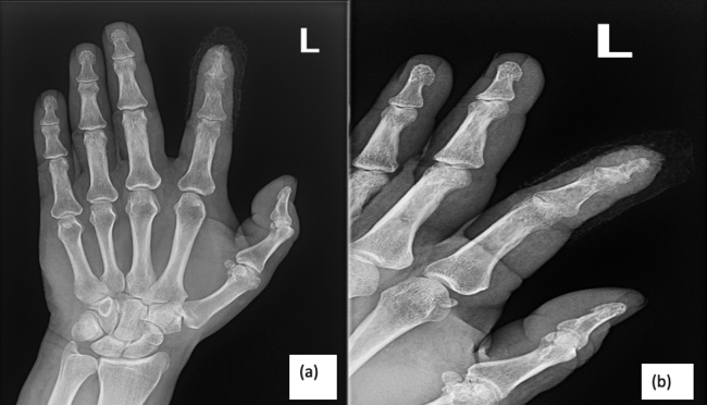
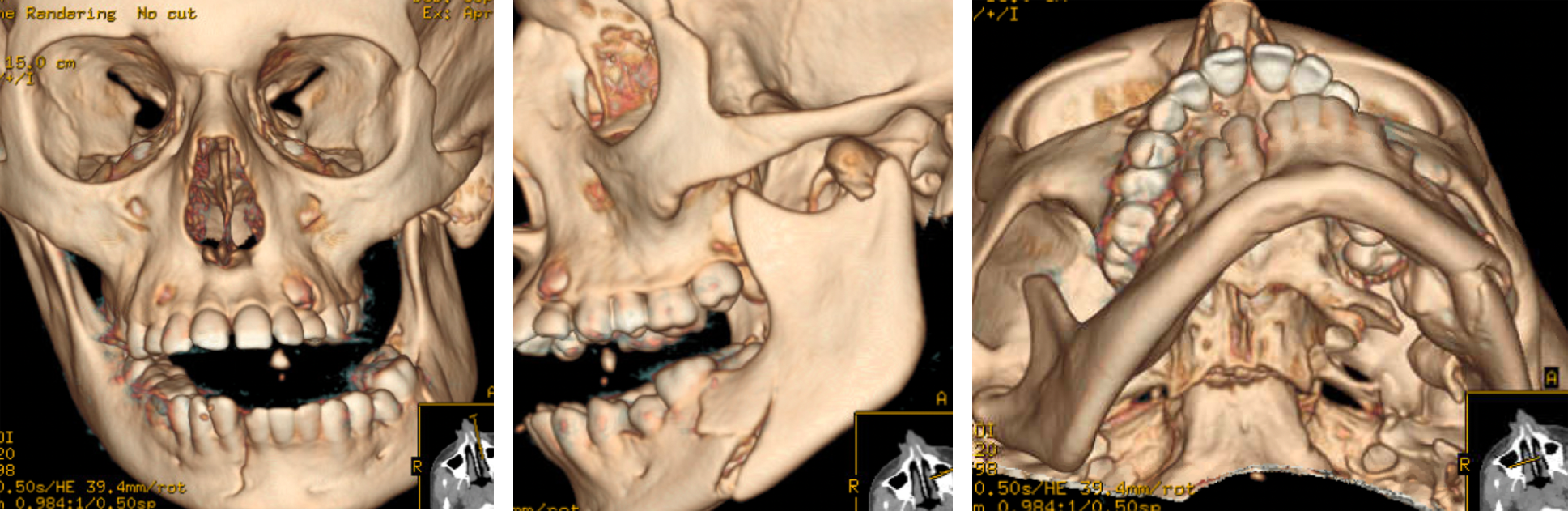
A 12-year-old boy with Angelman syndrome, developmental delay, and OI type I presented to the emergency department with malocclusion and jaw pain after an unwitnessed fall. Clinical examination showed mandibular deformity, gap in the lower front teeth, and malocclusion. Computed tomography (CT) of the face revealed displaced mandible fractures involving the right parasymphysis, left angle, and condylar neck (Figure 1). Medical history was significant for developmental delay, epilepsy, aortic root dilation, chronic kidney disease (single kidney), neurogenic bowel and bladder, imperforate anus, and tethered cord. The patient had previously undergone multiple operations for upper and lower extremity fractures.
Preoperative clearances were obtained from the patient’s cardiologist and nephrologist. After informed consent from parents, the patient was taken to the operating room and surgery was performed under general anesthesia with a nasotracheal tube. Intraoral buccal sulcus incisions were used to access the fracture sites at the parasymphysis and angle. The fractures were debrided of blood and loose fragments. Arch bars were placed and mandibulomaxillary fixation (MMF) was performed. Dental wear facets were used as guides to place the patient in native occlusion. This was done very gently so as not to cause an iatrogenic fracture. Open reduction internal fixation (ORIF) of the left angle fracture was performed with a 6-hole fracture plate with bicortical screws for the lower border and a 4-hole mini plate with monocortical screws for the upper border. ORIF of the parasymphysis fracture was performed with a 6-hole fracture plate with bicortical screws for the lower border, and the arch bar provided upper border fixation by acting as a tension band. The operation was without incident.
Results
The patient did not tolerate MMF due to his developmental delay. He was taken to the operating room on postoperative day 6 for arch bar removal. Remainder of the postoperative course was unremarkable.
Approximately 10 months after surgery, the patient presented to the office with indications of jaw pain. A definitive diagnosis of jaw pain was difficult to make because the patient was developmentally delayed and nonverbal; however, the patient pointed towards his lower face, indicating discomfort. There were no signs of facial trauma, occlusion was good, and mouth opening was normal. Imaging was not performed as it would have required general anesthesia and multiple medical clearances. The suspected cause of the pain was hardware palpability, looseness, or nerve impingement. It is a common practice to electively remove hardware in skeletally immature children to minimize any possible restriction of mandibular growth.16 The patient was therefore taken back to the operating room. Access to the plates was obtained by reopening the lower gingivobuccal sulcus incisions. There was stable bony union of the fracture segments with bony overgrowth over the plates and no signs of infection. All hardware was removed. Postoperative course was uneventful, and his pain completely resolved. Occlusion was normal at 1-year follow-up.
Discussion
OI is the most common inherited cause of bone fragility.17 The underlying cause is qualitative and/or quantitative defects of type 1 collagen, which is the most abundant protein in bone.2 This leads to low bone mineral density resulting in brittle bones that are susceptible to fractures with minor trauma. Most cases are autosomal dominant. The Sillence classification divides the disease into 4 types.18 Type I is the most common and mildest form, Type II is lethal, Type III is the most severe, and Type IV is of intermediate severity. This scheme has been expanded to include newer types based on more recently discovered genetic mutations.19 The hallmark of OI is generalized osteopenia.2 Extraskeletal manifestations include blue sclerae, hearing loss, joint hypermobility, valvular heart disease, and central nervous system abnormalities.19 Dentinogenesis imperfecta, a disorder of dentin production, commonly occurs in patients with OI.1 It is the most common genetic dental abnormality and is characterized by brittle, discolored teeth that are susceptible to decay and loss.20
Fractures are the most common presenting symptom of OI.1 They most commonly occur in the vertebrae and extremities due to their high mechanical load. Facial bone fractures are infrequently reported, accounting for 1 to 2% of fractures in OI patients.21 They occur either spontaneously or in response to mild trauma. Dental extractions can also lead to mandible fractures.14 Mastication and yawning have also been shown to cause mandible fractures.15 The definitive management is a reduction and stabilization of the fractured bone to restore normal occlusion and jaw opening. Bone healing is normal in OI patients, although the new bone is of poor quality just like the native bone.10 Regular dental evaluations are important for maintenance of oral health, treatment of damaged teeth, and correction of occlusal abnormalities.4 Prophylactic calcium and vitamin D supplementation help preserve bone mineral density.1 Bisphosphonates are the mainstay of medical therapy. These drugs decrease bone resorption by causing apoptosis of osteoclasts.2 The result is increased bone volume with decreased risk of fractures. Newer therapies that show promise are denosumab (RANKL inhibitor), romosozumab (sclerostin inhibitor), and anabolic agents.1,22
There are very few reported cases of mandible fracture management in OI patients. Feifel managed bilateral body and subcondylar fractures in an 8-year-old girl with microplates and mandibulomaxillary fixation.11 The maxillomandibular fixation was removed at 13 days. Intraoral spring-loaded orovestibular plates were placed in the posterior dentition for another 3 months to facilitate passive mouth opening and prevent foreshortening of the ramus. Long-term follow-up showed excellent mouth opening. Kim et al reported a case of a patient with OI type I and class III relationship who sustained multiple mandibular fractures that were treated with miniplates.12 Postoperative radiographs showed a new ramus fracture, thought to be due to Kocher clamp application for reduction. This iatrogenic fracture was successfully corrected with arch bars and 5 weeks of mandibulomaxillary fixation. Al-Osaimi et al reported on a spontaneous mandibular body fracture in an edentulous OI patient with an atrophic mandible who was on bisphosphonate therapy.5 The fracture was repaired with a miniplate, but the patient subsequently had another fracture distal to the hardware that required fixation with a reconstruction plate. Kobayashi et al treated multiple mandibular fractures in a 1-year-old child with open reduction internal fixation of the displaced symphysial fracture with resorbable plates and screws and nonoperative management of the minimally displaced angle and subcondylar fractures.13 The child healed well with normal occlusion and jaw opening. Gallego et al reported on a mandibular fracture after molar extraction. The fracture was repaired with a plate and mandibulomaxillary fixation for 15 days.14 Long-term follow-up showed good bone healing. Nilesh et al fabricated an arch bar retained thermoformed splint to treat multiple mandibular fractures in a 5-year-old child in mixed dentition.9 Hardware was avoided to prevent tooth bud injury. The splint was removed after 2 weeks, and the fractures healed with good occlusion.10
Patients with OI are at risk for several anesthetic and surgical complications that increase with disease severity. The following factors need to be considered when planning surgery:
1. A preoperative workup is essential. Pulmonary function can be compromised due to chest wall deformities (pectus excavatum or carinatum) and thoracic spine deformities (scoliosis, kyphosis), which may result in progressive restrictive pulmonary disease.8 Cardiac abnormalities include valvular heart diseases like aortic root dilation and mitral valve insufficiency.23 Echocardiography and pulmonary function tests can help diagnose these problems.
2. Anesthetic complications, although infrequent, increase with the severity of OI.24 Communication with the patient may be challenging as hearing loss is common. Intravenous catheter insertion may also be difficult due to distorted anatomic landmarks and multiple previous intravenous accesses.24 Patients may not be able to lie completely flat due to kyphoscoliosis, and therefore a molding mattress may be needed. Intubation can be challenging due to distorted anatomy from spinal and chest wall deformities. Force of the laryngoscope during intubation can fracture the brittle mandible or break fragile teeth. A short neck and craniocervical junction skull base abnormalities can limit neck extension. Excessive extension of the cervical spine can lead to cervical instability from ligamentous laxity and fracture of the brittle cervical spine. Use of fiberoptic bronchoscope, video laryngoscope, and intubating laryngeal mask airway can overcome some of these difficulties.24,25,26 In high-risk patients, endotracheal intubation may altogether be avoided by total intravenous anesthesia with an laryngeal mask airway.27 It has been suggested that succinylcholine should be avoided because the muscular fasciculations produced by the drug may cause bone fractures. However, there is no clear evidence for this, and this drug has been used without problems in OI patients.25 A retrospective chart review of 205 pediatric surgeries performed at an OI center of excellence revealed a 1.5% incidence of difficult airway, with the risk of anesthetic complications and difficult intravenous catheterization increasing with the severity of OI.24
3. Extremity fractures can occur with blood pressure cuffs and tourniquets.28,29 However, if care is taken, noninvasive blood pressure cuffs can safely be used in OI type I patients.30 Invasive blood pressure measurement may be considered in patients with OI types III and IV. Peripheral nerve blocks can be challenging as the altered anatomy distorts bony landmarks; ultrasound guidance may be helpful.24
4. Hyperthermia during surgery has been reported in patients with OI. Avoidance of inhalational anesthetics, which can trigger malignant hyperthermia, has been recommended in favor of total intravenous anesthesia.31 However, multiple studies have confirmed that this hyperpyrexia is not malignant hyperthermia but due to some another mechanism.32,33 Hypermetabolism has been postulated as a possible cause.34 Higher levels of thyroxine have been reported in OI patients; thyroxine uncouples oxidation, converting some of the energy for adenosine triphosphate generation to heat.34 The current consensus is to follow standard anesthetic practices of intraoperative temperature management.
5. Iatrogenic injuries may occur. Increased fragility makes the bones susceptible to fracture during fracture manipulation and reduction.12 Use of traction devices like Kocher clamps can place undue pressure on the osteopenic bone resulting in microfractures.10 Fractures of other bones can occur, eg, from inadvertently leaning on the patient, placing heavy instruments on the patient, or during transportation from the stretcher to the operating room table. All pressure points need to be well padded.
6. Patients with OI are prone to easy bruising and increased bleeding with surgical procedures.35,36 This is attributed to increased capillary fragility and abnormal platelet function.37 Abnormalities that have been identified include decreased von Willebrand factor, reduced platelet retention, impaired platelet factor 3 release, and abnormal platelet aggregation.37,38 Rothschild et al reported a 17% incidence of significant bleeding (blood loss > 10% of estimated total blood volume), with the risk increasing with the severity of OI.24 Platelet function tests may identify patients with impaired function. The reverse Trendelenburg position facilitates venous drainage and therefore may decrease bleeding. Mild hypotensive anesthesia may also decrease operative blood loss.
7. Monitoring for bleeding in the immediate postoperative period is critical as it may cause airway embarrassment and hypotension. Patients may have elevated body temperatures due to hypermetabolism, which may be confused with infection. Postoperative rehabilitation and pain management are crucial to improving recovery. The patient may experience painful spasms for which low-dose diazepam can be considered for short-term management.7 Untreated spasms can lead to fractures.
Conclusions
Surgical management of patients with OI may be challenging due to several factors such as difficult airway and increased bleeding. Important considerations include preoperative cardiopulmonary workup, careful handling of brittle bones, avoidance of pressure injuries during surgery, and postoperative monitoring for hemorrhage.
Acknowledgments
Affiliations: 1Aga Khan University, Karachi, Pakistan; 2Lurie Children’s Hospital, Northwestern University Feinberg School of Medicine, Chicago, IL.
Correspondence: Farooq Shahzad; fshahzad@luriechildrens.org
Disclosures: The authors have no relevant financial or non-financial interests to disclose.
References
1. Tournis S, Dede AD. Osteogenesis imperfecta - A clinical update. Metabolism. 2018;80:27-37. doi:10.1016/j.metabol.2017.06.001
2. Forlino A, Marini JC. Osteogenesis imperfecta. Lancet. 2016;387(10028):1657-1671. doi:10.1016/S0140-6736(15)00728-X
3. Gil JA, DeFroda SF, Sindhu K, Cruz AI Jr, Daniels AH. Challenges of fracture management for adults with osteogenesis imperfecta. Orthopedics. 2017;40(1):e17-e22. doi:10.3928/01477447-20161006-04
4. Rosén A, Modig M, Larson O. Orthognathic bimaxillary surgery in two patients with osteogenesis imperfecta and a review of the literature. Int J Oral Maxillofac Surg. 2011;40(8):866-873. doi:10.1016/j.ijom.2011.02.028
5. Al-Osaimi A, Samman M, Al-Shakhs M, Al-Suhaim F, Ramalingam S. An unusual case of atrophic mandible fracture in a patient with osteogenesis imperfecta and on oral bisphosphonate therapy: Case report. Saudi Dent J. 2014;26(2):68-73. doi:10.1016/j.sdentj.2013.12.008
6. Rodrigo C. Anesthesia for maxillary and mandibular osteotomies in osteogenesis imperfecta. Anesth Prog. 1995;42(1):17-20.
7. Esposito P, Plotkin H. Surgical treatment of osteogenesis imperfecta: current concepts. Curr Opin Pediatr. 2008;20(1):52-57. doi:10.1097/MOP.0b013e3282f35f03
8. Widmann RF, Bitan FD, Laplaza FJ, Burke SW, DiMaio MF, Schneider R. Spinal deformity, pulmonary compromise, and quality of life in osteogenesis imperfecta. Spine (Phila Pa 1976). 1999;24(16):1673-1678. doi:10.1097/00007632-199908150-00008
9. Nilesh K, Sawant A, Taur S, Parkar MI. Management of multiple mandibular fractures in a child with osteogenesis imperfecta using arch bar retained thermoformed splints: a novel technique. J Clin Pediatr Dent. 2016;40(4):322-327. doi:10.17796/1053-4628-40.4.322
10. Lewis MK, Stoker NG. Surgical management of the patient with osteogenesis imperfecta. J Oral Maxillofac Surg. 1987;45(5):430-437. doi:10.1016/0278-2391(87)90011-5
11. Feifel H. The surgical treatment of mandibular fractures in a child with osteogenesis imperfecta. Int J Oral Maxillofac Surg. 1996;25(5):360-362. doi:10.1016/s0901-5027(06)80030-5
12. Kim MY, Kim CH. Iatrogenic mandibular fracture after open reduction and internal fixation in a patient with osteogenesis imperfecta. Br J Oral Maxillofac Surg. 2017;55(9):971-973. doi:10.1016/j.bjoms.2017.09.003
13. Kobayashi Y, Satoh K, Mizutani H. Osteogenesis imperfecta diagnosed from mandibular and lower limb fractures: a case report. Craniomaxillofac Trauma Reconstr. 2016;9(2):141-144. doi:10.1055/s-0035-1550063
14. Gallego L, Junquera L, Pelaz A, Costilla S. Pathological mandibular fracture after simple molar extraction in a patient with osteogenesis imperfecta treated with alendronate. Med Oral Patol Oral Cir Bucal. 2010;15(6):e895-e897. Published 2010 Nov 1. doi:10.4317/medoral.15.e895
15. Ram H, Shadab M, Vardaan A, Aga P. Fracture of mandible during yawning in a patient with osteogenesis imperfecta. BMJ Case Rep. 2014;2014:bcr2013203385. Published 2014 Aug 7. doi:10.1136/bcr-2013-203385
16. Berryhill WE, Rimell FL, Ness J, Marentette L, Haines SJ. Fate of rigid fixation in pediatric craniofacial surgery. Otolaryngol Head Neck Surg. 1999;121(3):269-273. doi:10.1016/S0194-5998(99)70183-X
17. Mäkitie RE, Costantini A, Kämpe A, Alm JJ, Mäkitie O. New insights into monogenic causes of osteoporosis. Front Endocrinol (Lausanne). 2019;10:70. Published 2019 Feb 25. doi:10.3389/fendo.2019.00070
18. Sillence DO, Senn A, Danks DM. Genetic heterogeneity in osteogenesis imperfecta. J Med Genet. 1979;16(2):101-116. doi:10.1136/jmg.16.2.101
19. Rossi V, Lee B, Marom R. Osteogenesis imperfecta: advancements in genetics and treatment. Curr Opin Pediatr. 2019;31(6):708-715. doi:10.1097/MOP.0000000000000813
20. Kaur A, Kumar S, Karda B, Chibh R. Management of dentinogenesis imperfecta: a report of two cases. Int J Clin Pediatr Dent. 2019;12(5):464-466. doi:10.5005/jp-journals-10005-1681
21. Bergstrom L. Osteogenesis imperfecta: otologic and maxillofacial aspects. Laryngoscope. 1977;87(9 Pt 2 Suppl 6):1-42.
22. Marom R, Rabenhorst BM, Morello R. Osteogenesis imperfecta: an update on clinical features and therapies. Eur J Endocrinol. 2020;183(4):R95-R106. doi:10.1530/EJE-20-0299
23. Bonita RE, Cohen IS, Berko BA. Valvular heart disease in osteogenesis imperfecta: presentation of a case and review of the literature. Echocardiography. 2010;27(1):69-73. doi:10.1111/j.1540-8175.2009.00973.x
24. Rothschild L, Goeller JK, Voronov P, Barabanova A, Smith P. Anesthesia in children with osteogenesis imperfecta: Retrospective chart review of 83 patients and 205 anesthetics over 7 years. Paediatr Anaesth. 2018;28(11):1050-1058. doi:10.1111/pan.13504
25. Bojanić K, Kivela JE, Gurrieri C, et al. Perioperative course and intraoperative temperatures in patients with osteogenesis imperfecta. Eur J Anaesthesiol. 2011;28(5):370-375. doi:10.1097/EJA.0b013e3283459616
26. Karabiyik L, Parpucu M, Kurtipek O. Total intravenous anaesthesia and the use of an intubating laryngeal mask in a patient with osteogenesis imperfecta. Acta Anaesthesiol Scand. 2002;46(5):618-619. doi:10.1034/j.1399-6576.2002.460525.x
27. Erdoğan MA, Sanli M, Ersoy MO. Anesthesia management in a child with osteogenesis imperfecta and epidural hemorrhage. Braz J Anesthesiol. 2013;63(4):366-368. doi:10.1016/j.bjane.2012.07.008
28. Kostopanagiotou G, Coussi T, Tsaroucha N, Voros D. Anaesthesia using a laryngeal mask airway in a patient with osteogenesis imperfecta. Anaesthesia. 2000;55(5):506. doi:10.1046/j.1365-2044.2000.01425-28.x
29. Oliverio RM Jr. Anesthetic management of intramedullary nailing in osteogenesis imperfecta: report of a case. Anesth Analg. 1973;52(2):232-236.
30. Sullivan BT, Margalit A, Garg VS, Njoku DB, Sponseller PD. Incidence of fractures from perioperative blood pressure cuff use, tourniquet use, and patient positioning in osteogenesis imperfecta. J Pediatr Orthop. 2019;39(1):e68-e70. doi:10.1097/BPO.0000000000001105
31. Ogawa S, Okutani R, Suehiro K. Anesthetic management using total intravenous anesthesia with remifentanil in a child with osteogenesis imperfecta. J Anesth. 2009;23(1):123-125. doi:10.1007/s00540-008-0698-z
32. Porsborg P, Astrup G, Bendixen D, Lund AM, Ording H. Osteogenesis imperfecta and malignant hyperthermia. Is there a relationship?. Anaesthesia. 1996;51(9):863-865. doi:10.1111/j.1365-2044.1996.tb12619.x
33. Peluso A, Cerullo M. Malignant hyperthermia susceptibility in patients with osteogenesis imperfecta. Paediatr Anaesth. 1995;5(6):398-399. doi:10.1111/j.1460-9592.1995.tb00340.x
34. Cropp GJ, Myers DN. Physiological evidence of hypermetabolism in osteogenesis imperfecta. Pediatrics. 1972;49(3):375-391.
35. Morton ME. Excessive bleeding after surgery in osteogenesis imperfecta. Br J Oral Maxillofac Surg. 1987;25(6):507-511. doi:10.1016/0266-4356(87)90144-6
36. Cole NL, Goldberg MH, Loftus M, Kwok V. Surgical management of patients with osteogenesis imperfecta. J Oral Maxillofac Surg. 1982;40(9):578-584. doi:10.1016/0278-2391(82)90286-5
37. Evensen SA, Myhre L, Stormorken H. Haemostatic studies in osteogenesis imperfecta. Scand J Haematol. 1984;33(2):177-179. doi:10.1111/j.1600-0609.1984.tb02393.x
38. Hathaway WE, Solomons CC, Ott JE. Platelet function and pyrophosphates in osteogenesis imperfecta. Blood. 1972;39(4):500-509.