Spindle Cell Lipoma in the Posterior Neck
Case Description
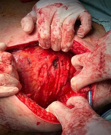
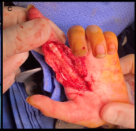
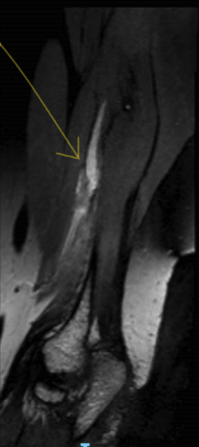
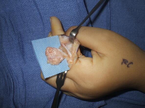
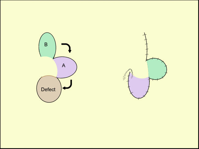
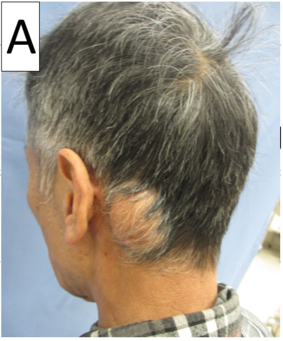
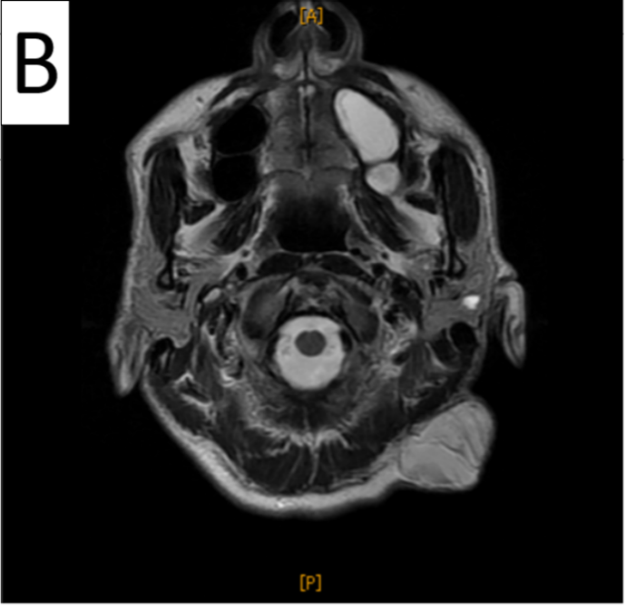
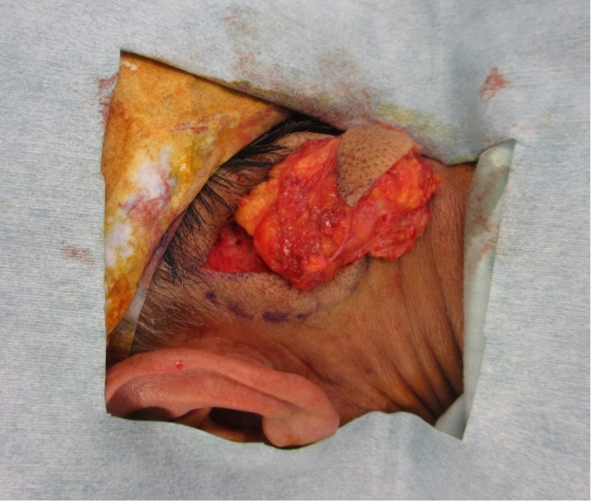
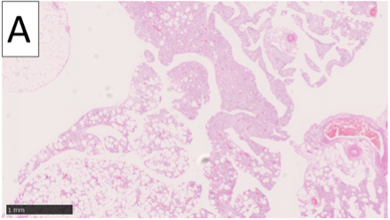
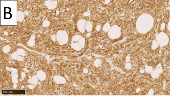
A 71-year-old man presented with a slow-growing subcutaneous tumor (prevalent since 6 years of age) on the left posterior neck. The patient had no previous relevant medical history. On physical examination, a 4- × 3-cm tumor that was elastic, soft, and somewhat hypermobile (Figure 1A) was revealed. There was no pain, redness, or elevated temperature in or around the tumor site. Magnetic resonance imaging (MRI) showed a heterogeneous internal structure on T1-weighted and T2-weighted images (Figure 1B). No significant lymphadenopathy was found. During surgery, an incision was made directly on the tumor, and the tumor was removed along the excised skin in a spindle shape (Figure 2). Histopathological examination using hematoxylin-eosin staining showed mature adipocytes, spindle cell proliferation (Figure 3A), and CD34 positivity (Figure 3B). No nuclear pleomorphism or mitotic figure was observed. The patient was diagnosed with spindle cell lipoma (SCL). There has been no pain or tumor recurrence 4 months postsurgery.
Questions
- How often does SCL occur?
- What are the various useful approaches to diagnose SCL?
- What are the characteristic pathological findings of SCL?
- What is the differential diagnosis of SCL?
Q1. How often does SCL occur?
SCL is a subtype of lipoma, a benign tumor composed mainly of mature adipocytes and spindle-shaped cells.1 SCL is rare and accounts for approximately 1.5% of all adipose tumors.2 It is more common in males aged 40 to 70 years, with a predilection for the shoulder and posterior neck.1,3
Q2. What are the various useful approaches to diagnose SCL?
MRI is useful in diagnosing SCL, which is characterized by a well-defined subcutaneous mass with high signal on T1- and T2-weighted images, low signal on fat-suppressed images, low-signal areas on T1- and T2-weighted images mixed with fatty components, and contrast effects only on nonfatty components.4
Q3. What are the characteristic pathological findings of SCL?
SCL is generally considered to be definitively diagnosed by histopathological examination. It is characterized by the proliferation of spindle-shaped cells, thick collagen bundles, mucous matrix in the interstitium, and scattered mast cells, which are composed of mature adipocytes.⁵ It is also characterized by CD34 positivity of spindle-shaped cells on immunohistochemical staining.6,7
Q4. What is the differential diagnosis of SCL?
Differential diagnoses of SCL include fibrolipoma, pleomorphic lipoma, and liposarcoma. Fibrolipoma is characterized by collagenous fibrosis with few cellular components between fibrous tissues. Pleomorphic lipoma is a malignant tumor that affects the back of the head, shoulders, and back of men aged 40-70 years and has histopathological similarities to SCL; however, it is differentiated by atypical multinucleated giant cells called floret cells. Liposarcoma is a malignant tumor of the thigh, shoulder, and retroperitoneum in men aged 50–60 years. Histologically, liposarcoma differs from SCL in the presence of atypical spindle-shaped cells with pleomorphic nuclei and lipoblasts.8 Simple resection is sufficient for the treatment of SCL. There is no recurrence or metastasis and the prognosis is good.1
Summary
We report the case of a 71-year-old man with SCL in the posterior neck. SCL is a relatively rare subtype of lipoma that predominantly affects the shoulder and posterior neck in men.
Acknowledgments
Affiliations: Department of Plastic and Reconstructive Surgery, Kansai Medical University, Osaka, Japan
Correspondence: Natsuko Kakudo, MD, PhD; kakudon@hirakata.kmu.ac.jp
Disclosures: The authors have no non-financial or commercial, proprietary, or financial interest in the products or companies described in the manuscript. The authors did not receive grants or a consultant honorarium to conduct the study, write the manuscript or otherwise assist in the development of the above-mentioned manuscript.
References
1. Enzinger FM, Harvey DA. Spindle cell lipoma. Cancer. 1975;36(5):1852-1859. doi:10.1002/1097-0142(197511)36:5<1852::aid-cncr2820360542>3.0.co;2-u
2. Fletcher CD, Martin-Bates E. Spindle cell lipoma: a clinicopathological study with some original observations. Histopathology. 1987;11(8):803-817. doi:10.1111/j.1365-2559.1987.tb01884.x
3. Billings SD, Folpe AL. Diagnostically challenging spindle cell lipomas: a report of 34 "low-fat" and "fat-free" variants. Am J Dermatopathol. 2007;29(5):437-442. doi:10.1097/DAD.0b013e31813735df
4. Binh MB, Sastre-Garau X, Guillou L, et al. MDM2 and CDK4 immunostainings are useful adjuncts in diagnosing well-differentiated and dedifferentiated liposarcoma subtypes: a comparative analysis of 559 soft tissue neoplasms with genetic data. Am J Surg Pathol. 2005;29(10):1340-1347. doi:10.1097/01.pas.0000170343.09562.39
5. Fletcher CDM, Bridge JA, Hogendoorn PCW and Mertens F. WHO Classification of Tumours of Soft Tissue and Bone. IARC Press; 2013:29-30.
6. Suster S, Fisher C. Immunoreactivity for the human hematopoietic progenitor cell antigen (CD34) in lipomatous tumors. Am J Surg Pathol. 1997;21(2):195-200. doi:10.1097/00000478-199702000-00009
7. Templeton SF, Solomon AR Jr. Spindle cell lipoma is strongly CD34 positive. An immunohistochemical study. J Cutan Pathol. 1996;23(6):546-550. doi:10.1111/j.1600-0560.1996.tb01447.x
8. Enzinger FM, Weiss SW. Soft Tissue Tumors. 2nd ed. C. V. Mosby Co; 1988:346.