Patient Presentation
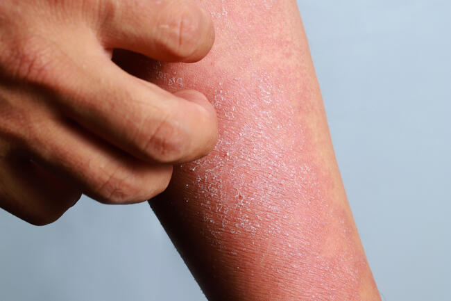
A 45-year-old Caucasian woman presented to the dermatology clinic complaining of a facial rash and knuckle lesions. She had developed a slightly violaceous erythematous eruption around her eyelids associated with periorbital swelling during the previous 2 years, and recently began feeling less energetic. She was noted to have edema of both hands, as well as periungual erythema and telangiectasias, in addition to violaceous dome-shaped papules on the dorsal knuckles. Laboratory work-up including muscle enzyme levels and electromyogram (EMG) were within normal limits, and an abdominal computed tomography (CT) scan performed several months earlier was unremarkable. A skin biopsy from her dorsal hand was performed, and she was sent for a muscle biopsy of her deltoid, which demonstrated no evidence of myositis.
What is Your Diagnosis?
Diagnosis: Amyopathic Dermatomyositis (Dermatomyositis Siné Myositis)
Amyopathic dermatomyositis (ADM) is a form of dermatomyositis (DM) in which patients exhibit exclusively the cutaneous features of DM, but lack clinically-evident muscle involvement. The term ADM was first suggested by Pearson in 19791 to classify patients who demonstrated the typical skin findings of DM without evidence of an inflammatory myopathy, and Krain was the first physician to evaluate such patients circa 1975.2 The characteristic and highly suggestive skin lesions in DM may be the sole manifestations at disease onset in up to 40% of patients,3 and may indicate a poorer prognosis and a greater challenge to treatment.4,5 ADM is considered a distinct clinical entity residing at one pole on a continuum of the idiopathic inflammatory myopathies (IIMs), which range from isolated cutaneous disease (ADM) at one end, to isolated muscle disease (polymyositis, PM) at the opposite end, with classic DM at an intermediate point.6
Clinicopathologic Features of DM and ADM
Dermatomyositis patients develop skeletal muscle inflammation and weakness as well as characteristic skin findings, which include Gottron’s papules on the dorsal aspects of the finger joints, and a classic violaceous macular periorbital heliotrope rash. Various organs may be involved in DM including the lungs and esophagus, as well as the heart.3 Patients suffer primarily from proximal muscle involvement and demonstrate elevation of the muscle-derived enzymes creatinine phosphokinase (CPK) and aldolase, as well as glutamic oxaloacetic acid transaminase and lactate dehydrogenase. The disease occurs in a biphasic age distribution, initially in children and later in the middle-aged population, and tends to affect women more often than men (~2-3:1).5,7,8 DM is associated with other collagen vascular diseases,4 with severe pulmonary complications,9,10 as well as with underlying internal maligancy.11 Skin manifestations were found to precede muscle weakness in more than 50% of patients with DM in a study by el-Azhary et al,7 and patients such as these may or may not go on to develop muscle symptoms several months to years later.5,8,12 Criteria for diagnosis of DM were proposed by Bohan and Peter in 197513 and included: symmetric proximal muscle weakness with or without dysphagia or respiratory muscle involvement, abnormal muscle biopsy, elevation of skeletal muscle-derived enzymes, abnormal EMG, and characteristic skin findings. Autoantibodies implicated in DM include ANA in up to 80% of patients; anti-tRNA synthetase antibodies including anti-Jo1, anti-EJ, anti-PL7, anti-PL12, anti-OJ and anti-KS; anti-SRP; and anti-Mi-2, anti-Scl and anti-KL6, which may be seen more frequently in patients with interstitial lung disease. Certain HLA alleles including HLA-B14 and B-40, and DLA-DR7 and DRw53, have also been associated with DM. Criteria for Diagnosis of ADM proposed by Euwer and Sontheimer in 19935 included the following:
• Presence of pathognomonic cutaneous changes of DM (Gottron’s papules, periungual nailfold erythema/ telangiectasias, violaceous facial erythema/edema including a periorbital distribution — the “heliotrope rash”).
• Skin biopsy compatible with DM.
• No clinical evidence of proximal muscle weakness within 2 years of skin findings.
• No elevation of CPK or aldolase levels within 2 years of skin findings.
Subsequent modifications to the above criteria were proposed,6,10 and to date the exact definition of ADM and its precise features are still under scrutiny.1,4,7,10,14 Exclusion criteria for ADM include systemic immunosuppressive therapy, which may blunt muscle disease from manifesting, as well as concurrent treatment with drugs known to induce a DM-like dermatosis.6 Currently, ADM is considered a distinct clinical entity comprising roughly 2% to 18% of all cases of DM,7 but its incidence has been reported as high as 25% in Chinese patients.15 In addition to CPK and aldolase levels, other diagnostic methods used to exclude subclinical myositis in suspected cases of ADM include EMG and muscle biopsy, as well as MRI and muscle ultrasound.10,16 Magnetic resonance imaging has also been found useful in locating relevant muscle biopsy sites and in screening for malignancy.15 The recent association of ADM with autoantibodies against a 155kD protein and Se autoantigen may be of use in specifically identifying patients with amyopathic DM.6
Cutaneous Findings in ADM
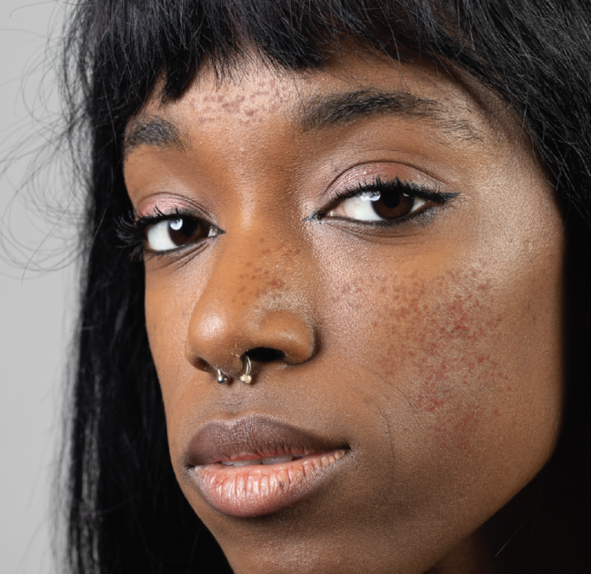
Skin findings are clinically identical in DM and ADM,7,10 and include primarily Gottron’s papules, which are erythematous to violaceous flat-topped papules found principally on bony prominences and extensor surfaces of the hand, as well as the violaceous macular periorbital heliotrope eruption. Gottron’s sign may also be seen as macular violaceous erythema on the extensor surfaces of knuckles, elbows, knees and medial malleoli. Patients typically complain of pruritus, scalp inflammation, photosensitivity, as well as violaceous facial or truncal erythema (“shawl sign”) and periungual erythema and telangiectasias. Other cutaneous lesions include macular violaceous erythema on the hips or thighs (“holster sign”), poikiloderma, nonscarring alopecia, and Raynaud phenomenon, as well as calcinosis cutis, vasculitis and sclerodermatous changes.3,6,8,16
Histopathologic Features
Skin biopsy in ADM is histologically consistent with DM,10 displaying hyperkeratosis, basal keratinocyte liquefactive degeneration, and lymphohistiocytic perivascular inflammation, with vacuolar interface changes ranging from focal vacuolar degeneration along the dermal-epidermal junction to extensive specimen involvement. Acanthosis, areas of epidermal hyperplasia to focal atrophy, increased dermal edema, and follicular plugging may also be noted.12,17 Cutaneous changes in ADM may not be microscopically appreciable at times, often necessitating serial sections.16 Muscle biopsy reveals a CD4+ T-cell-directed Th-1 pattern of microvascular injury, and a CD8+ T-cell-directed Th-2 pattern of myocyte cytotoxicity.10 Association with Malignancy DM and ADM may be considered paraneoplastic syndromes and have been reported to correlate with various malignant diseases in anywhere from 6% to 43% of adults,18 but not so in children.4 Cases of lymphoma, and breast, lung, colon, ovarian and renal carcinomas have been associated specifically with ADM,7,19 with DM having been linked to several other cancers.11 Nasopharyngeal carcinoma has been reported at high rates among Chinese ADM patients,20 and transitional cell carcinoma of the bladder presenting with hematuria has also been described.21 Leukocytoclastic vasculitis and skin necrosis have been suggested as potential markers of underlying malignancy,22,23 and risk factors identified by Sparsa et al23 include the presence of constitutional symptoms, lack of Raynaud phenomenon, and elevated ESR and CPK levels. Screening laboratory studies may include a complete blood count, liver function tests, chest X-ray, urinalysis, fecal occult blood test, and sigmoidoscopy, in addition to thorough history-taking and physical examination.24 Serum Epstein-Barr virus IgA level and laryngoscopy may be warranted particularly in Chinese patients,20 as may be a CA-125 level in women.21 Additional studies may include abdominal-thoracic CT scans in men and pelvic-abdominal-thoracic CT scans in women, due to the increased incidence of non-Hodgkin lymphoma and ovarian, pancreatic and lung cancer.23
Differential Diagnosis
One of the most common erroneous diagnoses made in ADM patients is lupus erythematosus, primarily the subacute cutaneous variant (SCLE), which may be clinically identical to ADM.3 The pruritus often experienced by ADM patients can help to distinguish the disease from lupus, as can a lower incidence of arthralgias and differing immunofluorescence features in ADM.17 Other conditions that may confound an accurate diagnosis include contact dermatitis, lichen planus, psoriasis and seborrheic dermatitis.5 Polymorphous light eruption, trichinosis, atopic dermatitis and erythroderma must also be excluded.17 The use of certain drugs including hydroxyurea, practolol, alfuzosin and phenytoin can occasionally induce DM-like skin changes and must be ruled out as well.10,24
Managing This Condition
While the principal therapy in DM is high-dose systemic corticosteroids (prednisone 1 mg/kg per day to 2 mg/kg per day) with a slow taper over 24 to 36 months, debate exists concerning their efficacy in managing ADM patients.4,7 Many ADM patients are recalcitrant to systemic corticosteroid treatment, particularly those with extensive skin involvement,3,16 but they may derive more benefit from topical steroids.3,4 Treatment with hyroxychloroquine and quinacrine may also be beneficial, even more so when combined with a protective sunscreen.12 Alternative agents include the immunosuppressives, methotrexate and mycophenolate mofetil, which are indicated particularly in refractory skin disease,10 but may cause various undesirable side effects. Azathioprine (Imuran), cyclophosphamide (Cytoxan, Neosar), and high-dose IV immunoglobulin (IVIG) have been employed as well,3,6 and patients who experience severe pruritus may benefit from sedating antihistamines to aid in sleeping.18 Anti-tumor necrosis factor-alpha agents including thalidomide, etanercept (Enbrel) and infliximab (Remicade) have also been reported to be successful.6
Patient Presentation
A 45-year-old Caucasian woman presented to the dermatology clinic complaining of a facial rash and knuckle lesions. She had developed a slightly violaceous erythematous eruption around her eyelids associated with periorbital swelling during the previous 2 years, and recently began feeling less energetic. She was noted to have edema of both hands, as well as periungual erythema and telangiectasias, in addition to violaceous dome-shaped papules on the dorsal knuckles. Laboratory work-up including muscle enzyme levels and electromyogram (EMG) were within normal limits, and an abdominal computed tomography (CT) scan performed several months earlier was unremarkable. A skin biopsy from her dorsal hand was performed, and she was sent for a muscle biopsy of her deltoid, which demonstrated no evidence of myositis.
What is Your Diagnosis?
Diagnosis: Amyopathic Dermatomyositis (Dermatomyositis Siné Myositis)
Amyopathic dermatomyositis (ADM) is a form of dermatomyositis (DM) in which patients exhibit exclusively the cutaneous features of DM, but lack clinically-evident muscle involvement. The term ADM was first suggested by Pearson in 19791 to classify patients who demonstrated the typical skin findings of DM without evidence of an inflammatory myopathy, and Krain was the first physician to evaluate such patients circa 1975.2 The characteristic and highly suggestive skin lesions in DM may be the sole manifestations at disease onset in up to 40% of patients,3 and may indicate a poorer prognosis and a greater challenge to treatment.4,5 ADM is considered a distinct clinical entity residing at one pole on a continuum of the idiopathic inflammatory myopathies (IIMs), which range from isolated cutaneous disease (ADM) at one end, to isolated muscle disease (polymyositis, PM) at the opposite end, with classic DM at an intermediate point.6
Clinicopathologic Features of DM and ADM
Dermatomyositis patients develop skeletal muscle inflammation and weakness as well as characteristic skin findings, which include Gottron’s papules on the dorsal aspects of the finger joints, and a classic violaceous macular periorbital heliotrope rash. Various organs may be involved in DM including the lungs and esophagus, as well as the heart.3 Patients suffer primarily from proximal muscle involvement and demonstrate elevation of the muscle-derived enzymes creatinine phosphokinase (CPK) and aldolase, as well as glutamic oxaloacetic acid transaminase and lactate dehydrogenase. The disease occurs in a biphasic age distribution, initially in children and later in the middle-aged population, and tends to affect women more often than men (~2-3:1).5,7,8 DM is associated with other collagen vascular diseases,4 with severe pulmonary complications,9,10 as well as with underlying internal maligancy.11 Skin manifestations were found to precede muscle weakness in more than 50% of patients with DM in a study by el-Azhary et al,7 and patients such as these may or may not go on to develop muscle symptoms several months to years later.5,8,12 Criteria for diagnosis of DM were proposed by Bohan and Peter in 197513 and included: symmetric proximal muscle weakness with or without dysphagia or respiratory muscle involvement, abnormal muscle biopsy, elevation of skeletal muscle-derived enzymes, abnormal EMG, and characteristic skin findings. Autoantibodies implicated in DM include ANA in up to 80% of patients; anti-tRNA synthetase antibodies including anti-Jo1, anti-EJ, anti-PL7, anti-PL12, anti-OJ and anti-KS; anti-SRP; and anti-Mi-2, anti-Scl and anti-KL6, which may be seen more frequently in patients with interstitial lung disease. Certain HLA alleles including HLA-B14 and B-40, and DLA-DR7 and DRw53, have also been associated with DM. Criteria for Diagnosis of ADM proposed by Euwer and Sontheimer in 19935 included the following:
• Presence of pathognomonic cutaneous changes of DM (Gottron’s papules, periungual nailfold erythema/ telangiectasias, violaceous facial erythema/edema including a periorbital distribution — the “heliotrope rash”).
• Skin biopsy compatible with DM.
• No clinical evidence of proximal muscle weakness within 2 years of skin findings.
• No elevation of CPK or aldolase levels within 2 years of skin findings.
Subsequent modifications to the above criteria were proposed,6,10 and to date the exact definition of ADM and its precise features are still under scrutiny.1,4,7,10,14 Exclusion criteria for ADM include systemic immunosuppressive therapy, which may blunt muscle disease from manifesting, as well as concurrent treatment with drugs known to induce a DM-like dermatosis.6 Currently, ADM is considered a distinct clinical entity comprising roughly 2% to 18% of all cases of DM,7 but its incidence has been reported as high as 25% in Chinese patients.15 In addition to CPK and aldolase levels, other diagnostic methods used to exclude subclinical myositis in suspected cases of ADM include EMG and muscle biopsy, as well as MRI and muscle ultrasound.10,16 Magnetic resonance imaging has also been found useful in locating relevant muscle biopsy sites and in screening for malignancy.15 The recent association of ADM with autoantibodies against a 155kD protein and Se autoantigen may be of use in specifically identifying patients with amyopathic DM.6
Cutaneous Findings in ADM
Skin findings are clinically identical in DM and ADM,7,10 and include primarily Gottron’s papules, which are erythematous to violaceous flat-topped papules found principally on bony prominences and extensor surfaces of the hand, as well as the violaceous macular periorbital heliotrope eruption. Gottron’s sign may also be seen as macular violaceous erythema on the extensor surfaces of knuckles, elbows, knees and medial malleoli. Patients typically complain of pruritus, scalp inflammation, photosensitivity, as well as violaceous facial or truncal erythema (“shawl sign”) and periungual erythema and telangiectasias. Other cutaneous lesions include macular violaceous erythema on the hips or thighs (“holster sign”), poikiloderma, nonscarring alopecia, and Raynaud phenomenon, as well as calcinosis cutis, vasculitis and sclerodermatous changes.3,6,8,16
Histopathologic Features
Skin biopsy in ADM is histologically consistent with DM,10 displaying hyperkeratosis, basal keratinocyte liquefactive degeneration, and lymphohistiocytic perivascular inflammation, with vacuolar interface changes ranging from focal vacuolar degeneration along the dermal-epidermal junction to extensive specimen involvement. Acanthosis, areas of epidermal hyperplasia to focal atrophy, increased dermal edema, and follicular plugging may also be noted.12,17 Cutaneous changes in ADM may not be microscopically appreciable at times, often necessitating serial sections.16 Muscle biopsy reveals a CD4+ T-cell-directed Th-1 pattern of microvascular injury, and a CD8+ T-cell-directed Th-2 pattern of myocyte cytotoxicity.10 Association with Malignancy DM and ADM may be considered paraneoplastic syndromes and have been reported to correlate with various malignant diseases in anywhere from 6% to 43% of adults,18 but not so in children.4 Cases of lymphoma, and breast, lung, colon, ovarian and renal carcinomas have been associated specifically with ADM,7,19 with DM having been linked to several other cancers.11 Nasopharyngeal carcinoma has been reported at high rates among Chinese ADM patients,20 and transitional cell carcinoma of the bladder presenting with hematuria has also been described.21 Leukocytoclastic vasculitis and skin necrosis have been suggested as potential markers of underlying malignancy,22,23 and risk factors identified by Sparsa et al23 include the presence of constitutional symptoms, lack of Raynaud phenomenon, and elevated ESR and CPK levels. Screening laboratory studies may include a complete blood count, liver function tests, chest X-ray, urinalysis, fecal occult blood test, and sigmoidoscopy, in addition to thorough history-taking and physical examination.24 Serum Epstein-Barr virus IgA level and laryngoscopy may be warranted particularly in Chinese patients,20 as may be a CA-125 level in women.21 Additional studies may include abdominal-thoracic CT scans in men and pelvic-abdominal-thoracic CT scans in women, due to the increased incidence of non-Hodgkin lymphoma and ovarian, pancreatic and lung cancer.23
Differential Diagnosis
One of the most common erroneous diagnoses made in ADM patients is lupus erythematosus, primarily the subacute cutaneous variant (SCLE), which may be clinically identical to ADM.3 The pruritus often experienced by ADM patients can help to distinguish the disease from lupus, as can a lower incidence of arthralgias and differing immunofluorescence features in ADM.17 Other conditions that may confound an accurate diagnosis include contact dermatitis, lichen planus, psoriasis and seborrheic dermatitis.5 Polymorphous light eruption, trichinosis, atopic dermatitis and erythroderma must also be excluded.17 The use of certain drugs including hydroxyurea, practolol, alfuzosin and phenytoin can occasionally induce DM-like skin changes and must be ruled out as well.10,24
Managing This Condition
While the principal therapy in DM is high-dose systemic corticosteroids (prednisone 1 mg/kg per day to 2 mg/kg per day) with a slow taper over 24 to 36 months, debate exists concerning their efficacy in managing ADM patients.4,7 Many ADM patients are recalcitrant to systemic corticosteroid treatment, particularly those with extensive skin involvement,3,16 but they may derive more benefit from topical steroids.3,4 Treatment with hyroxychloroquine and quinacrine may also be beneficial, even more so when combined with a protective sunscreen.12 Alternative agents include the immunosuppressives, methotrexate and mycophenolate mofetil, which are indicated particularly in refractory skin disease,10 but may cause various undesirable side effects. Azathioprine (Imuran), cyclophosphamide (Cytoxan, Neosar), and high-dose IV immunoglobulin (IVIG) have been employed as well,3,6 and patients who experience severe pruritus may benefit from sedating antihistamines to aid in sleeping.18 Anti-tumor necrosis factor-alpha agents including thalidomide, etanercept (Enbrel) and infliximab (Remicade) have also been reported to be successful.6