


Utility of Transradial Approach for Peripheral Vascular Interventions
From 1Apex Heart Institute, Ahmedabad, India; 2Department of Cardiology, Sheth V.S. General Hospital and Smt. N.H.L. Municipal Medical College, Ahmedabad-380 006, India; 3Department of Cardiology, Mercy Hospital and Community Medical Center, Scranton, Pennsylvania; 4Deborah Heart and Lung Institute, Browns Mills, New Jersey; 5Division of Cardiovascular Diseases, Mayo Clinic, Rochester, Minnesota; and 6NYU Langone Medical Center, Division of Cardiology, New York, New York.
Disclosure: The authors have completed and returned the ICMJE Form for Disclosure of Potential Conflicts of Interest. The authors report no conflicts of interest regarding the content herein.
Manuscript submitted July 8, 2014, provisional acceptance given July 28, 2014, final version accepted August 22, 2014.
Address for correspondence: Tejas Patel, MD, FACC, FSCAI, FESC, Chairman, Apex Heart Institute, Ahmedabad-380 054, Gujarat, India. Email: tejaspatel@apexheart.in
Abstract
This review demonstrates the feasibility of the transradial approach (TRA) to address different peripheral vascular lesions. The TRA has long been used to address practically all coronary artery lesion subsets. It has shown significant benefits when 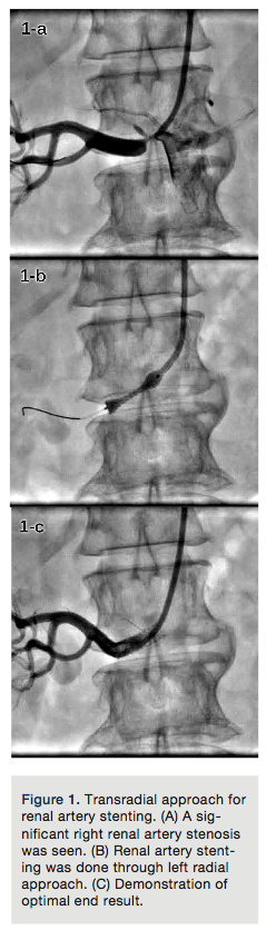 compared with transfemoral approach (TFA), particularly a reduction in puncture-site related bleeding complications. TRA can be utilized effectively to address peripheral vascular lesions, including the renal, iliac, subclavian, carotid, vertebrobasilar, and superficial femoral systems. Utility of TRA for addressing peripheral vascular lesions using different techniques has been discussed in detail. Advantages and limitations of the TRA are highlighted, and the results of different studies using TRA for peripheral interventions are discussed. The pros and cons of TRA versus TFA for peripheral procedures are also demonstrated. The TRA is an effective alternative for TFA to address most peripheral vascular lesion subsets. However, there is a need for the development of radial-specific hardware to track bulky devices.
Reprinted with permission from The Journal of Invasive Cardiology 2015; 27(6): 277-282.
Key words: transradial approach, peripheral vascular stenosis, renal artery stenosis, carotid artery stenosis, peripheral vascular intervention
The transradial approach (TRA) for coronary angiography and interventions is a well-established technique.1-10 It has shown its superiority over the transfemoral approach (TFA) and transbrachial approach (TBA) to establish itself as an effective alternative technique for most coronary intervention subsets.1-10 We now have a genuine reason to assume that most advantages associated with TRA may be reproduced while addressing peripheral vascular lesions, in spite of the absence of supportive 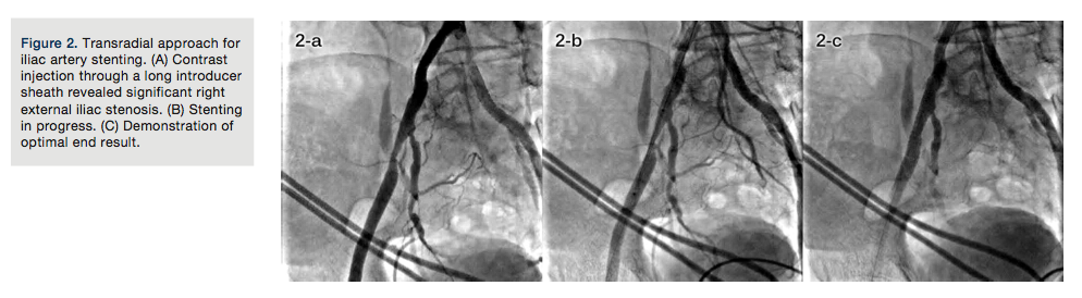 evidence, including randomized comparisons. A few observational studies, feasibility studies, technical reports, case series, and case reports have demonstrated successful application of this technique for addressing peripheral vascular lesions, including internal carotid, vertebral and basilar, subclavian and innominate, renal, iliac, celiac, mesenteric, and superficial femoral artery lesions.11-23 TFA is sometimes precluded by severe local atherosclerosis. In these cases, brachial artery access has been used as an alternative route.24-27 Nonetheless, TBA is also associated with a sizable risk of local complications, especially if performed by operators with limited experience with this arterial entry site.1,28 With this background, TRA for peripheral vascular interventions is gradually emerging as an effective alternative route for TFA.
General TRA benefits
-
Available literature on TRA for coronary interventions has proven beyond a doubt that this approach practically eliminates risk of access-site related vascular complications, such as big hematomas and the need for vascular repair or blood transfusion.1-10,29-33 We now have a genuine reason to assume that this advantage associated with TRA may be reproduced for peripheral vascular lesions, although it needs to be validated by further supportive evidence, including
 randomized comparisons.
 -
For most peripheral vascular interventions, early ambulation is possible using TRA. Early ambulation provides potential cost reduction and economic benefit through various avenues, including expedited room turnover/increased throughput (both through the catheterization laboratory and same-day/recovery unit); decreased intensity of care required by nursing and support staff; shorter length of stay; enhanced ability to perform same-day percutaneous coronary intervention (PCI); and a more rapid return to productivity for working patients.34-37
-
TRA provides an alternative access to TFA in patients with severe lower-limb and/or aortoiliac vascular disease.
General TRA limitations
-
Small radial artery (RA) diameter, RA spasm, significant RA calcification, radial and brachial tortuosities, loops, subclavian tortuosities, arteria lusoria, as well as a dilated and distorted aortic arch may pose technical challenges for advancement of a guide catheter or long introducer sheath. However, there are methods to overcome these issues to a large extent.38-42
-
A lack of dedicated hardware may increase the difficulty of addressing mesenteric or superficial femoral lesions, particularly in very tall patients.16,18,21,22,42 A left radial approach and high puncture should solve this issue in most
 cases.21,22,43
Important considerations
TRA for renal artery stenting. Renal artery stenting techniques have evolved over time, shifting from the use of larger 8 French (Fr) sheaths or guide catheters required to allow the passage of a bulky stent system over .035-inch wires to contemporary 6Fr compatible devices over .014-inch PTCA wires.12,20-22,44 This evolution in hardware actually increased the safety and efficacy of renal artery stenting, leading to its performance in coronary-like fashion and the evolution of TRA as an effective alternative approach for TFA.
Specific benefits.
-
The renal artery most commonly points downward after its origin; thus, TRA offers more co-axial and less traumatic guide catheter cannulation as compared with TFA (Figure 1A).
-
Renal artery diameter usually ranges from 4 to 7mm; 6Fr (or at most, 7Fr) guide-catheter compatible balloons and stents can comfortably be used via TRA (Figures 1B and 1C).
-
A large number of patients undergoing renal artery stenting have uncontrolled hypertension despite high doses of antihypertensive medications. TRA is likely to reduce access-site related bleeding and related complications significantly in this
 situation.
Technical tips.
-
A left radial approach should be preferred for renal artery stenting, because the distance from access to renals is shorter, and less catheter manipulation is usually required, since there is no need to traverse the aortic arch. Thus, practically all renal artery stenting procedures can be performed using regular-length multipurpose (MP) or Judkins Right (JR) guide catheters.
-
However, a right radial approach can also be effectively and safely used for renal artery stenting by using 125cm-long, 6Fr MP or JR guide catheters.
TRA for iliac artery stenting. Approximately one-third of obstructive lesions in peripheral arterial occlusive disease affect the aortoiliac segment.17,45 Iliac artery obstructions have traditionally been treated with open surgery. However, percutaneous transluminal angioplasty (PTA) with stenting has emerged as a less invasive alternative treatment and has proven an effective technique for the treatment of patients with focal iliac artery stenosis.17,20-22 Although TFA is time tested and preferred, TRA is a good alternative approach.17,20-22,45
Technical tips.
-
The left radial is a preferred approach, because it eliminates extra length and tortuosity of the aortic arch, allowing relatively easy tracking and deployment of a long 6Fr sheath.

-
After obtaining left radial access with a 5Fr introducer sheath, an internal mammary artery catheter is advanced over a long (300cm) .035-inch guidewire into the descending aorta and then the entire system is exchanged for a long (usually 100cm or 110cm) 6Fr introducer sheath. The sheath is positioned at the ostium of the culprit common iliac artery or in the distal aorta just above the bifurcation; the procedure is then performed in the usual fashion (Figure 2A).
-
Depending on operator experience and hardware availability, balloon-expandable or self-expandable stents can be chosen (Figures 2B and 2C).
Specific limitations.
-
Deployment of a long sheath in the descending aorta or iliac system via right TRA is sometimes more difficult and cumbersome.
Future directions.
-
Low-profile iliac stents that are compatible with 6Fr or 7Fr guide catheters shall eliminate the need for a long introducer sheath, making this procedure even more simple.
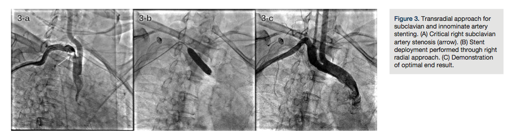
TRA for subclavian and innominate artery stenting. Occlusive lesions of the innominate and subclavian arteries might cause serious morbidity and should be treated if symptomatic. Endovascular treatment is recommended because it has a lower complication rate compared with surgical methods.46-48 Most studies have used the femoral approach; however, some have used brachial and axillary approaches.46,47 Ipsilateral TRA is a feasible and reproducible approach (Figures 3A, 3B, and 3C).
Technical tips.
-
45cm-long, 6Fr or 7Fr introducer sheaths should be used for ipsilateral TRA, and the distal tip should be kept near the lesion.
-
While addressing innominate artery stenosis, care should be exercised in placing the stent, so that it does not protrude much into the aorta or into the orifice of the right common carotid artery.
-
For the bovine arch variation, care should be taken not to stent across the origin of the left common carotid artery.
-
Avoid stenting across the vertebral artery origin, as it could increase the likelihood of vertebral artery occlusion and/or brain embolization.
-
Avoid deploying the stent across the origin of the internal mammary artery.
Future directions.
-
Further miniaturization of hardware will increase procedural safety and operator comfort level.
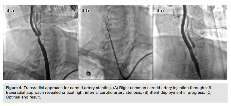
TRA for carotid artery stenting. Carotid artery stenting is an accepted treatment modality as an alternative to carotid endarterectomy for revascularization of atherosclerotic internal carotid artery stenosis in patients who are at high risk for surgery.14,15 Traditionally, the femoral approach is utilized with acceptable success rates. Small feasibility studies using ipsilateral and contralateral TRA have been published.14,15,32,49
Technical tips.
-
The 5Fr Simmons-1 catheter (Terumo) is a workhorse catheter to cannulate the common carotid artery using both ipsilateral and contralateral TRA. For contralateral TRA, the 5Fr Tig-1 Optitorque catheter (Terumo) is an alternative catheter.
-
A .035-inch Amplatz super-stiff wire is parked in the external carotid artery or deep in the common carotid artery away from the origin of the internal carotid artery.
-
Aim at optimal co-axial positioning of a 6Fr or 7Fr carotid sheath of your choice in the common carotid artery (Figure 4A).
-
The remaining steps of the procedures are the same as with TFA (Figures 4B and 4C).
Future directions.
-
Further miniaturization of hardware is necessary.
-
Development of carotid-specific curves of large-bore sheathless guide catheters may resolve advancement-related and cannulation-related issues.14
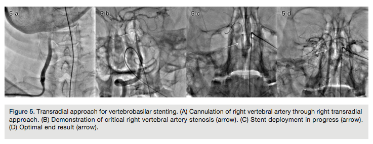
TRA for vertebrobasilar stenting. Endovascular intervention of vertebrobasilar stenosis is a relatively new alternative modality of management, supported by very few case reports and studies.13,50 TFA has been the preferred approach in most studies. However, a recent study has documented the feasibility and reproducibility of the TRA in chronic as well as acute occlusions of the vertebrobasilar system.13
Technical tips.
-
A 6Fr internal memory artery guide catheter through ipsilateral TRA easily cannulates the vertebral artery ostium. The same guide catheter with side-holes should be used to address ostial vertebral artery stenosis (Figures 5A and 5B).
-
Regular PTCA guidewires, balloon catheters, and coronary stents should be used (Figures 5C and 5D).
-
Predilation of the lesion should be done with low pressure (between 4 and 6atm).
-
The stent should be deployed at low pressure (8-10atm).
-
The procedure should be divided into two stages for acute intracranial vertebral or basilar artery occlusions. In the first stage, the lesion should be dilated using a 1.5x10mm or 1.5x12mm PTCA catheter at 4-6atm in order to establish distal flow. In the second stage, the patient should be brought to the catheterization laboratory after 24 hours and the lesion should be stented using a coronary stent at 8-10atm. This strategy should be used to prevent hyperperfusion brain injury.13
Future directions.
-
Studies involving larger patient populations are necessary to establish safety, efficacy, and reproducibility of this approach, and also to standardize balloon dilatation and stent implantation pressures.
-
More data will be generated to establish prevention strategies of hyperperfusion brain injury.
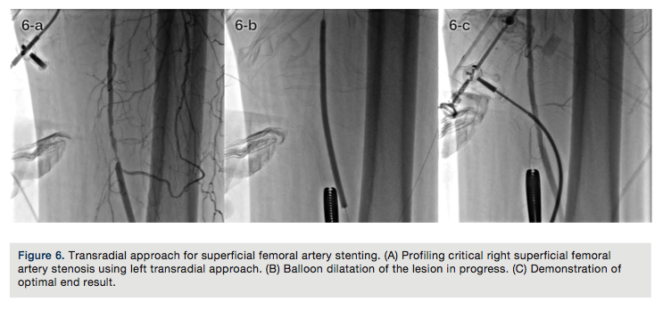
TRA for stenting of other important peripheral vascular lesions. Endovascular management of celiac, mesenteric, and superficial femoral arterial systems has been described.16,18-22 TRA is an emerging approach for addressing vascular lesions of these systems, as evident by a few case reports.16,18
Technical tips.
-
Left TRA should be the preferred approach to avoid the aortic arch and related issues.
-
The celiac trunk is usually at a similar distance as the renal artery from radial puncture site; hence, its lesions can be addressed using regular length guide catheters. This distance is longer for mesenteric arteries, necessitating the use of long guide catheters (125cm).
-
Mesenteric arteries arise at a very acute downward angle, especially in very thin patients, leading to easy co-axial cannulation with diagnostic or guide catheters via TRA.22
-
Sometimes, when mesenteric artery diameter is very large, a long (100cm or 110cm) 6Fr or 7Fr introducer sheath should be brought to the descending aorta. A 6Fr guide catheter should be used to selectively cannulate the culprit mesenteric system. After crossing the lesion with an extra-support .014-inch PTCA or .018-inch PTA wire, the guide catheter can be carefully removed and a self-expandable large diameter stent can be negotiated on the same wire and deployed across the lesion.
-
A lateral view is useful for celiac and mesenteric systems because they most often arise anteriorly from the aorta.
-
Superficial femoral artery lesions should be approached via the left TRA with 125cm-long, 6Fr MP or JR guide catheters, 300cm-long .014-inch PTCA guidewires, and long-shaft PTA catheters (Figures 6A, 6B, and 6C).18,20-22
Future directions.
-
There is a need for the development of dedicated hardware to address lesions in these areas.
Conclusion
TRA is emerging as a useful tool for most peripheral vascular interventions, offering the advantages of very low local vascular and bleeding complications, higher patient and staff comfort, rapid turnover, and lower hospital costs.
Once you have an established transradial program for percutaneous coronary intervention, you have a good reason to develop a transradial program for peripheral vascular lesions. Having a good reason is essential to launching a successful program, because it sparks the urge to get training, fosters support in the catheterization laboratory, and encourages operators to overcome a new “learning curve” that is more challenging for neuro-interventions and easier for other vascular interventions.51 Although technical refinements of TRA have been done to a great extent, technological breakthroughs are awaited. Industry needs to focus on the development of dedicated hardware to address specific peripheral vascular lesions, so the technique becomes more simple and easier to reproduce.
Acknowledgment. The authors are grateful to Mr Yash Soni and Mr Chidambaram Iyer for their extremely valuable support during preparation of this manuscript.
References
-
Kiemeneij F, Laarman GJ, Odekerken D, et al. A randomized comparison of percutaneous transluminal coronary angioplasty by the radial, brachial and femoral approaches: the ACCESS study. J Am Coll Cardiol. 1997;29:1269-1275.
-
Philippe F, Larrazet F, Meziane T, et al. Comparison of transradial vs transfemoral approach in the treatment of acute myocardial infarction with primary angioplasty and abciximab. Catheter Cardiovasc Interv. 2004;61:67-73.
-
Cooper CJ, El-Shiekh RA, Cohen DJ, et al. Effect of transradial access on quality of life and cost of cardiac catheterization: a randomized comparison. Am Heart J. 1999;138(3 Pt 1):430-436.
-
Hildick-Smith DJ, Walsh JT, Lowe MD, Shapiro LM, Petch MC. Transradial coronary angiography in patients with contraindications to the femoral approach: an analysis of 500 cases. Catheter Cardiovasc Interv. 2004;61:60-66.
-
Louvard Y, Lefèvre T, Allain A, Morice MC. Coronary angiography through the radial or the femoral approach: the CARAFE study. Catheter Cardiovasc Interv. 2001;52:181-187.
-
Louvard Y, Benamer H, Garot P, et al; the OCTOPLUS study group. Comparison of transradial and transfemoral approaches for coronary angiography and angioplasty in octogenarians (the OCTOPLUS study). Am J Cardiol. 2004;94:1177-1180.
-
Mann T. Stenting in acute coronary syndromes: a comparison of radial versus femoral access sites. J Am Coll Cardiol. 1998;32:572-576.
-
Slagboom T, Kiemeneij F, Laarman GJ, Wieken R, Odekerken D. Actual outpatient PTCA: results of the OUTCLAS pilot study. Catheter Cardiovasc Interv. 2001;53:204-208.
-
Jolly SS, Amlani S, Hamon M, Yusuf S, Mehta SR. Radial versus femoral access for coronary angiography or intervention and the impact on major bleeding and ischemic evens: a systematic review and meta-analysis of randomized trials. Am Heart J. 2009;157:132-140.
-
Agostoni P, Biondi-Zoccai GG, de Benedictis ML, et al. Radial versus femoral approach for percutaneous coronary diagnostic and interventional procedures: systematic overview and meta-analysis of randomized trials. J Am Coll Cardiol. 2004;44:349-356.
-
Sanghvi K, Kurian D, Coppola J. Transradial intervention of iliac and superficial femoral artery disease is feasible. J Interv Cardiol. 2008;2:385-387.
-
Trani C, Tommasino A, Burzotta F. Transradial renal stenting: why and how. Catheter Cardiovasc Interv. 2009;74:951-956.
-
Patel T, Shah S, Malhotra H, Radadia R, Shah L, Shah S. Transradial approach for stenting of vertebro basilar stenosis: a feasibility study. Catheter Cardiovasc Interv. 2009;74:925-931.
-
Patel T, Shah S, Ranjan A, Malhotra H, Pancholy S, Coppola J. Contralateral transradial approach for carotid artery stenting: a feasibility study. Catheter Cardiovasc Interv. 2010;75:268-275.
-
Folmar J, Sachar R, Mann T. Transradial approach for carotid artery stenting: a feasibility study. Catheter Cardiovasc Interv. 2007;69:355-361.
-
Patel T, Kuladhipati I, Shah S. Successful percutaneous endovascular management of acute post-traumatic superior mesenteric artery dissection using a transradial approach. J Invasive Cardiol. 2010;22:E61-E64.
-
Staniloae CS, Korabathina R, Yu J, Kurian D, Coppola J. Safety and efficacy of transradial aortoiliac interventions. Catheter Cardiovasc Interv. 2010;75:659-662.
-
Trani C, Burzotta F, Tommasino A, et al. Transradial approach to treat supportive femoral artery in-stent stenosis. Catheter Cardiovasc Interv. 2009;74:494-498.
-
Yamashita T, Tmai S, Tamada T, et al. Transradial approach for non-coronary angiography and interventions. Catheter Cardiovasc Interv. 2007;70:303-308.
-
Patel T, Shah S, Ranjan A. Appendix 1. In: Patel’s Atlas of Transradial Intervention: The Basics. Garzona C (ed). Hobe Sound, FL: Seascript Company; 2007:191-194.
-
Patel T, Shah S, Ranjan A. Chapter 13. In: Patel’s Atlas of Transradial Intervention: The Basics. Garzona C (ed). Hobe Sound, FL: Seascript Company; 2007:177-180.
-
Louvard Y, Chawla K, Lefevre T. Transradial approach for non-coronary diagnosis and interventions. Indian Heart J. 2008;60(Suppl A):45A-50A.
-
Staniloae CS, Korabathina R, Coppola JT. Transradial access for peripheral vascular interventions. Catheter Cardiovasc Interv. 2013;81:1194-1203.
-
Lorenzoni R, Roffi MJ. Transradial access for peripheral and cerebrovascular interventions. J Invasive Cardiol. 2013;25:529-536.
-
Sievert H, Ensslen R, Fach A, et al. Brachial artery approach for transluminal angioplasty of the internal carotid artery. Cathet Cardiovasc Diagn. 1996;39:421-423.
-
Kaukanen ET, Manninen HI, Matsi PJ, Soder HK. Brachial artery access for percutaneous renal artery interventions. Cardiovasc Intervent Radiol. 1997;20:353-358.
-
Ernst S, Fischbach R, Brochhagen HG, Heindel W, Landwehr P. Transbrachial thrombolysis, PTA and stenting in the lower extremities. Cardiovasc Intervent Radiol. 2003;26:516-521.
-
Hildick-Smith DJ, Khan ZI, Shapiro LM, Petch MC. Occasional-operator percutaneous brachial coronary angiography: first, do no arm. Catheter Cardiovasc Interv. 2002;57:161-165.
-
Rao SV, Ou FS, Wang TY, et al. Trends in the prevalence and outcomes of radial and femoral approaches to percutaneous coronary intervention: a report from the National Cardiovascular Data Registry. JACC Cardiovasc Interv. 2008;1:379-386.
-
Yatskar I, Selzer F, Feit F, et al. Access site hematoma requiring blood transfusion predicts mortality in patients undergoing percutaneous coronary intervention: data from the National Heart, Lung, and Blood Institute Dynamic Registry. Catheter Cardiovasc Interv. 2007;69:961-966.
-
Chase AJ, Fretz EB, Warburton WP, et al. Association of the arterial access site at angioplasty with transfusion and mortality, the MORTAL study (Mortality benefit Of Reduced Transfusion after percutaneous coronary intervention via the Arm or Leg). Heart. 2008;94:1019-1025.
-
Vavalle JP, Rao SV. The association between the transradial approach for percutaneous coronary interventions and bleeding. J Invasive Cardiol. 2009;21:21A-24A.
-
Etxegoien N, Rhyne D, Kedev S, et al. The transradial approach for carotid artery stenting. Catheter Cardiovasc Interv. 2012;80:1081-1087.
-
Ziakas A, Klinke P, Fetz E, et al. Same-day discharge is preferred by the majority of the patients undergoing radial PCI. J Invasive Cardiol. 2004:16:562-565.
-
Kiemeneij F. My home is my castle. J Invasive Cardiol. 2004:16:566.
-
Caputo R. Transradial arterial access: economical considerations. J Invasive Cardiol. 2009;21:18A-20A.
-
Chen JP. All roads (even those less traveled) lead to Rome: benefits of transradial coronary interventions. JACC Cardiovasc Interv. 2010;3:259.
-
Coppola J, Patel T, Kwan T, et al. Nitroglycerin, nitroprusside, or both, in preventing radial artery spasm during transradial artery catheterization. J Invasive Cardiol. 2006;18:155-158.
-
Louvard Y, Lefevre T. Loops and transradial approach in coronary diagnosis and intervention. Catheter Cardiovasc Interv. 2000;51:250-252.
-
Gilchrist IC. Transradial technical tips. Catheter Cardiovasc Interv. 2000;49:353-354.
-
Pancholy SB, Coppola J, Patel T. Subcutaneous administration of nitroglycerin to facilitate radial artery cannulation. Catheter Cardiovasc Interv. 2006;68:389-391.
-
Patel T, Shah S, Ranjan A. Chapters 6-11. In: Patel’s Atlas of Transradial Intervention: The Basics. Garzona C (ed). Hobe Sound, FL: Seascript Company; 2007:40-154.
-
Saito S. Right or left side? Catheter Cardiovasc Interv. 2003;58:301-304.
-
Sanghvi K, Coppola J, Patel T. Cranio-caudal (transradial) approach for renal artery intervention. J Interv Cardiol. 2013;26:530-535.
-
Gandini R, Fabiano S, Chiocchi M, et al. Percutaneous treatment in iliac artery occlusion: long-term results. Cardiovasc Intervent Radiol. 2008;31:1069-1076.
-
Brountzos E, Petersen B, Binkert C, et al. Primary stenting of subclavian and innominate artery occlusive disease: a single center’s experience. Cardiovasc Intervent Radiol. 2004;27:616-623.
-
Brountzos E, Malagari K, Kelekis D. Endovascular treatment of occlusive lesions of the subclavian and innominate arteries. Cardiovasc Intervent Radiol. 2006;29:503-510.
-
Yu J, Korabathina R, Coppola J, et al. Transradial approach to subclavian artery stenting. J Invasive Cardiol. 2010;22:204-206.
-
Turgeman Y, Suleiman K, Feldman A, et al. Transradial diagnosis and intervention of supraaortic vessels. J Invasive Cardiol. 2013;25:300-303.
-
Jinkins JS, Patel SN, White CJ, et al. Endovascular stenting of vertebral artery stenosis. J Am Coll Cardiol. 2010;55:538-542.
-
Trammel J. Launching a successful transradial program. J Invasive Cardiol. 2009;21:3A-8A.


