


Finalists From CRT’s 2024 Nurses and Technologists Interesting Cases Competition
The Cardiovascular Research Technologies (CRT) meeting was held March 9-12 in Washington, D.C.
The Cardiovascular Research Technologies (CRT) meeting was held March 9-12 in Washington, D.C.
© 2024 HMP Global. All Rights Reserved.
Any views and opinions expressed are those of the author(s) and/or participants and do not necessarily reflect the views, policy, or position of Cath Lab Digest or HMP Global, their employees, and affiliates.
[Editor's Note: A free pdf is available for download to the right of this article - look for the red PDF icon]
New at the CRT meeting in 2024 was a competition focused on cases submitted by nurses and technologists, with four finalists asked to present their cases March 10th during CRT’s Nurses and Technologists session.
 “This debut session at CRT garnered substantial praise and positive feedback from attendees,” said Dionne Ross, MSN, RN, NE-BC, MedStar Heart and Vascular Institute Cath Lab Nurse Director, who introduced the session. “The presenters demonstrated remarkable skill, especially considering it was their first time presenting in this setting. Excitement is palpable for next year’s presenters.”
Cath Lab Digest partnered with CRT to celebrate the finalists and share the four cases with readers.
The winners (in alphabetical order) are:
• Arteria Lusoria: Fluoroscopic Recognition From the Right Radial Approach and Tips for Selective Coronary Engagement and Guide Support for PCI
Richard Casazza, MAS, RT(R)(CI)
• Supersaturated Oxygen Therapy: The Next Frontier in STEMI Treatment
Tahnee M. Clark, MSN, AGACNP-BC
• Peripheral Stent Embolization: What is the Best Treatment Approach?
Chenoa Flanagan, RCIS, BA
• Not Another Straight Fix: Stenting a Kinked LVAD Outflow Graft
Brandy D. Smith, RCIS, LMRT
Read the four cases below and plan to submit your own case next year! CRT 2025 is scheduled to take place March 8-11, 2025, at the Washington Hilton Hotel in Washington, D.C.
Learn more at CRTmeeting.org
Arteria Lusoria: Fluoroscopic Recognition From the Right Radial Approach and Tips for Selective Coronary Engagement and Guide Support for PCI
Arteria lusoria (Figure 1) or aberrant right subclavian artery is the most common embryologic abnormality of the aortic arch. It occurs in 0.2%-1.7% of the population.1 When an aberrant subclavian is identified, the brachiocephalic trunk is absent and four great vessels arise from the arch of the aorta: the right common carotid artery, the left common carotid artery, the left subclavian artery, and lastly, the right subclavian, which has a distal left-sided origin. Arteria lusoria, literally translating to “arterial freak of nature”, can be a nightmare for an interventional cardiologist to deal with from the right radial approach. In a perfect world, all patients would have previous computed tomography (CT) scans identifying this quagmire beforehand. Unfortunately, that is not the case. Anatomic variation of the arterial pathway has an adverse impact on transradial coronary procedural outcomes.
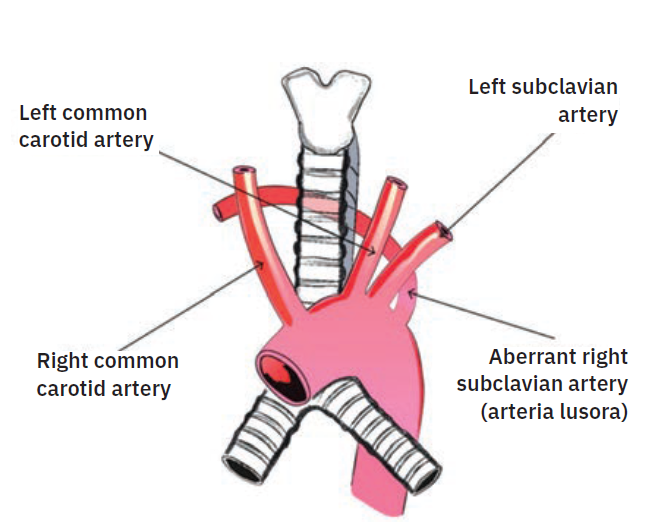
aberrant right subclavian artery (arteria lusoria).
The “Shark’s Tooth”
Radiographically, when encountering right subclavian tortuosity, catheters for the most part cross to the left side of the trachea, and can have an “S”-shaped rounded curve or resemble the profile view of an elephant’s head. Right subclavian tortuosity in and of itself can make an easy procedure difficult and laborious. Encountering an aberrant right subclavian amplifies the difficulties further. Patients with an aberrant subclavian done from the radial approach are shown to only have a procedural success rate of 60%2, although this source from Valsecchi et al is somewhat antiquated and techniques dealing with tortuous anatomy have evolved significantly. However, an aberrant subclavian can still put a significant damper on a proceduralist’s day.
Although encountering an aberrant right subclavian during cardiac catheterization is quite infrequent, operators still do encounter it. For the most part, this congenital anomaly is unknown prior to the cath and is typically identified when a right radial approach is utilized. Upon entering the aorta, catheters frequently go into the descending aorta (due to the orientation and location of the junction of the right subclavian and aorta), which may be misconstrued as severe tortuosity.
With “standard” right subclavian tortuosity, the catheter crosses the right to left side of the trachea and has a rounded-out shape. Catheters also move inferiorly for a few centimeters before moving back right towards the trachea again, prior to reaching the sinotubular junction. In contrast, with an aberrancy, the catheter crosses the trachea and travels significantly left past the trachea and abruptly turns back to the right, towards the transverse and ascending aorta that is delineated quite well with an left anterior oblique (LAO)/caudal view. The catheter may also follow a superior trajectory before reaching and/or cannulating the coronary ostia.
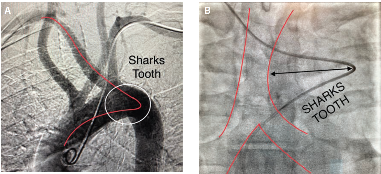
sign in patient with arteria lusoria.
Radiographically, all have a similar appearance (Figure 2A-B). They move left across the trachea, well into the lung field, and make an acute trajectory change back to the right, towards the transverse and ascending aorta. This abrupt course of the catheter change resembles a “shark’s tooth” and as a radiographic sign, may offer the clue of an aberrant subclavian. Different wires may enable easier negotiation into the ascending aorta. Even if operators get close to the coronary ostia, tension and energy built up in the catheter can disallow selective engagement and force a bailout to the contralateral radial or the femoral approach. Conversely, there are times in the presence of this ominous anatomy when catheters will engage quicker than the operator can do a “time out.” A skillful operator might display some “catheter legerdemain” to negotiate these problems; however, a little bit of luck also goes a long way. One doesn’t necessarily have to be a funambulist to recognize and selectively cannulate the coronaries.
References
1. Patel T, Shah S, Pancholy S, et al. Working through challenges of subclavian, innominate, and aortic arch regions during transradial approach. Catheter Cardiovasc Interv. 2014 Aug 1; 84(2): 224-235. doi:10.1002/ccd.25418
2. Valsecchi O, Vassileva A, Musumeci G, et al. Failure of transradial approach during coronary interventions: anatomic considerations. Catheter Cardiovasc Interv. 2006 Jun; 67(6): 870-878. doi:10.1002/ccd.20732
Note: This case was originally published as an expanded version in the April 2022 issue of Cath Lab Digest as “Fluoroscopic Recognition of Arteria Lusoria When Utilizing the Right Radial Approach During Cardiac Catheterization: ‘Shark’s Tooth’ Sign”.
Supersaturated Oxygen Therapy: The Next Frontier in STEMI Treatment

A 50-year-old gentleman with a prior medical history significant for tobacco use, ceased 15 years ago, presented to an outside medical facility following a witnessed cardiac arrest while vacationing in the Florida Keys. Cardiopulmonary resuscitation was initiated by the patient’s spouse until emergency medical services arrival, during which the patient was found to be in ventricular fibrillation. Following the second defibrillation attempt, the patient successfully converted to sinus rhythm. The patient was subsequently transferred to the Miami Cardiac and Vascular Institute (MCVI), intubated, sedated, hemodynamically stable, and devoid of the need for vasopressor or inotropic support. Laboratory analysis indicated leukocytosis, acute kidney injury, and elevated high-sensitivity troponin levels peaking at 71,050. Electrocardiogram findings revealed sinus rhythm with marked anterior ST-elevations and reciprocal changes in lead III.
The patient was emergently taken to the cardiac catheterization lab where a coronary angiogram revealed a total occlusion to the mid left anterior descending artery. A cardiac stent was successfully deployed, followed by a one-hour infusion of supersaturated oxygen (SSO2) therapy (ZOLL Medical), maintaining an activated clotting time >300 seconds. Additionally, due to cardiogenic shock, an Impella CP device (Abiomed) was inserted. Transthoracic echocardiogram (TTE) findings revealed apical and antero-apical akinesis with an ejection fraction (EF) of 25%-30%. On hospital day one, the Impella was explanted due to hemolysis and treatment with a milrinone infusion was initiated. On hospital day two, the patient was successfully extubated and the milrinone infusion was notably discontinued. On hospital day three, the patient was transferred out of the intensive care unit and cardiac magnetic resonance imaging revealed near transmural late gadolinium enhancement involving the anterior and anterolateral wall of the mid ventricle, extending to involve the anterior apical, lateral apical, and inferior apical segments and apex, illustrating the impact of SSO2 therapy.
A repeat TTE on hospital day four demonstrated mild apical hypokinesis with an improved EF of 45%-50% and the patient was discharged home on guideline-directed medical therapy.
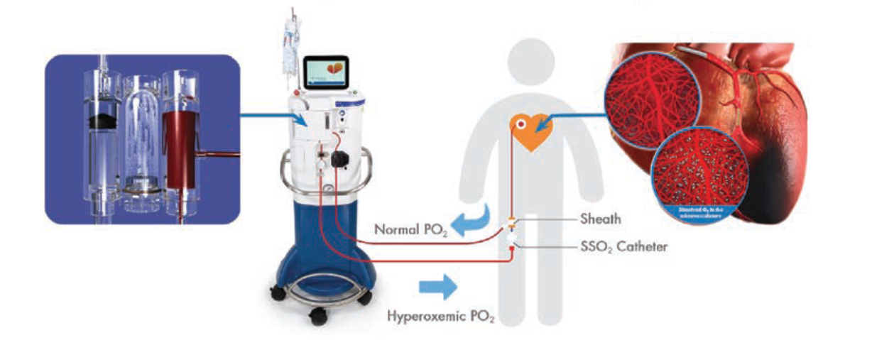
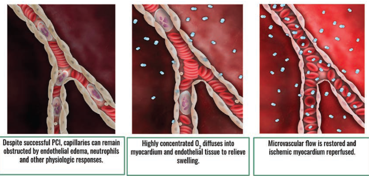
The aforementioned clinical scenario represents 1 of 33 patients since May 2021 who have received SSO2 therapy at MCVI. SSO2 therapy is indicated for ST-elevated myocardial infarction (STEMI) patients undergoing primary percutaneous coronary intervention (PCI) for anterior myocardial infarction (MI) presenting within six hours of symptoms onset. It is a one-time, 60-minute localized hyperoxemic oxygen infusion to the coronaries following an acute MI, and has been found to help reduce infarct size post PCI and prevent heart failure in this population of patients. The therapy delivers 7-10 times the normal oxygen level to the heart to enhance microvascular blood flow post PCI, with no impact on door-to-balloon times. In a broad context, the time taken to deliver SSO2 therapy with the ability to augment patient outcomes is minute. SSO2 therapy is the first FDA-approved, catheter-based therapy to demonstrate a significant reduction in infarct size of approximately 26%.1
Despite successful PCI, endothelial edema, neutrophils, and blebs caused by hypoxia continue to restrict microvascular flow in the infarct zone. SSO2 therapy helps to restore microvascular flow by delivering hyperoxemic levels of dissolved oxygen via the patient’s own plasma to focally reduce endothelial swelling, thereby improving flow. When microvascular flow is restored, the myocardium is reperfused, nourishing and healing endothelial tissue.
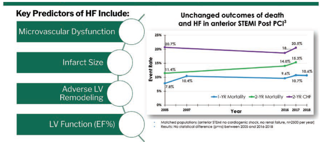
Despite current advances in PCI, a strong need for improving patient outcomes in acute MIs still exists. Heart attacks lead to heart failure: 20%-30% at one year, and 71% within five years. Mortality and heart failure rates after an MI treated with PCI have remained unchanged or even increased since 2005.2 What has been observed in terms of blood flow in this population of patients is surface level; 90% of myocardial flow is in the microvasculature.3,4 SSO2 therapy can be utilized to reduce the development of heart failure, as it has shown a clinically significant median reduction in infarct size, left ventricular remodeling, and improved microvasculature flow.5,6
References
1. Stone GW, Martin JL, de Boer MJ, et al; AMIHOT-II Trial Investigators. Effect of supersaturated oxygen delivery on infarct size after percutaneous coronary intervention in acute myocardial infarction. Circ Cardiovasc Interv. 2009 Oct; 2(5): 366-375.
2. Zandecki Ł, Sadowski M, Janion M, et al. Survival benefit from recent changes in management of men and women with ST-segment elevation myocardial infarction treated with percutaneous coronary interventions. Cardiol J. 2019; 26(5): 459-468.
3. Kaul S, Ito H. Microvasculature in acute myocardial ischemia: part I: evolving concepts in pathophysiology, diagnosis, and treatment. Circulation. 2004 Jan 20; 109(2):146-149.
4. Morris PD, Gosling R, Zwierzak I, et al. A novel method for measuring absolute coronary blood flow and microvascular resistance in patients with ischaemic heart disease. Cardiovasc Res. 2021 May 25; 117(6): 1567-1577.
5. Warda HM, Bax JJ, Bosch JG, et al. Effect of intracoronary aqueous oxygen on left ventricular remodeling after anterior wall ST-elevation acute myocardial infarction. Am J Cardiol. 2005 Jul 1;96(1): 22-24.
6. David SW, Khan ZA, Patel NC, et al. Evaluation of intracoronary hyperoxemic oxygen therapy in acute anterior myocardial infarction: The IC-HOT study. Catheter Cardiovasc Interv. 2019 Apr 1; 93(5): 882-890.
Read more on SSO2 in CLD:
Baptist Health’s Miami Cardiac & Vascular Institute Experience With SuperSaturated Oxygen (SSO2) Therapy to Improve Outcomes in STEMI Patients
Peripheral Stent Embolization: What is the Best Treatment Approach?

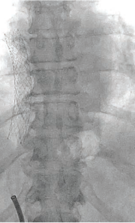
A 56 year-old-male with hypertension, type II diabetes mellitus, and hyperlipidemia was referred from an outside hospital for endovascular percutaneous catheterization retrieval. He had recently undergone right iliac vein intervention using an Abre 14.0 mm x 120 mm nitinol self-expanding stent (Medtronic) and was following up for an iliac vein extension. On fluoroscopy, the iliac stent could not be seen, a finding which confirmed the suspicion of a stent embolism. It was ultimately found to have migrated to the junction of the inferior vena cava and the right atrium.
Two options were considered, the first being a surgical approach, which would require the patient to undergo open-heart surgery. The other option was to treat it by percutaneous catheterization, either with complete capture and retrieval or by relocation and trapping. A major concern was preventing the stent from embolizing into the right ventricle, which would be detrimental to the patient. A strategy was devised for the complete capture and retrieval of the stent.
Initially, we attempted to snare the stent from both sides via two vascular accesses. To snare the upper portion of the stent, a 20 mm Amplatz Goose Neck snare (Medtronic) was introduced through a 23 cm 8 French (F) sheath in the right internal jugular vein. Simultaneously, a 30 mm Amplatz Goose Neck snare was introduced through a 30 cm 14F sheath in the right femoral vein to snare the bottom part of the stent. This method of stent retrieval was not successful; therefore, we had to rethink our strategy.
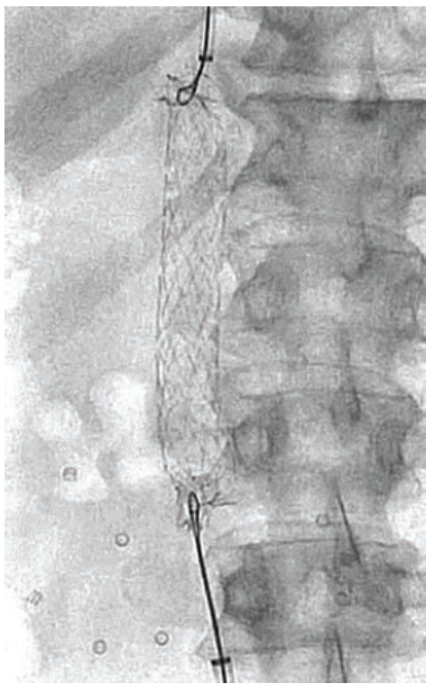
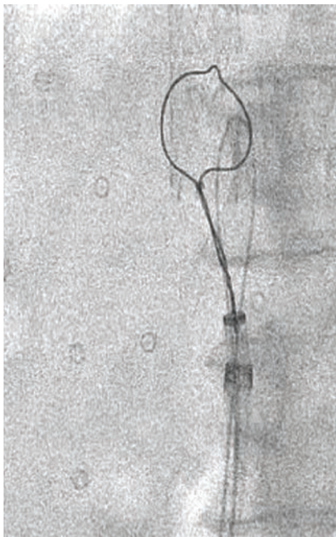
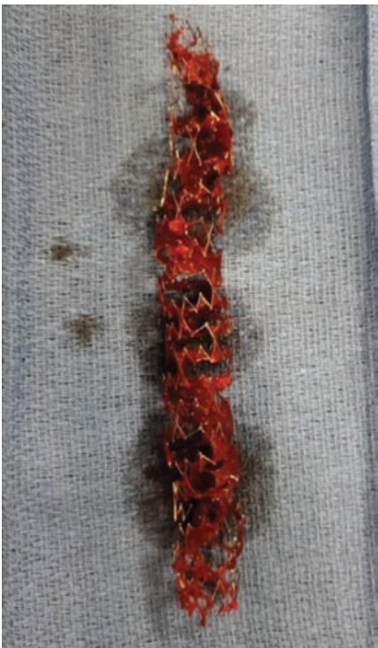
We upsized the 14F sheath to 24F and placed a second 20F sheath within it. We put a third 16F sheath inside these two previous 24F and 20F sheaths. Dr. Nelson Bernardo, an interventional cardiologist at Medstar Washington Hospital Center, practices this technique, known as mother-child-grandchild. The two snares described previously were used to capture both sides of the stent with the top snare pulling from the top and the bottom snare pulling from below, preventing the stent from embolizing into the right ventricle and slowly bringing it down into the inferior vena cava. The traction of the stent’s edge elongated it and made it smaller, which allowed us to capture its middle portion with a third 35 mm Amplatz Goose Neck snare. The stent was then pulled as a unit into the 16F sheath. Then the stent was pulled together with the 16F and 20F sheaths, taking them out of the 24F sheath as a single unit. A repeat fluoroscopy was performed to ensure that no residual pieces were left behind, and an angiogram was performed to confirm that there was no evidence of dissection. Upon examination, the stent was found to be intact and to have been endothelialized much earlier than expected. In the common femoral vein, hemostasis was achieved using a Perclose (Abbott). While in the internal jugular vein, hemostasis was achieved by manual compression.
The biggest lesson I learned from this case was that if you want to change the function, you have to change the structure. Changing the structure of the stent allowed for successful retrieval, and endovascular percutaneous catheterization proved to be the best option for this patient.
Not Another Straight Fix: Stenting a Kinked LVAD Outflow Graft

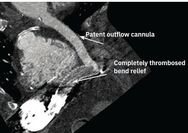
Providing high-quality care in novel situations is one of the most rewarding aspects of working in interventional cardiology. A 62-year-old woman with a HeartMate 3 Left Ventricular Assist Device (LVAD) (Abbott) implanted two years prior presented with a drive-line infection leading to pneumonia and intubation. She had low flows despite three pressors and LVAD controller adjustments. Due to the unclear etiology of persistent low flows, a 4-dimensional heart scan was ordered. It showed a kinked, possibly thrombotic outflow graft, with decreased flow proximally, patent flow distally, and concern for thrombus within the graft.
The patient was not a surgical candidate. The multidisciplinary team discussed her case. The team included heart failure, surgery, interventional cardiology, palliative care physicians, an LVAD coordinator, and the patient’s social worker. Together with the patient’s family, they agreed that percutaneous intervention of the outflow graft to relieve the obstruction and improve LVAD flow was the best course of action. The team’s technical considerations included using a cerebral protection device to prevent distal embolization and ensuring the wire did not advance into the pump housing.
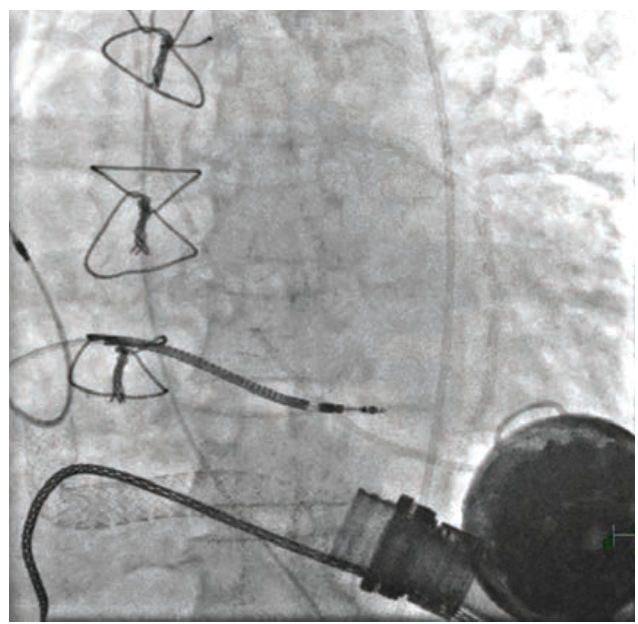
The cath lab team began by deploying the cerebral protection device filters in the right brachiocephalic and left common carotid arteries via a 6 French (F) radial sheath. Access to the right common femoral artery was obtained with ultrasound and micropuncture. Following a pre-closure device, a 12F sheath was inserted. The LVAD speed was 0.8 L/min, and the patient’s mean arterial pressure (MAP) was 75 mmHg with pressors. The outflow graft was cannulated with a Stiff Glidewire and a 5F GlideCath (both Terumo Interventional Systems). The shorter sheath was exchanged for a Shuttle sheath (Cook Medical), and an injection was performed through the GlideCath. This image showed a clear obstruction at the bend relief space, causing the kink and low flows.
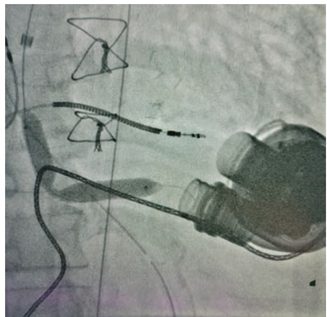
An 11 mm x 16 mm covered stent was placed, followed by an .035-inch 10 mm x 40 mm balloon, but the kink remained. A 12 mm x 40 mm percutaneous transluminal angioplasty (PTA) balloon was inserted and the kink gave. However, the flows did not improve and, in fact, temporarily dropped.
At this point, it was clear that the patient did not have thrombus inside the graft. Typically, thrombus formation occurs early post LVAD implantation. Pannus formation outside the LVAD outflow graft caused the kink. Pannus formation is a chronic process marked by tissue ingrowth containing proteinaceous particles that form outside grafts.
The precipitous drop in flow was caused by the pannus shifting toward the proximal outflow graft near the pump housing. The team quickly deployed an 11 mm x 16 mm stent to support the proximal graft, followed by a 12 mm x 40 mm PTA balloon. The patient’s LVAD flow improved to a remarkable 4.5 L/min.
All equipment was removed, and closure was completed using the single pre-closure device and a radial band. The patient did not have any bleeding or hematomas. Her flows remained stable, and all pressors were weaned. She continued to improve and was discharged to a skilled nursing facility.
LVADs are valuable tools in the treatment of end-stage heart failure. The MOMENTUM 3 study demonstrated that continuous flow HeartMate 3 LVAD patients had a 79% survival rate at two years and 54% at five years.1 The expectation is that patients will live longer and have a higher quality of life with LVADs. Complications that arise, such as device thrombus or pannus, need to be recognized and managed appropriately. Pannus formation is not a widely cited cause of LVAD failure; however, possible pannus etiology should be considered with LVAD low flows. PCI of the LVAD outflow graft is a reasonable treatment option, particularly for patients who are not surgical candidates.
Reference
1. Mehra MR, Goldstein DJ, Cleveland JC, et al. Five-year outcomes in patients with fully magnetically levitated vs axial-flow left ventricular assist devices in the MOMENTUM 3 randomized trial. JAMA. 2022 Sep 27; 328(12): 1233-1242.
