

Role of Diastology in Transcatheter Aortic Valve Implantation
J INVASIVE CARDIOL 2018;30(3):E23-E24.
Key words: aortic regurgitation, Doppler echocardiography, transcatheter aortic valve implantation, diastolic function, mitral regurgitation
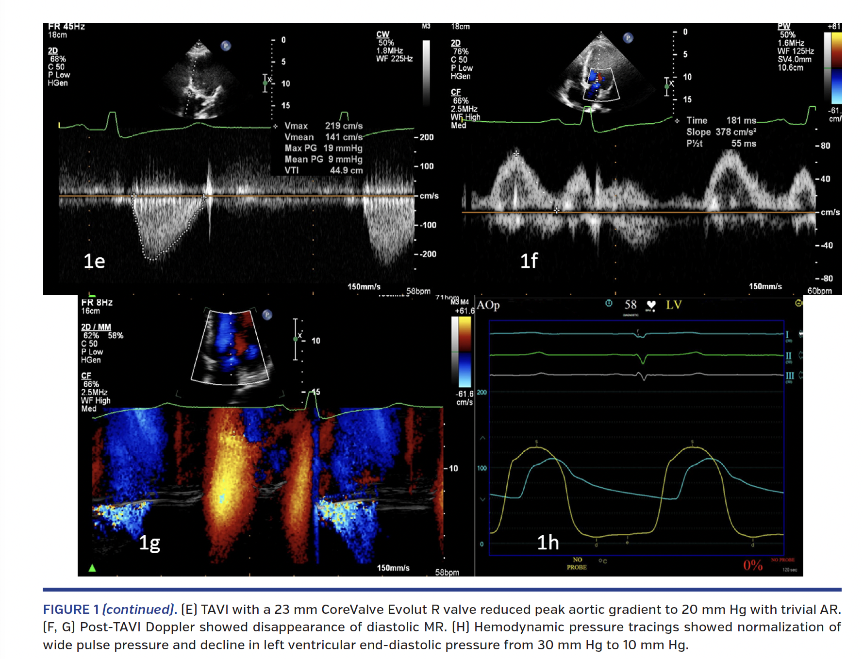
A 57-year-old gentleman was evaluated for symptomatic aortic bioprosthetic valve dysfunction 10 years after aortic valve replacement for aortic stenosis. Doppler echocardiography (Figure 1A) showed severe aortic regurgitation with pressure half-time of 170 ms and peak aortic gradient of 50 mm Hg. Mitral pulsed Doppler and color M-mode Doppler showed diastolic mitral regurgitation in the diastasis between the E and A waves (Figures 1B and 1C, blue arrows). Cardiac catheterization confirmed severe aortic regurgitation with elevation of left ventricular end-diastolic pressure (Figure 1D). Transcatheter aortic valve implantation (TAVI) with a 23 mm CoreValve Evolut R self-expanding valve (Medtronic) reduced the peak aortic gradient to 20 mm Hg with trivial aortic regurgitation (Figure 1E). Post-TAVI Doppler evaluation showed disappearance of diastolic mitral regurgitation (Figures 1F and 1G). Hemodynamic pressure tracings showed normalization of wide pulse pressure and decline in left ventricular end-diastolic pressure from 30 mm Hg to 10 mm Hg (Figure 1H).
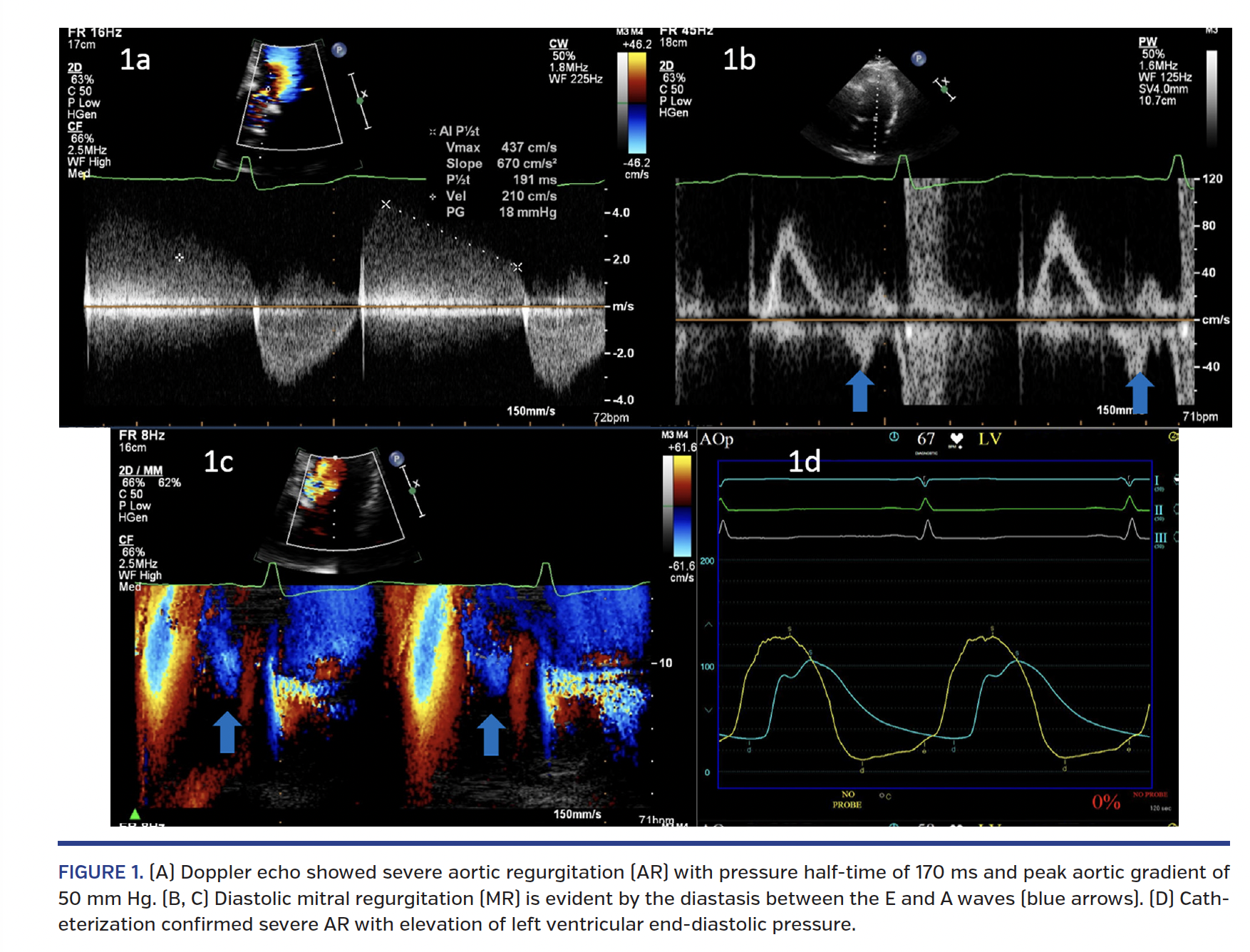
Transient elevation of left ventricular diastolic pressure above left atrial pressure can occur in severe aortic regurgitation in the diastasis phase, reflected as the flow reversal at the end of the mitral propagation E wave. This also may be associated with a relatively abrupt deceleration of the E wave as the diastolic mitral regurgitation may eschew part of mid-diastolic inflow to the left ventricle. Post thoracotomy pericardial restraint and persistent left ventricular hypertrophy could also have contributed to the elevation of left ventricular end-diastolic pressure in this patient besides severe aortic regurgitation. The recognition of mitral E wave reversal in such a situation could serve as non-invasive evidence of elevated left ventricular diastolic pressures, thereby guiding therapeutic decision making.
From the Department of Cardiology, Sree Chitra Tirunal Institute for Medical Sciences and Technology, Thiruvananthapuram, Kerala, India.
Disclosure: The authors have completed and returned the ICMJE Form for Disclosure of Potential Conflicts of Interest. The authors report no conflicts of interest regarding the content herein.
Manuscript accepted June 26, 2017.
Address for correspondence: Dr Arun Gopalakrishnan, Assistant Professor, Department of Cardiology, Sree Chitra Tirunal Institute for Medical Sciences and Technology, Thiruvananthapuram, Kerala, India – 695011. Email: arungopalakrishnan99@gmail.com


