


Surgical Offloading of the Diabetic Foot
Patients with neuropathy are at an increased risk of developing ulcerations. The pathway to tissue loss is often related to repetitive microtrauma to the insensate foot, particularly at areas of bony prominence and peak pressures, including the plantar metatarsal head, medial first metatarsal head, lateral fifth metatarsal head, base of the fifth metatarsal or the pulp of the toes.1,2
Given that the most common cause of peripheral neuropathy in the United States is diabetes, the majority of the neuropathic foot ulcers occur among patients with uncontrolled diabetes.3 Neuropathic foot ulcerations are closely related to recurrence. The recurrence rate of diabetic foot ulcers is estimated to be as high as 60% within 3 years.3 Causes for ulcer recurrence are multifactorial, including peripheral neuropathy, callus, foot deformity, elevated peak pressure, vascular disease, poorly fitted footwear, and puncture wounds.4 Of note for this article, an unaddressed underlying biomechanical deformity is a major contributor to chronic and recurring neuropathic foot ulcers.
We will present case studies to briefly demonstrate different surgical techniques to address the underlying deformities in an effort to resolve neuropathic foot ulcers and mitigate recurrence.
Evaluation and Treatment Selection Considerations
Numerous authors advocate for correction of biomechanical deformities to mitigate neuropathic foot ulcers (ie, surgical offloading). Previous studies proposed a treatment-based classification and found that prophylactic diabetic foot surgery was overall safe and effective.5 In a recent systematic review, the qualitative analyses concluded that surgical offloading was an effective method to treat recurrent neuropathic foot ulcers and to reduce plantar pressure.6,7 Nonetheless, one should note that the level of evidence surrounding surgical offloading may be weak due to study designs that are subject to bias with limited sample sizes. Additionally, while the pathophysiology of peripheral neuropathy, particularly among patients with diabetes, is beyond the scope of this review, it certainly adds another layer of complexity to the postoperative healing process, as the evidence has shown that compromised peripheral neural function can alter bone healing.8
Therefore, the risks of perioperative and postoperative complications are always a concern even if a patient’s diabetes is well controlled. Due to this reason, we and many podiatric surgeons recommend optimizing patients for surgical offloading, and reserving surgery for when conservative offloading measures fail.
Wound evaluation, especially in patients with neuropathy, should include a thorough biomechanical workup. Specifically, the determination of the presence of a rigid versus a reducible deformity is essential for procedural selection. In the case of rigid deformity especially around the joints, arthroplasty, arthrodesis or exostectomy can remove the bony prominence in an effort to reduce the peak pressure point. Addressing the center of rotation of angulation (CORA) is paramount to achieve a plantigrade foot and involves internal and/or external fixation. When the patient’s deformity is reducible, tendon balancing may be sufficient to achieve the correction. We present various approaches to different deformities based on the deformity from forefoot to hindfoot. Although the topic of wound care is beyond the scope of this review it is essential to continue appropriate wound care for an optimal outcome.
What You Should Know About Digital Deformities
Rigid hallux malleus. Hallux malleus is a common hallux deformity that leads to increased pressure at the pulp of the great toe. As described above, the repetitive microtrauma that results in callus formation often turns into a chronic and/or recurrent neuropathic foot ulcer in the setting of peripheral neuropathy (Figure 1a). The rigidity of the hallux interphalangeal joint creates pressure distally through the gait cycle (Figure 1b). A procedure to address this deformity is the hallux interphalangeal arthroplasty. This procedure restores the range of motion at this joint and allows the pressure to dissipate proximally away from the tip of the toe. Although correcting the rotational deformity is not the primary goal, plication of the joint capsule may help rotate the distal hallux in addition to the arthroplasty (Figure 2a). The patient pictured has had no recurrence of ulceration or callus 1-year status post right hallux interphalangeal arthroplasty (Figure 2b).
Semi-reducible hammertoe. Recurring neuopathic ulcers on the toes may often result from digital contractures. These ulcers may be dorsal at the interphalangeal joint or distal at the pulp of the digit. While a dorsal digital wound may respond to extra-depth shoes and a wide toe box, wounds at the pulp of the toe may be difficult to treat conservatively. In our experience, a neoprene top cover and offloading insert can mitigate the shearing force at the pulp of the digit. In the case of a recurring wound and callus at the pulp of the digit, a percutaneous flexor tendon release can help correct a semi-reducible digital contracture. One can employ an 18-gauge needle for minimal tissue loss. to minimize the incision and trauma. There is accumulating evidence suggesting this minimally invasive procedure is safe to be performed in outpatient clinics and well-tolerated under digital block.9
Surgical Offloading for Metatarsal Deformities
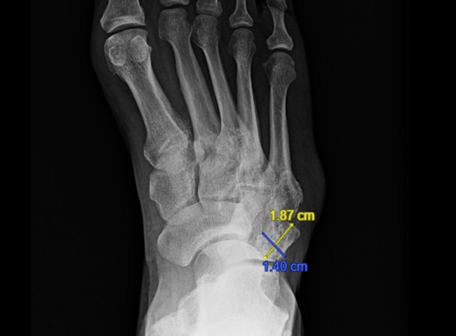
Central metatarsal heads. The most common location of neuropathic foot ulcerations is under the lesser metatarsal heads. More than 55% of the neuropathic foot ulcers are found to be under the second to fourth metatarsal head.4 The etiology of these ulcerations is multifactorial and may be related to structure (ie, anatomical misalignment) and/or dynamic function (ie, biomechanical imbalance). As with hallux valgus and concomitant hammertoe deformities, shortened first metatarsals, elongated second metatarsals or hypermobile first rays are subject to develop a hammertoe deformity, which leads to a retrograde force on the lesser metatarsal heads (Figure 3). Additionally, ankle equinus due to contracture of the posterior compartment of the leg can increase the peak pressure at the lesser metatarsal heads.10
A detailed musculoskeletal and biomechanical examination is imperative in order to formulate an appropriate surgical offloading procedure. A number of surgical offloading procedures are available to address recurring neuropathic foot ulcers under the lesser metatarsal heads. A metatarsal head resection may have indication for a chronic neuropathic foot ulcer with underlying osteomyelitis. However, without osteomyelitis, elective metatarsal head resection can lead to a development of a new neuropathic foot ulcer due to the transfer of the peak pressure to the adjacent metatarsal head. As a result, an alternative approach is the floating osteotomy.11 The advantage of this procedure includes a minimally invasive approach, quick recovery time, and immediate weight-bearing in protective footwear (Figure 4).
Since this osteotomy does not utilize internal fixation, nonunion and malunion are potential complications. Therefore, it is worth noting that long-term follow-up on patients who had floating osteotomies to address neuropathic foot ulcers will help determine the procedure’s efficacy and long-term outcome. Nonetheless, in our experience, a small subset of patients with recurring neuropathic foot ulcers may benefit from the floating osteotomy which permits minimal disruption of a patient’s work life and reduces the plantar peak pressure at the respective metatarsal head as the cause of a recurring neuropathic foot ulcer.
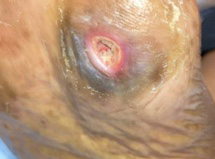
The base of the fifth metatarsal is also subject to developing neuropathic foot ulcers. Due to the protuberance of the styloid process, ill-fitting footwear can cause increased shearing force to the styloid process. Therefore, ensuring appropriate width and sufficient padding of the waist of the shoe should be the top priority in conservative management. Aside from the anatomical predisposition, gait patterns that increase pressure to the lateral column can further exacerbate the peak plantar pressure to the styloid process. Therefore, biomechanical evaluation to include any hindfoot varus, metatarsus adductus, and weakness of eversion are essential as the findings will guide the appropriate surgical offloading approach.
When one cannot address the neuropathic foot ulcer at the base of the fifth metatarsal through shoe modification, achieving a plantigrade foot is the primary goal of surgical offloading. In our experiences, many recurring plantar fifth metatarsal ulcers are due to the weak eversion of the foot (Figure 5). To address this deficit, anterior tibialis tendon transfer may correct the underlying deformity (Figure 6).
Transmetatarsal amputation. One common limb salvage procedure is transmetatarsal amputation (TMA). Although typically the surgeon will aim to maintain the parabola and will use the plantar flap to increase the soft tissue padding at the distal stump, soft tissue loss due to infection and/or prior wounds may lead to less than ideal soft tissue coverage. Additionally, the loss of the attachment of the extensor tendons weakens the dorsiflexion of the foot. As a result, we assess the amount of dorsiflexion preoperatively and intraoperatively. If there is less than 5 degrees of dorsiflexion from neutral position, we perform percutaneous Achilles tendon lengthening to mitigate peak pressure at the distal stump preemptively. For a patient experiencing a recurring neuropathic ulcer at the distal TMA stump, a thorough assessment of the parabola and ankle dorsiflexion is paramount for surgical offloading planning. In the case of a revision TMA due to ulceration at the distal stump, it is not uncommon to further resect the residual metatarsals, which shortens the foot and potentially sacrifices the attachment of the anterior tibialis. To offset the weakened dorsiflexory power, we find Achilles tendon lengthening is often necessary.
Insights on Midfoot Deformities
Charcot neuroarthropathy with rocker bottom deformity. Deformity at the midfoot is frequently caused by Charcot neuroarthropathy among neuropathic patients. When the debilitating condition occurs at the midfoot, subluxation of the midfoot joints can lead to a rocker bottom deformity, which causes increased peak pressure at the apex of the deformity plantarly. If conservative offloading measures fail, and there is an unbraceable deformity, recurrent ulcerations will occur (Figure 7a). As a result, surgical reconstruction to recreate a plantigrade foot by means of surgical offloading is paramount to prevent reulceration.
Charcot foot reconstruction aims to create stability by utilizing external and internal fixation. A plantar approach to the midfoot rocker bottom deformity constitutes resection of the apex of the deformity utilizing a plantar based wedge (Figure 8). The resected area is reduced via a tension band wire technique (Figure 7b). The plantigrade foot is fixed with a circular external fixator during a staged procedure when there is a concern of infection (ie, osteomyelitis) which prevents us from placing internal fixation.
Surgically Offloading the Hindfoot
Ankle Charcot neuroarthropathy. The risk of limb loss and the complexity of surgical correction of a deformity increase as the deformity moves from the forefoot to the hindfoot. Reconstruction of ankle Charcot neuroarthropathy is challenging and can have an extended recovery period. There is a higher rate of amputation in patients with ankle Charcot. Goals of reconstruction are to ensure that patients can ambulate in a supportive brace or shoe following reconstruction. An example of a lateral ankle ulceration secondary to a varus dislocation of the ankle is illustrated in Figure 10. The ulceration developed a sinus tract due to chronic osteomyelitis and was first managed via surgical curettage and resection of the lateral malleolus, antibiotic beads, bone cultures and long-term antibiotics (Figure 10). After controlling infection, the ankle was stabilized and reduced by a static circular external fixator (Figure 11). At the 6-month follow-up, the lateral ankle wound healed as the varus correction “offloaded” the pressure to the lateral ankle (Figure 12). The plantigrade foot and rectus ankle were fitted in the brace to allow the patient to ambulate and rehabilitate.
In Conclusion
The idea of surgical offloading may seem intuitively straightforward as a powerful arsenal to heal neuropathic foot ulcers and mitigate the recurrence. However, each approach of surgical offloading may not be the same as each deformity and individual biomechanical considerations are different. Patients with peripheral neuropathy can complicate the postoperative course and make surgical intervention more difficult. The lack of sensation and pain can allow patients to ambulate during the postoperative period without any proper sensory feedback and can alter the proper healing process.12 Therefore, it is imperative for foot and ankle reconstructive surgeons to provide patient education about the potential risks and complications related to surgical reconstruction, perform meticulous biomechanical evaluation and carry out careful perioperative management to ensure optimal outcomes.
Chia-Ding Shih, DPM, MPH, MA (Med. Sci.) is an Assistant Professor of Clinical Surgery at Keck School of Medicine of USC and an Adjunct Assistant Professor at the California School of Podiatric Medicine at Samuel Merritt University. Dr. Shih is also the chair for the Public Health and Podiatric Preventive Medicine Committee (PHPPMC) of the American Podiatric Medical Association (APMA) and the vice chair for the Public Health Committee of California Podiatric Medical Association. He is the Early Career Representative for the American Diabetes Association Foot Care Interest Group.
Laura Shin, DPM, PhD is an Assistant Professor of Clinical Surgery at Keck School of Medicine. She is currently the Chair-Elect of the American Diabetes Association Foot Care Interest Group and liaison for the American Limb Preservation Society. She is a reconstructive surgeon and physician-scientist who specializes in podiatry, including foot and ankle deformities, Charcot neuroarthropathy, limb salvage, wound care and diabetic feet and ankles.
References
1. Eastman DM, Dreyer MA. Neuropathic Ulcer. In: StatPearls. StatPearls Publishing; 2022.
2. Boulton AJM. Pressure and the diabetic foot: clinical science and offloading techniques. Am J Surg. 2004;187(5A):17S–24S.
3. Armstrong DG, Boulton AJM, Bus SA. Diabetic foot ulcers and their recurrence. N Engl J Med. 2017;376(24):2367–2375.
4. Lavery LA, Peters EJG, Armstrong DG. What are the most effective interventions in preventing diabetic foot ulcers? Int Wound J. 2008;5(3):425-433.
5. Armstrong DG, Lavery LA, Stern S, Harkless LB. Is prophylactic diabetic foot surgery dangerous? J Foot Ankle Surg. 1996;35(6):585–589.
6. Bus SA, van Deursen RW, Armstrong DG, et al. Footwear and offloading interventions to prevent and heal foot ulcers and reduce plantar pressure in patients with diabetes: a systematic review. Diabetes Metab Res Rev. 2016;32 Suppl 1:99–118.
7. Yammine K, Assi C. Surgical offloading techniques should be used more often and earlier in treating forefoot diabetic ulcers: an evidence-based review. Int J Low Extrem Wounds. 2020;19(2):112–119.
8. Beeve AT, Brazill JM, Scheller EL. Peripheral neuropathy as a component of skeletal disease in diabetes. Curr Osteoporos Rep. 2019;17(5):256–269.
9. Bonanno DR, Gillies EJ. Flexor tenotomy improves healing and prevention of diabetes-related toe ulcers: a systematic review. J Foot Ankle Surg. 2017;56(3):600–604.
10. Searle A, Spink MJ, Ho A, Chuter VH. Association between ankle equinus and plantar pressures in people with diabetes. A systematic review and meta-analysis. Clin Biomech. 2017;43:8–14.
11. Tamir E, Tamar M, Ayalon M, Koren S, Shohat N, Finestone AS. Effect of mini-invasive floating metatarsal osteotomy on plantar pressure in patients with diabetic plantar metatarsal head ulcers. Foot Ankle Int. Published online December 17, 2020:1071100720976099.
12. Jeffcoate W, Game F. The Charcot foot reflects a response to injury that is critically distorted by preexisting nerve damage: an imperfect storm. Diabetes Care. 2022;45(7):1691–1697.


