


Skin of Color in Podiatric Dermatology
A Roundtable Discussion
A Roundtable Discussion
Q: What does the literature tell us about lower extremity dermatology in skin of color?
A: There is not nearly enough information available on this topic, says Tracey C. Vlahovic, DPM, FFPM, RCPS (Glasg). Having written chapters in books to assist clinicians, Dr. Vlahovic, a Clinical Professor in the Department of Podiatric Medicine at Temple University School of Podiatric Medicine, feels there is much more education that is necessary.1,2
G. Dock Dockery, DPM, FACFAS, agrees that there is a paucity of literature, including clinical guidelines, textbooks, and peer-reviewed articles dedicated to lower extremity dermatology in patients with skin of color. He says he has observed some general dermatology publications addressing skin of color, but mostly regarding conditions of the head, neck, and trunk. He notes a few good books on dermatology for skin of color,3-5 but that lower extremity information is still sparse in these examples.
“There is a concerted movement to correct the problem,6-8 but there is still a need for more focused reporting of lower extremity skin conditions in patients with darker skin,” he adds.
Dermatology textbooks, per the panelists, do not always reflect an accurately diverse patient population, in their experience depicting too high a ratio of Fitzpatrick skin phototypes 1 and 2. William P. Scherer, DPM, MS, points to a 2021 report that cited only 15% of images in common medical student resources depicted skin of color.9 This is in spite of 2020 census data showing that persons of color comprise 40% of the US population.10 He stresses that this is significant medical concern, since by 2042, this number is expected to rise to more than 50%.11
“Becoming skillful in dermatology requires pattern recognition to determine conditions, and that can only be accomplished by training your brain and eyes to recognize certain colors, shapes, and elevations that have tremendous variability among a potential of 1,000 different skin and nail conditions that can present in the lower extremity,” he explains. Without an in-depth curriculum, and comprehensive resources (such as accurately represented textbooks) as a requirement, Dr. Scherer expresses concern about a resultant gap in diagnostic knowledge.
However, Alton R. Johnson, Jr., DPM, DABPM, FACPM, FASPS, CWSP, sees improvement on the horizon. In preparing a publication for submission a few years ago,12 he notes that the available literature to review was not voluminous. However, over the last few years, Dr. Johnson, a Clinical Assistant Professor of Orthopaedic Surgery at the University of Michigan Medical School, says he notices an uptick in evidence and discussion, specifically in dermatological manifestations of infection in persons of color, in addition to systemic dermatological presentations.
Lesly D. Robinson, DPM, CWSP, DABPM, Chair of the Department of Podiatric Medicine at Temple University School of Podiatric Medicine, cites an example of this evidence as it relates to measuring transepidermal water loss.13 “Significantly higher in patients with psoriasis, eczema, and other skin disorders, this is considered a marker of skin hydration and skin barrier function. More objective measurement will improve research in skin of color.”
Q: What important history-taking points should clinicians focus on when addressing lower extremity dermatology concerns in patients with highly pigmented skin?
A: Several panelists stress that a patient’s personal and family history is vital when addressing skin issues in people of color. They mention that this is particularly important when the concerns relate to melanonychia, pigmented skin lesions, rashes, and skin cancer.
These history items can help avoid delays in diagnosis, explains Dr. Scherer, a former Adjunct Professor at Barry University School of Podiatric Medicine and current frequent podiatric dermatology lecturer practicing in South Florida. He adds that such delays likely contribute to worse prognoses for skin cancer in those with highly pigmented skin.14 Differences in prevailing types and locations of skin cancer among races may also delay diagnosis or result in misdiagnosis. Of note, the 5-year survival rate for malignant melanoma is approximately 66% for non-Hispanic Black patients, compared with 90 percent for non-Hispanic White patients.14,15
Dr. Robinson adds that it is important to know the patient’s healing and scarring history, along with feedback on keloid formation and skin hyperpigmentation. She says that other important history points include itchiness and tightness that can result from transepidermal water loss. Dr. Dockery also advocates for a precise history of past and current skin treatments and medications.
The history of present illness (HPI), specifically, might provide important data in these cases, says Dr. Johnson. He shares that clinical manifestations of certain lower extremity dermatologic conditions may not appear with the same timeline between varying skin pigmentations. He uses poison ivy as an example, where, in his experience, redness and purpura may be delayed or look different in higher-number Fitzpatrick skin phototypes, but a good history of present illness may reveal a hiking trip in a wooded area and pruritus that began shortly thereafter.
Q: What findings on a physical exam are important to know about when evaluating lower extremity skin of color?
A: Dr. Johnson stresses that signs of infection are a key consideration on a lower extremity dermatological exam for patients with skin of color. Dr. Robinson echoes this thought, sharing that erythema may not show as clearly on varying skin pigmentations. She notes that hyperpigmentation might be a sign of irritation and decreased skin barrier function. “Moreover, flakes on the topmost surface usually occur when the skin is severely dry and could be caused by a damaged skin barrier and may be the only sign of irritation or severe dryness for those with darker skin,” she explains.
“You definitely have to be very diligent in your clinical exam when looking for infection when it comes to lower extremity dermatological manifestations in patients of high pigmentation,” agrees Dr. Johnson. In the recent study he coauthored, he says they found that based on clinical examination alone, clinicians were more likely to miss infections in patients with higher skin pigmentation, irrespective of if the examiner was a person of color attributing to likely unconscious bias.12
Variations in pigmentation in general are also important to evaluate, adds Dr. Vlahovic. “I am always looking for pigmentation (changes),” she says. “Some of it is natural variation, like what I see on the plantar foot, but sometimes it can be hyperpigmentation from a skin rash or the periwound (area).”
Since visual skin assessment is still the gold standard of evaluating dermatologic conditions, Dr. Dockery recommends optimizing the light source in the exam area, using natural light or even a halogen lamp, rather than typical clinic fluorescent lighting. “Fluorescent lights often give the illusion of a bluish tint rather than showing an accurate color,” he explains.
He adds that in his experience, standard diascopy skin blanch testing may have very limited value in skin of color, especially when it comes to differentiating erythema and purpura. Greater amounts of melanin in darker skin may alter the blanch test, making the color change hard to evaluate, says Dr. Dockery, the Founder, Chairman, and President of The International Foot and Ankle Foundation for Education and Research. Other important clinical responses of the skin, he says, like cyanosis, paleness, jaundice, and flushing, may appear dramatically different on skin of color as compared to lighter skin.3,4,16
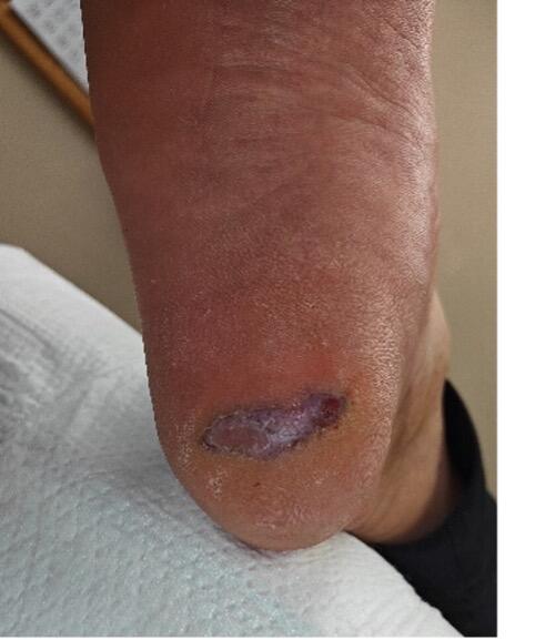
Common skin conditions like eczema can vary in appearance for different skin complexions, says Dr. Scherer. Typically described as red and itchy skin, he says that this tends to appear as small bumps in Black patients. Psoriasis may also go undetected in patients of color, as the condition may manifest as scales and patches that appear purple or gray, as opposed to the more classically described red or pink. He stresses that this can lead to delays in diagnosis and treatments.17,18
Q: Are there any important diagnostic tools or resources that DPMs should learn more about in order to best serve this patient population?
A: There are multiple devices and modalities the panelists shared as valuable. Dr. Robinson advocates for more objective measurement of skin irritation, such as that of transepidermal water loss, may demonstrate the extent of impaired skin barrier function and reduced skin hydration. An open-chamber, unventilated-chamber small device, such as the VapoMeter (Delfin Technologies) may assist in this assessment. Dr. Vlahovic cautions providers to be aware of tools and treatments that may cause hypopigmentation or hyperpigmentation. “For example, cryotherapy has potential to cause hypopigmentation, and I discuss this with the patient before treatment,” she explains. Fluorescence imaging and thermography are among the resources Dr. Johnson feels can have a positive impact on care and differential diagnoses for dermatologic conditions and wounds in highly pigmented skin.
Dr. Scherer lists a Wood’s lamp and having readily available 2mm and 3mm punch biopsy supplies as vital for all patient populations, but especially to help mitigate diagnostic challenges in patients of color. “Podiatrists historically have been under-biopsying soft tissue and nail lesions and that is a significant problem in our profession,” he says. “Performing a skin or nail biopsy is quick and easy and will provide a definitive laboratory diagnosis to help physicians narrow their differential diagnosis.”
The panel overwhelmingly cites a high-quality dermatoscope as a tool of value in this population and across the board. Dr. Dockery says that understanding the dermatoscopic features of various cutaneous malignancies versus their benign counterparts is an important, potentially life-saving skill set.19 Dr. Vlahovic adds that she finds the dermatoscope particularly helpful when evaluating longitudinal melanonychia. The panel shared that multiple courses exist to assist providers in learning dermoscopy skills.
Dr. Scherer points out that there are organizational and social media movements that may provide education and resources for clinicians. At least 15 facilities exist in the US dedicated specifically to medical excellence for skin of color, he says. He adds that patients and physicians alike have created social media accounts dedicated to educating providers and public on the presentation of skin symptoms in various skin pigmentations, such as @brownskinmatters on Instagram. Also, the Skin of Color Society provides education and support for health care providers and the public at-large on dermatologic health issues for patients of color. The Skin of Color Society hosts a special session each year at the American Academy of Dermatology annual meeting, adds Dr. Dockery, that is an option to enhance one’s learning on the topic.
Q: What one thing do you think DPMs can incorporate into their practices today to enhance outcomes for dermatologic issues in patients of color?
A: Dr. Johnson encourages clinicians to update their dermatologic reference materials and atlases to include those that focus on photos and illustrations of manifestations in varying skin pigmentations. He also stresses the importance of having a low clinical threshold for biopsy to assist in definitive diagnosis.
In addition to actively seeking out educational programs and resources to better one’s knowledge on lower extremity dermatology in skin of color, Dr. Dockery hopes that the resources themselves will continue to improve. He lists atopic dermatitis, eczema, allergic contact dermatitis, psoriasis, lichen planus, dermatophytosis, infections, postinflammatory hyperpigmentation, keloid scarring, and dyschromia as pathologies he noted in his career for which it was difficult to source clear photos demonstrating the condition in darkly pigmented skin. However, he notes that current literature is addressing the issue, such as in a recent article on improving photodocumentation in skin of color.8
Dr. Vlahovic hopes providers exhibit a willingness to look deeper for skin color changes and refer out when needed. “For instance, psoriasis looks so different in skin of color than in a White patient,” she says. “It’s important to understand that inflammation will look different than in a textbook.”
Dr. Scherer urges DPMs to improve patient education on sun protection and combat misconceptions that less propensity for sunburn means less risk for skin cancer. “Everyone, regardless of skin tone, should limit prolonged sun exposure, seek shade, wear protective UV clothing, and use a broad-spectrum sunscreen with an SPF of 30 or higher to help prevent skin cancer and premature skin aging,” he says.
Dr. Robinson wants clinicians to know about ceramides, which not only keeps the fluid in the skin but also protects it from allergens.20 Ceramide levels decline as one ages, and exist in the skin in this order from most to least: Hispanic, Asian, White, and Black patients. Doctors should recommend ceramide-containing emollients and encourage a healthy diet containing wheat, soy, eggs, and dairy. These contain large amounts of sphingolipids, which can boost the body’s ceramide production.
The panelists disclose the following relationships relevant to this discussion: Dr. Dockery, Co-Chair, Podiatric Clinical Affairs Board, Sagis Diagnostics; Dr. Johnson, Moleculight; Dr. Robinson, no disclosures; Dr. Scherer, Bako Diagnostics; Dr. Vlahovic, Sagis Diagnostics, Bako Diagnostics, and Ortho Dermatologics.
References
1. Schleicher SM, Vlahovic TC. Skin Disease of the Lower Extremities: A Photographic Guide. HMP Communications; 2012.
2. Vlahovic TC, Schleicher SM. Atlas of Lower Extremity Skin Disease. Springer; 2022.
3. Kelly AP, Taylor SC, Lim HW, Serrano AMA. Taylor and Kelly’s Dermatology for Skin of Color. (2nd ed) McGraw-Hill;2008.
4. Kinai M. Dark Skin Dermatology Color Atlas: Clinical Dermatology. CreateSpace;2012.
5. Shah MK, Sheth PK. Pediatric Dermatology in Skin of Color: A Practical Guide. CRC Press;2021.
6. Mhlaba JM, Pontes DS, Patterson SS, Kundu RV. Evaluation of a skin of color curriculum for dermatology residents. J Drugs Dermatol. 2021;20(7):786-789.
7. Onasanya J, Liu C. Dermatology education in skin of colour: where we are and where do we go? Can Med Educ J. 2021;12(6):124-125.
8. Alvarado SM, Feng H. Representation of dark skin images of common dermatologic conditions in educational resources: a cross-sectional analysis. J Am Acad Dermatol. 2021;84(5):1427-1431.
9. Perlman KL, Williams NM, Egbeto IA, Gao DX, Siddiquee N, Park JH. Skin of color lacks representation in medical student resources: A cross-sectional study. Int J Women’s Dermatol. 2021;7(2):195-196.
10. US Census Bureau. Quick Facts – United States. Available at: https://www.census.gov/quickfacts/fact/table/US/PST045222 . Accessed July 25, 2023.
11. Colby SL, Ortman JM. Projections of the size and composition of the US population: 2014 to 2060. United States Census Bureau. Available at: https://www.census.gov/content/dam/Census/library/publications/2015/demo/p25-1143.pdf . Published March 2015. Accessed July 20, 2023.
12. Johnson J, Johnson AR Jr, Andersen CA, Kelso MR, Oropallo AR, Serena TE. Skin pigmentation impacts the clinical diagnosis of wound infection: imaging of bacterial burden to overcome diagnostic limitations. J Racial Ethn Health Disparities. 2023. doi: 10.1007/s40615-023-01584-8.
13. Alexis AF, Woolery-Lloyd H, Williams K, et al. Racial/ethnic variations in skin barrier: implications for skin care recommendations in skin of color. J Drugs Dermatol. 2021;20(9):932.
14. Gupta AK, Bharadwaj M, Mehrotra R. Skin cancer concerns in people of color: risk Factors and prevention. Asian Pac J Cancer Prev. 2016;17(12):5257-5264.
15. Culp MB, Lunsford NB. Melanoma among non-Hispanic Black Americans. Prev Chronic Dis. 2019;16:180640.
16. MedLine Plus. Blue discoloration of the skin. Available at: https://medlineplus.gov/ency/article/003215.htm#:~:text=When%20the%20oxygen%20level%20has,anemia%20(low%20blood%20count). Accessed July 25, 2023.
17. Sangha AM. Dermatological conditions in skin of color: managing atopic dermatitis. J Clin Aesthet Dermatol. 2023;14(3 Suppl 1):S20-S22.
18. Alexis AF, Blackcloud P. Psoriasis in skin of color: epidemiology, genetics, clinical presentation, and treatment nuances. J Clin Aesthet Dermatol. 2014;7(11):16-24.
19. Ezenwa E, Stein JA, Krueger L. Dermoscopic features of neoplasms in skin of color: a review. Int J Women’s Dermatol. 2021;7(2):145-151.
20. Taylor SC. Skin of color: biology, structure, function, and implications for dermatologic disease. J Am Acad Dermatol. 2002;46:S41-S62.


