A Novel Approach to Fifth Metatarsal Osteomyelitis
A Poster From SAWC Spring
A Poster From SAWC Spring
Hi everyone, my name is Shrunjay Patel, I am a podiatric surgeon practicing at the University of North Carolina in Chapel Hill and practice within the Department of Surgery. Today I would like to discuss two very interesting and challenging cases. Both of these patients presented with gangrenous changes to the lateral foot with associated osteomyelitis.
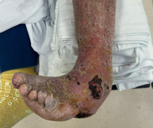
These patients had complex comorbidities including diabetes, peripheral vascular disease, history of renal transplant with immunosuppressive status, peripheral neuropathy, and congestive heart failure. Both of them opted for limb salvage options and wanted to avoid below-knee amputation. Patient 1 also presented with hallux gangrene and calcaneus gangrenous changes.
After he was revascularized by our vascular colleagues, we performed a hallux amputation, partial resection of the calcaneus with resection of the gangrenous tissue and bone, and partial fifth ray amputation. The partial fifth ray amputation and calcaneus resection sites could not be closed primarily due to large soft tissue loss. In order to cover the exposed fifth metatarsal base and peroneus braves tendon attachment, we harvested an abductus digits minimi muscle flap and inset it into the wound to cover the bone and vital structures.
We covered the muscle flap with SEFM, which is short for synthetic electrospun fiber matrix, which is a skin substitute. substitute. SEFM was also applied at the posterior calcaneus and a wound vac was initiated at the posterior calcaneus.
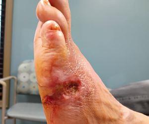
The patient was discharged with IV antibiotics and close follow-up. After a few weeks, once SEFM was absorbed into the wound bed, a second application of SEFM was applied over the lateral foot and posterior heel. The patient achieved complete wound closure at around week 14 without needing any additional surgeries or skin grafting.
Similarly, patient 2 had fifth metatarsal base gangrene with osteomyelitis. After debridement of the gangrenous tissue and bone, we opted for preservation of the 5th metatarsal base and peroneus braves tendon attachment. Patient also had large soft tissue loss due to gangrene, and in order to cover the wound bed we performed a proximally based abductor digiti minimi muscle flap.
We applied SEFM skin substitute to cover the flap. Patient was then discharged with IV antibiotics and close follow-up. The skin substitute had absorbed into the wound bed and infection had resolved. The flap was healthy and granulating well. And once 100% granulation tissue was achieved, we applied an autogenous split-thickness skin graft to the flap and the wound healed completely at around week 14.
Both of these patients went on to ambulating independently and have remained ulcer and infection free. for over two years. Abductor digiti minimi is a type 2 muscle with one dominant pedicle and one to two minor pedicles.
We can harvest either distally-based or proximally-based muscle flap to cover the vital structure such as fifth metatarsal base or proximal lateral heel. SEFM is a skin substitute which mimics extracellular matrix and can help with wound healing and can be used in conjunction with muscle flaps. It is important to preserve the peroneus brevis attachment to preserve its eversion function because if you reject and lose the tendon then the posterior tibial tendon becomes unopposed and this can lead to severe various deformities and abnormal gait which can cause further ulceration, infection and amputation.
We as surgeons can perform such orthoplastic techniques and achieve successful limb salvage. Hope you enjoy SAWC conference and thank you for listening.