Comprehensive Genomic Testing: Tissue Stewardship and Best Practices
Abstract
Biomarker-driven targeted therapies have shaped the oncology treatment landscape for patients with advanced solid tumors over the past decade. Comprehensive genomic profiling (CGP) has played a key role in precision medicine as it enables simultaneous identification of multiple biomarkers to guide cancer diagnosis, therapy selection, and prognostication. As a result, tissue stewardship for successful CGP testing is paramount. In addition, widespread adoption of less invasive sampling techniques leaves less diagnostic tissue for additional or future testing as smaller biopsies are acquired. To help oncology care practitioners overcome these challenges, this paper provides an overview of current genomic testing methodologies and offers guidelines on best practices for tissue stewardship and preanalytic practices for successful CGP testing and efficient turnaround times for laboratory tests.
Introduction
Over the past decade, the oncology treatment landscape for patients with advanced solid cancers has significantly evolved toward biomarker-driven targeted therapies.1,2 To date, patients with advanced non–small cell lung cancer have the highest number of well characterized genomic alterations with matching biomarker-directed therapies while an increasing number of treatment-associated biomarkers are emerging in other solid tumors.3 In a paradigm shift away from disease-specific treatment algorithms, there are also a growing number of targeted therapies that rely on tumor-type agnostic biomarkers.
Comprehensive genomic profiling (CGP) is a powerful precision medicine tool as it enables simultaneous identification of multiple biomarkers to guide cancer diagnosis, therapy selection, and prognostication. CGP is standard of care for many cancer types and depending on the tumor type, guidelines suggest CGP for advanced stage tumors at diagnosis and recurrence or progression to evaluate biomarkers with matched standard-of-care and investigational therapies.
This paper provides an overview of genomic testing methodologies, details guidelines on the best preanalytic practices for CGP testing, highlights tissue stewardship best practices, and offers guidance to enable efficient turnaround times for laboratory tests.
Overview of Genomic Testing and Methodologies
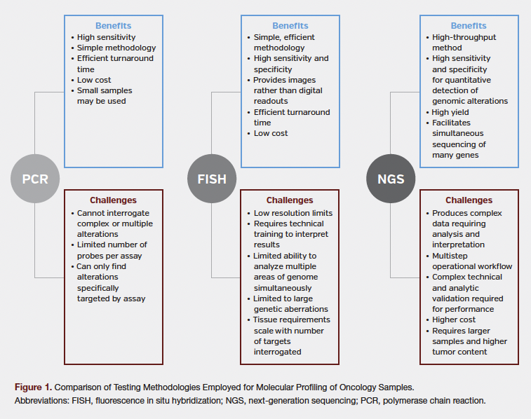
Genomic testing enables characterization of oncogenic mutations driving a specific tumor and can be performed as a series of single gene tests or as a comprehensive multigene test.4 Single gene genomic or protein-specific biomarker testing of tissue specimens employs immunohistochemistry (IHC), fluorescence in situ hybridization, and/or polymerase chain reaction, whereas multigene testing typically utilizes next generation sequencing (NGS) of DNA and/or RNA (Figure 1).
Best Practices for CGP Preanalytic Process
The following recommendations, focused on key steps of preanalytic CGP testing, increase the likelihood of successful testing.
Sample Acquisition
To minimize downstream issues, careful attention to specimen procurement and advance preparation is necessary.
- Planning: Consider multidisciplinary discussions between surgical oncologists, medical oncologists, pathologists, and proceduralists to develop an upfront plan for tissue specimen procurement and prioritization for diagnostic, prognostic, and therapeutic testing.5
- Patient Status: Before biopsy, the overall status of the patient, including level of tolerance to surgery and tumor location impacting the method of biopsy, should be considered.
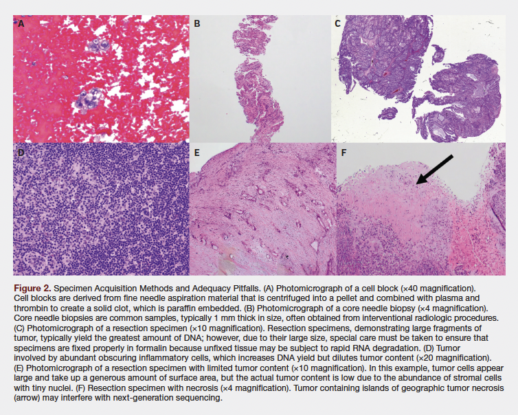
- Acquisition Method: Fine needle aspirations, biopsies, and fluid collection are the main sample acquisition techniques for obtaining tissue for CGP (Figure 2A-C).6 In choosing the most appropriate method, factors such as patient condition, tumor location and size, equipment availability, likely yield, and potential downstream uses of the sample should be considered.7,8
- Origin of Specimen: When possible, tissue with obvious necrotic areas and bone metastases should be avoided, as both lead to lower success rates.4 For metastatic disease, prioritize collecting a specimen from metastatic site rather than primary location, as it may contain more clinically relevant genomic alterations.9 Consult with pathologists rendering the diagnosis about the quality, quantity, and character of the specimen undergoing CGP testing. Advance notification to pathologists about the need for ancillary testing is recommended.
Sample Adequacy
As the size and quality of specimens are operationally determined by the surgeon, interventional radiologist, or proceduralist obtaining the sample, it may be necessary to establish a standard workflow for multidisciplinary communication before and/or during surgical procedures.6 This workflow should provide guidance from the testing laboratory and be revisited if changes occur in the workflow or assays of interest.
The primary goal of the sample acquisition should be to collect sufficient material for all purposes, including accurate diagnosis and molecular testing. If safe and technically feasible, additional specimens should be collected up front. For example, when using endobronchial ultrasound-guided transbronchial needle aspiration, the bronchoscopist should perform at minimum 3, and up to 5, passes, with the goal of collecting sufficient material for a tissue block for ancillary tests, including multiple IHC markers.10 When performing core needle biopsies, obtaining 3 core samples will help achieve optimal diagnostic yield.
Tumor purity or content is an important factor to consider, as presence of non-neoplastic cell types and necrotic material in submitted specimens (Figure 2D-F) may confound test results.11 To reduce failure and limit expense, obtaining accurate estimations of the fraction of cancer cells within specimens in consultation with an anatomic or molecular pathologist is recommended.12 Note that more than one specimen may be submitted if tumor content in either one seems inadequate.6
Sample Processing
It is helpful to establish specimen-specific workflows to minimize methodologic variability during specimen processing.
Sample Grossing and Fixation
The following points should be considered to preserve biomarker fidelity and minimize artifacts that may lead to inaccurate or failed test results.
- Maintain strict precautions to prevent cross-contamination. Handle one gross specimen at a time, properly label each container, clean or replace supplies between samples, and avoid serial numbering of similar specimen types.
- Fix samples immediately after receipt or place them in an appropriate nonfixative solution that prevents cell breakdown.
- Specimens should be completely submerged in formalin, with a preferred formalin volume to tissue mass of 10:1 and a minimum ratio of 4:1.
- Larger samples should be sliced such that the thickest sections are < 1 cm to facilitate diffusion of formalin throughout the tissue.
- As the type of fixative used impacts nucleic acid integrity and may be associated with higher failure rates, optimize and validate fixatives per tissue type before using it in samples.13 Most specimens can safely be fixed in 10% neutral-buffered formalin, which also minimizes nucleic acid damage.14,15
- Optimize and validate fixation time by tissue type, fixative solution, and fixation temperature. In general, 6-12 and 8-18 hours is enough to fix small biopsy and large surgical samples, respectively.
- The fixation time should be recorded in the pathology report, especially for cases in which time in formalin is longer than 24 hours.
Sample Embedding and Histologic Sectioning
Consider the following factors for preparation of samples prior to shipping:
- Embed specimen in paraffin within 1 hour of removal from fixative.16,17
- Strictly adhere to standardized protocols for reagent processing, processor function, and the use and maintenance of high-quality reagents—particularly alcohols—during processing of formalin fixed tissue. Avoid “topping off” processor chambers with nonstandard solutions, monitor total time in formalin to not exceed target fixation time, and use low-melting point (< 60 °C) paraffin wax to ensure adequate deparaffinization.
- Use a dedicated microtome, and if possible, have a dedicated laboratory. Use new disposable blades for each specimen and a clean water bath.
- To maximize tissue availability from formalin-fixed paraffin-embedded (FFPE) blocks, use specialized microtomes that make use of the whole specimen.7 If this is not feasible, slides may be cut to specifications.
- Cut extra sections to avoid tissue loss through recutting or refacing of FFPE blocks. Hematoxylin and eosin stain intervening sections to ensure specimen adequacy.
- Avoid using the superficial portion of an FFPE block taken from storage, as oxidation may result in biomarker degradation.
Storage of Blocks and Slides
Stored paraffin blocks are valuable for future testing in patient care, particularly when molecular testing is not available at the time of diagnosis, the tumor recurs, or new testing becomes available. Therefore, attention must be paid to duration, temperature, and condition of sample storage to minimize nucleic acid degradation.
- It is best to store samples in dry, pest-free conditions at room temperature (25 °C).
- Unstained slides older than 3 months should not be used for genomic testing. The block should be pulled, and fresh slides prepared.
- Label samples properly and log storage metadata to ensure data specificity and accuracy.6
- Before shipping, ensure that the labels on specimen blocks/slides and paperwork are correct (including accession number and patient identifiers).
The Future
The evolution of oncology treatment over the past decade has been markedly shaped by the advancement of genomic testing methodologies, with a shift toward biomarker-driven targeted therapies for various types of solid cancers. Integration of the multigene methodologies into current diagnostic paradigms and guidelines emphasize the impact of precision oncology on clinical care and improved patient outcomes. As a result, tissue stewardship for comprehensive and successful genomic testing is paramount. Moreover, widespread adoption of less invasive sampling techniques leaves less diagnostic tissue for additional or future testing as smaller biopsies are acquired. For patients with recurrent or progressive tumors, additional testing may be required to identify resistance alterations and/or guide next therapy. Enrollment of patients into clinical trials that require tissue-based or repeat testing poses another challenge. Finally, adherence to patient-informed consent in clinical trials, federal and state laws prohibiting exhaustion of the tissue during storage, and different tissue-retention requirements across states further complicate the issue.18
The key to addressing these challenges starts with understanding the ever-changing diagnostic and therapeutic landscape. This may start with biopharmaceutical companies partnering with diagnostic companies in the early phases of the drug development pipeline to evaluate companion diagnostics and timing of their implementation for the intended indication. Also, evaluating whether there is an optimal age for a tissue type subjected to analysis for a specific biomarker would be an important consideration. Equipped with this knowledge, partnerships among regulators, investigators, clinical trial sponsors, and pathology authorities can help improve existing recommendations and establish new guidelines for testing prioritization and informing patients.19
As the field continues to advance, collaboration among key stakeholders and care providers will be essential to align practices, optimize the use of precious tissue samples, and ensure that genomic testing remains at the forefront of personalized cancer treatment.
Author Information
Authors: Rebecca A. Previs, MD, MS1; Maureen E. Cooper, BS1; Kyle C. Strickland, MD, PhD1-4; Heidi Ko, DO1; Michelle F. Green, PhD1; Faezeh Koohestani, PhD5; Stephanie Hastings, PhD1; Zachary Wallen, PhD1; Sarabjot Pabla, PhD1; Jeffrey M. Conroy, BS1; Mary Nesline, MS1; Shengle Zhang, MD1; Durga Prasad Dash, PhD1; Brian Caveney, MD, JD, MPH1; Marcia Eisenberg, PhD1; Eric Severson, MD, PhD1; Shakti H. Ramkissoon, MD, PhD1
Affiliations: 1Labcorp, Burlington, NC; 2Foundation Medicine, Inc, Boston, MA; 3Almac Pharmaceuticals, Craigavon, UK; 4OncoQuest Pharmaceuticals, Edmonton, AB, Canada; 5Covance, Princeton, NJ
Address correspondence to:
Rebecca A. Previs
Labcorp Oncology
10 Moore Dr.
Durham, NC 27709
Email: rebeccaann.previs@labcorp.com
Disclosures: R.A.P., M.E.C., Z.W., J.M.C., M.N., M.E., and S.H.R. reported being an employee of Labcorp and owning Labcorp stock. K.C.S. reported being an employee of Foundation Medicine, Inc, Almac Pharmaceuticals, OncoQuest Pharmaceuticals, and Labcorp. H.K. reported being an employee of Labcorp and owning Gilead Sciences and Labcorp stock. M.F.G., S.H., S.P., D.P.D., and B.C. reported being an employee of Labcorp. F.K. reported being an employee of Covance. S.Z. reported no financial or other conflicts of interest. E.S. reported being an employee of Labcorp and receiving a speaker honorarium from the Cancer Care Business Exchange.
References
1. Kerr KM, Bibeau F, Thunnissen E, et al. The evolving landscape of biomarker testing for non-small cell lung cancer in Europe. Lung Cancer. 2021;154:161-175. doi:10.1016/j.lungcan.2021.02.026
2. Majeed U, Manochakian R, Zhao Y, Lou Y. Targeted therapy in advanced non-small cell lung cancer: current advances and future trends. J Hematol Oncol. 2021;14(108). doi:10.1186/s13045-021-01121-2
3. Chakravarty D, Johnson A, Sklar J, et al. Somatic genomic testing in patients with metastatic or advanced cancer: ASCO provisional clinical opinion. J Clin Oncol. 2022;40(11):1231-1258. doi: 10.1200/JCO.21.02767
4. Navani N, Butler R, Ibrahimo S, et al. Optimising tissue acquisition and the molecular testing pathway for patients with non-small cell lung cancer: A UK expert consensus statement. Lung Cancer. 2022;172:142-153. doi:10.1016/j.lungcan.2022.08.003
5. Nicholson AG, Kerr K, Gosney J. The Royal College of Pathologists: Standards and datasets for reporting cancers. Dataset for histopathological reporting of lung cancer. 2018. Available at https://www.rcpath.org/static/265cdf74-3376-40b0-b7d0e3ed8a588398/G048-Dataset-for-histopathological-reporting-of-lung-cancer.pdf
6. Ascierto PA, Bifulco C, Palmieri G, Peters S, Sidiropoulos N. Preanalytic variables and tissue stewardship for reliable next-generation sequencing (NGS) clinical analysis. J Mol Diagn. 2019;21(5):756-767. doi:10.1016/j.jmoldx.2019.05.004
7. Gan Q, Roy-Chowdhuri S. Small but powerful: the promising role of small specimens for biomarker testing. J Am Soc Cytopathol. 2020;9(5):450-460. doi:10.1016/j.jasc.2020.05.001
8. Sung S, Heymann JJ, Crapanzano JP, et al. Lung cancer cytology and small biopsy specimens: diagnosis, predictive biomarker testing, acquisition, triage, and management. J Am Soc Cytopathol. 2020;9(5):332-345. doi:10.1016/j.jasc.2020.04.014
9. Mata DA, Harries L, Williams EA, et al. Method of tissue acquisition affects success of comprehensive genomic profiling in lung cancer. Arch Pathol Lab Med. 2023;147(3):338-347. doi:10.5858/arpa.2021-0313-OA
10. Roy-Chowdhuri S, Dacic S, Ghofrani M, et al. Collection and handling of thoracic small biopsy and cytology specimens for ancillary studies: guideline from the College of American Pathologists in Collaboration with the American College of Chest Physicians, Association for Molecular Pathology, American Society of Cytopathology, American Thoracic Society, Pulmonary Pathology Society, Papanicolaou Society of Cytopathology, Society of Interventional Radiology, and Society of Thoracic Radiology. Arch Pathol Lab Med. 2020;9(4):286-290. doi:10.5858/arpa.2020-0119-CP
11. Kim L, Tsao MS. Tumour tissue sampling for lung cancer management in the era of personalised therapy: what is good enough for molecular testing? Eur Respir J. 2014;44:1011-1022. doi:10.1183/09031936.00197013
12. Haider S, Tyekucheva S, Prandi D, et al. Cancer genome Atlas Research Network. Systematic assessment of tumor purity and its clinical implications. JCO Precis Oncol. 2020;4:995-1005.PO.20.00016. doi:10.1200/PO.20.00016
13. Jain D, Allen TC, Aisner DL, et al. Rapid on-site evaluation of endobronchial ultrasound-guided transbronchial needle aspirations for the diagnosis of lung cancer: a perspective from members of the pulmonary pathology society. Arch Pathol Lab Med. 2018;142(2):253-262. doi:10.5858/arpa.2017-0114-SA
14. Bass BP, Engel KB, Greytak SR, Moore HM. A review of preanalytical factors affecting molecular, protein, and morphological analysis of formalin-fixed, paraffin-embedded (FFPE) tissue: how well do you know your FFPE specimen? Arch Pathol Lab Med. 2014;138(11):1520-1530. doi:10.5858/arpa.2013-0691-RA
15. Srinivasan M, Sedmak D, Jewell S. Effect of fixatives and tissue processing on the content and integrity of nucleic acids. Am J Pathol. 2002;161(6):1961-1971. doi:10.1016/S0002-9440(10)64472-0
16. Roy-Chowdhuri S, Dacic M, Ghofrani PB, et al. Collection and handling of thoracic small biopsy and cytology specimens for ancillary studies: guideline from the College of American Pathologists in collaboration with the American College of Chest Physicians, Association for Molecular Pathology, American Society of Papanicolaou Society of Cytopathology, Society of Interventional Radiology, and Society of Thoracic Radiology. Arch Pathol Lab Med. 2020;144 (8):933-958. doi:10.5858/arpa.2020-0119-CP
17. Wolff AC, Hammond ME, Hicks DG, et al. Recommendations for human epidermal growth factor receptor 2 testing in breast cancer: American Society of Clinical Oncology/College of American Pathologists clinical practice guideline update. J Clin Oncol. 2013;31(31):3997-4013. doi:10.1200/JCO.2013.50.9984
18. McCall SJ, Dry SM. Precision pathology as part of precision medicine: are we optimizing patients’ interests in prioritizing use of limited tissue samples? JCO Precis Oncol. 2019;3:1-6. doi:10.1200/PO.18.00238
19. De Las Casas LE, Hicks DG. Pathologists at the leading edge of optimizing the tumor tissue journey for diagnostic accuracy and molecular testing. Am J Clin Pathol. 2021;155(6):781-792. doi:10.1093/ajcp/aqaa212