Slideshow: Ankle Impingement Syndrome in Athletes
-
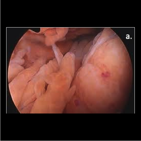
Figure 1a. Pigmented villonodular synovitis (PVNS).
-

Figure 1b. Tram lesion
-
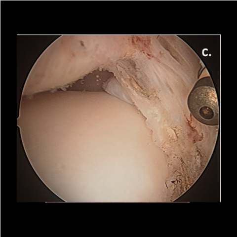
Figure 1c. Lateral gutter fibrosis
-
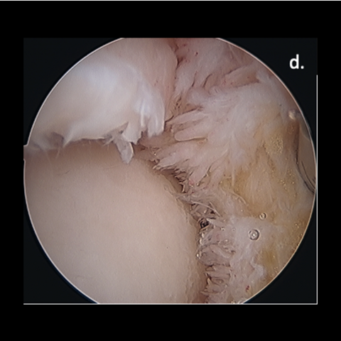
Figure 1d. Synovitis
-
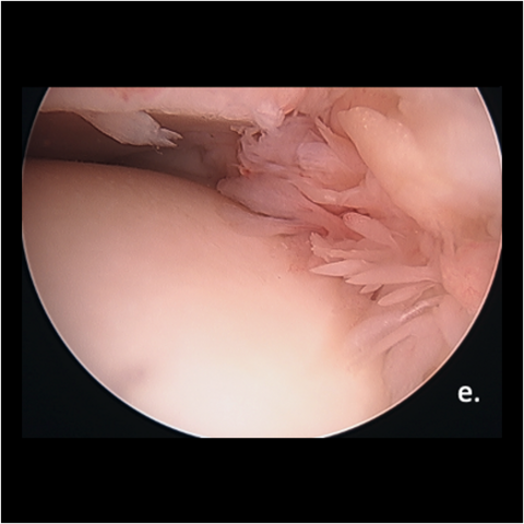
Figure 1e. Synovitis
-
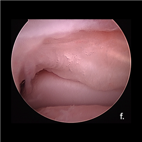
Figure 1f. Posterior tib-fib accessory ligament (posterior scope)
-
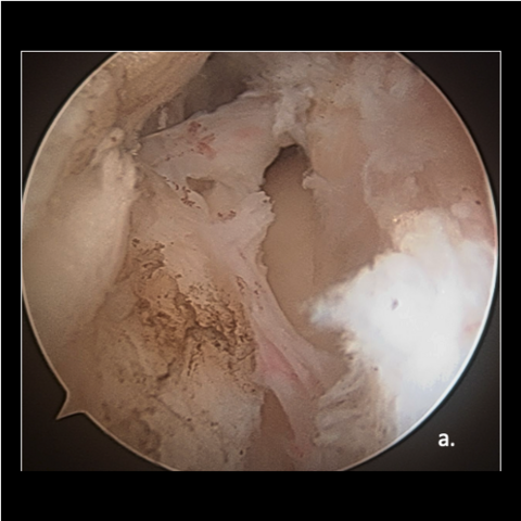
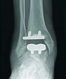
Figure 2. Postoperative scarring typically seen post–ankle ORIF
a. Initial entry into the joint after limited debridement -
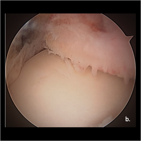
Figure 2. Postoperative scarring typically seen post–ankle ORIF
b. Post-debridement -
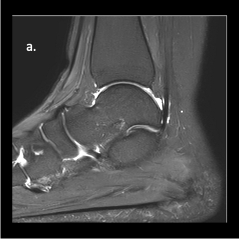
Figure 3. Anterior ankle
a. Normal fluid pattern -
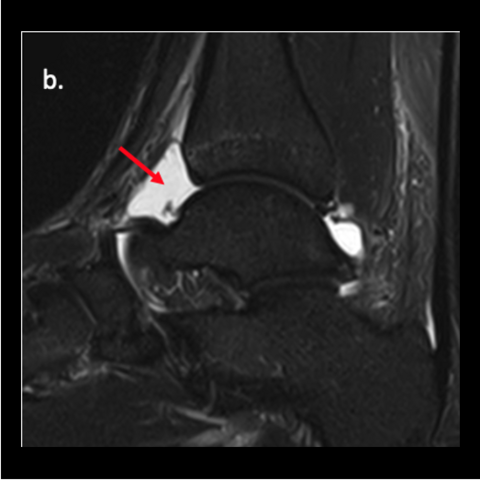
Figure 3. Anterior ankle
b. Joint effusion anteriorly, posterior fluid is within normal range
-
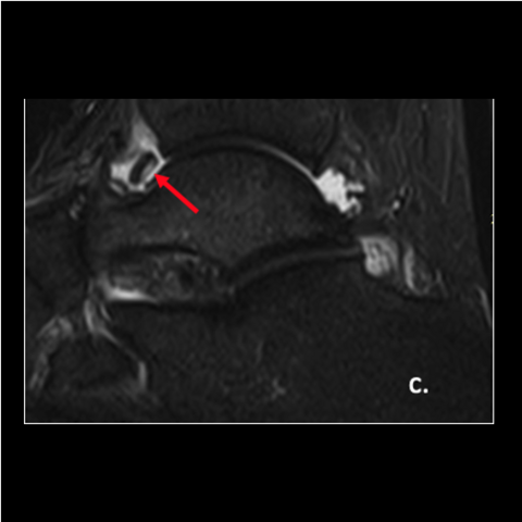
Figure 3. Anterior ankle
c. Osteochondral fragment
-
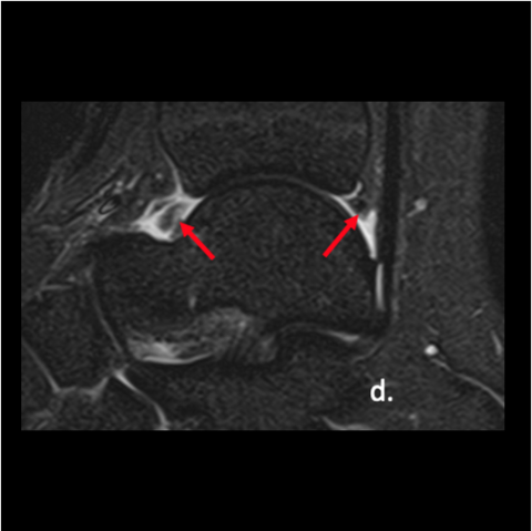
Figure 3. Anterior ankle
d. Multiple structures anteriorly and posteriorly, all abnormal -
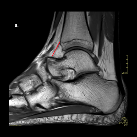
Figure 4. A 36-year-old soldier. Of note the soldier’s MRI was read as a normal MRI by the radiologist. Despite a normal reading, there are significant findings both anteriorly and posteriorly that warrant a combination scope.
a. T1 image—red line shows the anterior joint capsule -
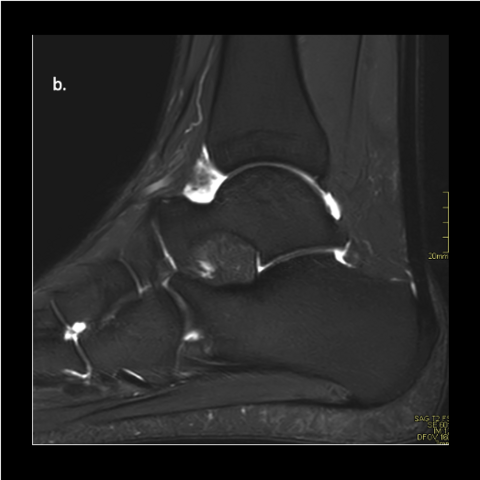
Figure 4. A 36-year-old soldier. Of note the soldier’s MRI was read as a normal MRI by the radiologist. Despite a normal reading, there are significant findings both anteriorly and posteriorly that warrant a combination scope.
b. T2 image—anterior capsule is filled with heterogenous material which is more than likely synovitis -
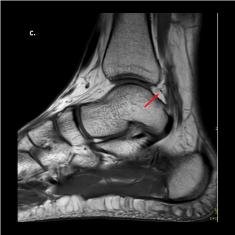
Figure 4. A 36-year-old soldier. Of note the soldier’s MRI was read as a normal MRI by the radiologist. Despite a normal reading, there are significant findings both anteriorly and posteriorly that warrant a combination scope.
c. Dense object sitting in the posterior ankle joint.
-
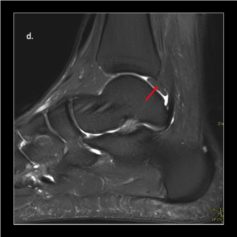
Figure 4. A 36-year-old soldier. Of note the soldier’s MRI was read as a normal MRI by the radiologist. Despite a normal reading, there are significant findings both anteriorly and posteriorly that warrant a combination scope.
d. More than likely a meniscoid band that is well mature -
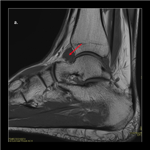
Figure 5. 35-year-old male soldier. MRI report was focused on the cartilage lesion and ignored all the soft tissue pathology, which was noted anterior and posteriorly.
a. Red arrow shows significant joint effusion anteriorly
-
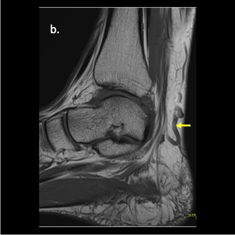
Figure 5. 35-year-old male soldier. MRI report was focused on the cartilage lesion and ignored all the soft tissue pathology, which was noted anterior and posteriorly.
b. Varicose veins -
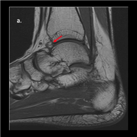
Figure 6. 30-year-old female soldier. Anterior fragment in the anterior joint capsule. Radiology report missed the fragment. Of note, the fragment was not visible on X-ray.
a. Small ossification in anterior joint
-
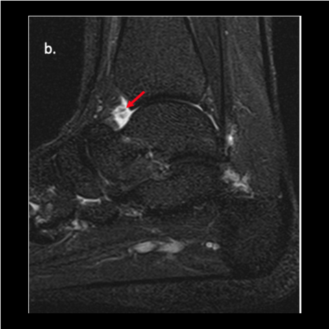
Figure 6. 30-year-old female soldier. Anterior fragment in the anterior joint capsule. Radiology report missed the fragment. Of note, the fragment was not visible on X-ray.
b. Fragment is surrounded by synovitis -
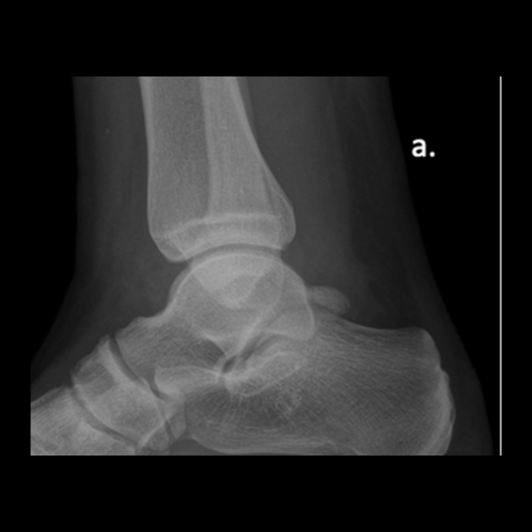
Figure 7. 25-year-old male soldier. Radiology report was normal. One only has to look at the normal X-ray and see how obvious the pathology was on the MRI. Cartilaginous pieces or non-osseous pieces often are exposed on MRI versus CT or X-ray.
a. X-ray shows no anterior fragments
-
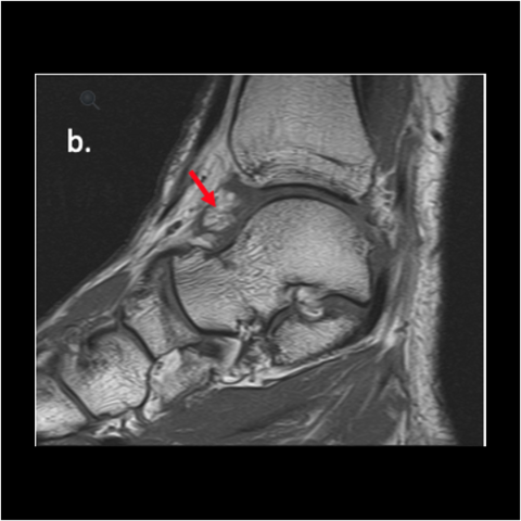
Figure 7. 25-year-old male soldier. Radiology report was normal. One only has to look at the normal X-ray and see how obvious the pathology was on the MRI. Cartilaginous pieces or non-osseous pieces often are exposed on MRI versus CT or X-ray.
b. T1 image shows anterior fragment that is dense
-
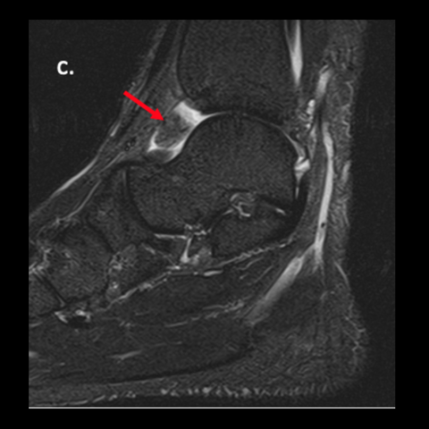
Figure 7. 25-year-old male soldier. Radiology report was normal. One only has to look at the normal X-ray and see how obvious the pathology was on the MRI. Cartilaginous pieces or non-osseous pieces often are exposed on MRI versus CT or X-ray.
c. T2 image shows the dense lesion
-
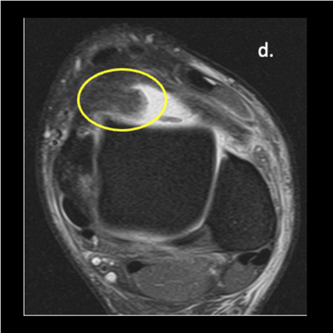
Figure 7. 25-year-old male soldier. Radiology report was normal. One only has to look at the normal X-ray and see how obvious the pathology was on the MRI. Cartilaginous pieces or non-osseous pieces often are exposed on MRI versus CT or X-ray.
d. T2 coronal shows how large the lesion is coming off the medial capsule
Click here for a related Sports Medicine column.