High-scoring SAWC Spring Abstracts
Case Series/Study
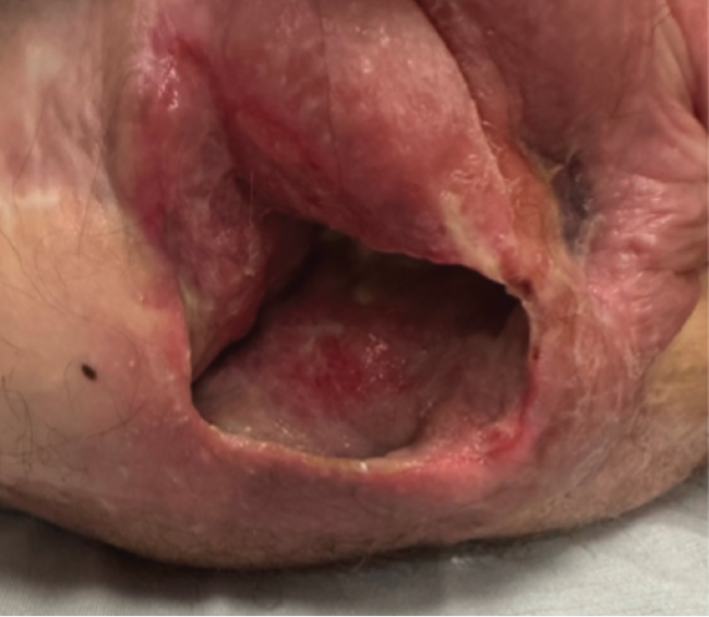
Observations on the Use of Fish Skin Graft (FSG) for the Rapid Coverage of Exposed Bone With Granulation Tissue in Mohs Micrographic Surgical Wounds of the Scalp
Ryan P. O’Quinn, MD; and Courtney Aldridge, PA-C
Introduction. Large and deep non-melanoma skin cancers treated with Mohs micrographic surgery can result in exposure of bone, especially on the skin of the scalp. These large areas of bone exposure can be a difficult management problem as there may be insufficient laxity to repair the wound with a primary closure or flap. Skin grafts may also not be practical because the avascular nature of the outer table of the bony calvarium often cannot support the nutritional needs of a graft. Complications associated with such wounds can include failed reconstructive surgical procedures (grafts and flaps), infection, slow healing, and progression of an acute post-surgical wound to a chronic nonhealing wound. FSGs have shown effectiveness in rapid coverage of exposed bone in Mohs surgical wounds of the scalp, which can expedite second-intention healing or facilitate a delayed skin graft once the bone is covered with granulation tissue. Methods. A 65-year-old female on an immunosuppressive medical regimen for renal transplantation presented with a large untreated squamous cell carcinoma (SCC) of the central top of scalp measuring 5.1 × 3.5 cm. The SCC was cleared by Mohs surgery in 2 stages, resulting in a wide wound measuring 8.3 × 5.7 cm that extended down to the bony calvarium. The wound was partially closed, but a wound measuring 3.0 × 3.5 cm remained with exposed bone at the wound base. The patient then underwent 4 applications of FSG with debridement before each application. Results. There was complete coverage of the 3.0 × 3.5 cm area of exposed calvarium with healthy granulation tissue after 4 applications of FSG. There was no infection or surrounding inflammation of the wound. Once granulation had been achieved the wound could have been grafted, but the choice was made to allow the wound to continue to heal by second intention to allow wound contracture to minimize alopecia at the site of the scar. The final scar healed nicely with excellent contour and measured 2.5 × 3.2 cm. Discussion. FSG is a biologic that has been proven to promote rapid granulation tissue formation in wounds. It has proven to be very effective in rapid coverage of wounds of the scalp with calvarium bone exposure that would be difficult to manage otherwise. Use of FSG is safe and effective in management of difficult scalp wounds, and its use may expedite healing and reduce complications in these challenging cases.
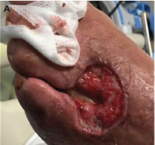
Definitive Wound Closure of a Large Scalp Mohs Defect With Exposed Cranium in an Irradiated Field Utilizing a Fish Skin Xenograft
Mark Suski, MD, FACS
Introduction. Mohs micrographic surgery is a tissue-sparing technique used to treat nonmelanoma skin cancers. Reconstruction of soft tissue defects after Mohs surgery can be a substantial challenge. Factors that exacerbate the clinical challenge include gender, modifiable risk factors, diabetes mellitus, size and severity of the defects, and the complexity of the reconstructive modality.1 In wounds with exposed underlying bone, there are limited reconstructive options. The closure could be further complicated if the area received prior radiation therapy due to insufficient blood supply, fibrosis, and attenuated cellular function.2 To this end, advanced biologics, including a fish skin graft, are often warranted. This investigation aimed to determine the clinical efficacy of fish skin in this subset of challenging patients. Methods. The patient was a 78-year-old White female who was status post-resection of a right parietal scalp sarcoma with a titanium plate cranioplasty and rotation flap reconstruction approximately 10 years ago. Her surgery was followed by adjuvant radiation therapy. Past medical history was pertinent for noninsulin-dependent diabetes mellitus and venous thromboembolism, requiring active anticoagulation. In July 2022, the patient underwent Mohs excision for a poorly differentiated squamous cell carcinoma of her vertex scalp within the prior radiation field. It required multiple passes for definitive oncological clearance. The final excision resulted in a 7 × 5 cm defect with an exposed cranium. The following day, she was taken to the operating room for bone burring and omega-3 fish skin xenograft placement. A nonadherent compressive postoperative dressing was applied, and the patient’s anticoagulation was restarted on postoperative day 1. Results. After 8 applications of the fish skin xenograft at weekly or biweekly intervals, the wound completely healed without needing a staged surgical reconstruction. Discussion. Omega-3 fish skin xenografts are FDA approved for treating chronic and acute surgical wounds.3,4 The product is an acellular dermal matrix harvested from Icelandic cod with a porous microstructure like human skin. Characteristics of the xenograft include bacterial resistance, angiogenesis, and inflammatory cytokine mitigation.5 This case report shows that a fish skin xenograft is suitable for complex wound closure, including difficult Mohs reconstruction.
References
1. Miller MQ, David AP, McLean JE, Park SS, Christophel J. Association of Mohs reconstructive surgery timing with postoperative complications. JAMA Facial Plast Surg. 2018;20(2):122-127. doi:10.1001/jamafacial.2017.1154
2. Hoang TT, Vu VQ, Trinh DT. Management of radiation-induced ulcers by singlestage reconstructive surgery: a prospective study. Ann Burns Fire Disasters. 2019;32(4):294-300.
3. Baldursson BT, Kjartansson H, Konrádsdóttir F, Gudnason P, Sigurjonsson GF, Lund SH. Healing rate and autoimmune safety of full-thickness wounds treated with fish skin acellular dermal matrix versus porcine small-intestine submucosa: a noninferiority study. Int J Low Extrem Wounds. 2015;14(1):37-43. doi:10.1177/1534734615573661
4. Lullove EJ, Liden B, Winters C, McEneaney P, Raphael A, Lantis II JC. A multicenter, blinded, randomized controlled clinical trial evaluating the effect of omega-3-rich fish skin in the treatment of chronic, nonresponsive diabetic foot ulcers. Wounds. 2021;33(7):169-177. doi:10.25270/wnds/2021.169177
5. Magnusson S, Baldursson BT, Kjartansson H, Rolfsson O, Sigurjonsson GF. Regenerative and antibacterial properties of acellular fish skin grafts and human amnion/chorion membrane: implications for tissue preservation in combat casualty care. Mil Med. 2017;182(S1):383-388. doi:10.7205/MILMED-D-16-00142
Clinical Research
Comparative Effectiveness Analyses for Medicare Beneficiaries With Diabetic Foot Ulcers Treated With and Without Hypothermically Stored Amniotic Membrane (HSAM)
Urvi Desai, PhD; Bradford Rice, PhD; Jian-Yu E, ScD; Serena Kongara, MPH; Justin Chun, MHS; and Robert Kirsner, MD, PhD
Introduction. HSAM is intended for use as a placental allograft wound covering for the management of acute and chronic wounds, including diabetic foot ulcers (DFUs). However, there is limited information about the real-world clinical and economic outcomes associated with using HSAM for people with DFUs. Methods. The 100% Medicare Fee-For-Service data in Standard Analytic File (Q1 2015-Q4 2021) were used to identify 2 mutually-exclusive cohorts of beneficiaries with DFUs: (i) beneficiaries who received HSAM (first claim considered as index date), and (ii) beneficiaries who received advanced wound care (eg, debridement, offloading, negative pressure wound therapy) but not HSAM (index date selected at random). Beneficiaries were required to have continuous enrollment in Medicare Parts A and B at least 6 months before and after the index date. Beneficiaries receiving HSAM were matched 1:1 to those not receiving HSAM using propensity score matching. Outcomes over 6 months post-index were compared between matched cohorts using statistical tests for paired data. Standardized difference (SD) >10% and P values < .05 were considered statistically significant. Results. Among all beneficiaries meeting the selection criteria, 3499 received HSAM and 589 330 did not receive HSAM. Compared to beneficiaries not receiving HSAM, those receiving HSAM had greater disease severity before treatment initiation as indicated by longer duration of active ulceration (6.5 vs 3.7 months), higher rates of DFU-related infections (62.8% vs 49.1%), more non-traumatic lower-limb amputations (11.3% vs 6.9%), and higher total medical costs ($31 855.9 vs $24 132.4). During a 6-month follow-up period, matched beneficiaries receiving HSAM (N = 3465) had lower rates of non-traumatic lower-limb amputations (9.6% vs 11.1%; SD = -4.64%; P =.05), as well as shorter stays and lower costs for hospitalization (4.5 vs 6.0 days; $9727.6 vs $12 161.4) and skilled nursing facility (3.7 vs 7.1 days; $1634.8 vs $2776.8; SD >10% and P <.001 for all comparisons) than similar beneficiaries not receiving HSAM. Discussion. The study suggests that HSAM is disproportionately used to treat more complex beneficiaries with more severe DFUs. After adjusting for baseline differences, beneficiaries receiving HSAM had lower rates of amputations and costs for hospitalization and skilled nursing facility visits than those not receiving HSAM.
Trends in Pressure Injury Prevalence Rates and Average Days to Heal Associated With Adoption of a Comprehensive Wound Care Program and Technology in a National Skilled Nursing Organization
Heba Tallah Mohammed, PhD; David Mannion, BA; Amy Cassata, RN, WCC; and Robert Fraser, MN, RN
Introduction. A large skilled nursing facility (SNF) in the United States adopted a holistic wound care model that included an artificial intelligence (AI) wound care management solution (WCMS) to improve pressure injury (PI) care. This descriptive evaluation study aimed at comparing the trend in PI point prevalence rates and average days to heal linked to adopting technology in practice from 2021 to 2022 for the same 8-month period (Jan-Aug). The study also assessed the rate of fines associated with PIs (F686 citations) in facilities that adopted the technology compared to those that did not. Methods. The study used the WCMS database to compare anonymous PI wound evaluations assessed in 2021 (15 583 patients) vs 2022 (30 657 patients) from all SNF facilities that adopted the technology in 2021 (69 centers) and 2022 (128 centers). Results. There was a 13.1% reduction in PI prevalence from 2021 to 2022 across all stages of PIs. For facilities that adopted the technology for more than 1 year, the reduction in prevalence reached 15.9%. The adopted branches in 2022 experienced a 37.4% reduction in average days to heal a PI, which equals an average of 17.7 days saved per PI compared to 2021 (P <.001). All stages showed a significant reduction in the average days to heal, with the most significant savings observed for stages 3 and 4, with an average of 35 days per stage 3 and 85 days per stage 4 PI in 2022 vs 2021 (P <.001). Overall, there was a 12.8% reduction in F686 citations reported in facilities that adopted the technology compared to the control group in 2021 to 2022. Only 1.8% of facilities using the technology in 2022 received a citation, compared to 16.5% in the control group. Discussion. With the estimated decrease in the prevalence of PI, there are up to $940 000 in savings in additional out-of-pocket expenditures. Reducing the average number of days to heal is linked to a potential savings of 29 872 hours of clinicians’ time caring for and applying dressings for PI. These savings highlight the necessity of following a model where digital tools are fully integrated into the health care system.
Evidence-based Practice
The Role of Photos as Supporting Documentation in Substitutes for the Treatment of Diabetic Foot Ulcers and Venus Leg Ulcers
Douglas A. Wright, DO; H.A. Wyatt, MD, PhD; Francisco Perez-Clavijo, DPM; Keith Van Meter, MD; and Francis G. James, SOC
Introduction. As part of the comments to the proposed LCD DL35041 and DA 54117 changes for Substitutes for the Treatment of Diabetic Foot Ulcers and Venous Leg Ulcers, the authors presented the case for photo documentation as a strategic part of the medical record that can greatly improve validation and transparency in medical assessment as well as serve as a deterrent to abuse by raising the burden of proof to meet the standard of practice. Methods. First, the authors reviewed the methods utilized by CMS to establish efficacy of skin substitutions in the area and found that they relied decisively on photo documentation in their review and conclusions. Second, the authors reviewed the current best practices regarding photo documentation in wound care in general and for lower extremity skin substitutes and found that it agreed with CMS methods stated above. However, photo documentation is not a required supporting document in submitting for reimbursement. Results. Proper and accurate photo documentation provides transparency and validation of the medical assessment for skin substitutes and improves the burden of proof, which can deter abuse. The proposed solution is to adopt photo documentation for skin substitutes as a means of defending proper use from unwarranted regulatory restrictions by providing clear, established evidence that is already part of the accepted medical record. Discussion. The poster would pose discussions on the benefits of the approach to stakeholders and the exploration of more standardization around photo documentation.
Implementation of Hospital-acquired Pressure Injury (HAPI) Preventative Bundle (HAPIPB) in Critical/Intensive Care Environment: A Quality Improvement (QI) Project
Red Tumang, DNP, MSOL, MSN, RN, CCRN, NEA-BC, CPHQ, WTA-C
Introduction. National Database of Nursing Quality Indicators had estimated that the average HAPI costs about $50 000 to $150 000 per injury, and mortality increases by 12%. Roughly 2.5 million patients with a HAPI are treated in US health care facilities annually, and 60 000 patients in US hospitals die each year from complications related to HAPI. There are 322 946 reported cases of Centers for Medicare & Medicaid Services (CMS) patients with HAPU/HAPI as a secondary diagnosis in each case. With an average charge of $40 381 per case, the annual total cost is $13 billion. Sixty-two percent of HAPI are surgical patients, while 76% are ICU patients and 81% are admitted patients. CMS offers no reimbursement for HAPI, and there are considerable fines and litigation for the development of HAPI. Patient safety, comfort, morbidity, and mortality are at risk. Methods. The objective was to investigate the reduction of HAPI incidence and prevalence in the adult critical care or intensive care patient population environment using HAPIPB and address any clinical knowledge gaps for change management of HAPI. Does a HAPIPB assist in reducing HAPI rates in the adult critical care and/or intensive care patient population of a level 1 academic trauma medical center within a 5-week timeframe? This was a pre-post comparative study analysis design using the HAPIPB and AHRQ toolkit questionnaires (N = 1120; pre = 560, post = 560 in 8 weeks). Results. There were 3 confirmed HAPI cases prior to HAPIPB implementation. Post-implementation resulted in zero HAPI cases on December 2021. A total of 1120 charts were audited in 8 weeks, and indicators included: 1) 42.3% increase in documentation of skin monitoring; 2) 69.9% increase in positioning documentation; and 3) 88.9% increase of nursing staff documenting upon admission. Z scores of each behavior are as follows: -15.201, -19.723, and -22.271, which is less than the standard alpha and asymp Sig (2-tailed) of less than .05, which concludes a difference between 2 sets of data that is statistically significant. Discussion. Positive clinical management of HAPI behavior changes factored into the overall reduction of HAPI rates. Pilot ICU had 3 HAPI cases reported in November 2021. After HAPIPB, follow-up data extraction revealed zero HAPI cases as of December 2021 and zero HAPI quarterly in 2022. These findings support the HAPIPB on reducing HAPI rates in adult inpatient critical/intensive care.
Health Economics
Short-stretch Cohesive 2-layer Compression System Improves Venus Leg Ulcer Clinical, Safety, and Resource Use Outcomes in Real World Clinical Settings
Christine Bongards, PhD; and Leah P. Griffin, MS
Introduction. The study aim was to evaluate short-stretch cohesive 2-layer compression system (C2L) for venous leg ulcer (VLU) care at US wound clinics. Methods. The VLUs were documented between January 2018 and August 2021 in the US Wound Registry, with a minimum 2 weeks of compression care. VLUs treated with greater than 50% C2L (>50%C2L) applications were compared to any other compression care (AOC). Outcomes analyzed included: healing rates, time-to-heal, complication rates, and resource use. Results. VLUs were managed with >50%C2L (n=2744) or AOC (n=27 055). There was no baseline difference in VLU Wound Healing Index.1 Compared to AOC, VLUs managed with >50%C2L had significantly higher healing rates at 4, 8, 12, and 16 weeks (P <.0001). At week 4, wound healing rates with >50%C2L and AOC were 22.0% vs 17.9%, respectively, and 78.4% vs 69.9% at week 16. Time-to-heal was 68 days with >50%C2L vs 81 days with AOC, and the duration of wound clinic care was 77 days vs 93 days, respectively (P <.0001). The weekly wound visits were reduced significantly (P <.0001) from 3.0 visits per week with AOC to 2.1 visits per week with >50%C2L. Patients in the >50%C2L group experienced significantly lower rates of adverse events (P <.0001), additional VLUs (20% lower), hospitalizations (88% lower), VLU infections (35% lower), and fewer patients required antibiotic prescriptions (29% lower; P <.0001). With >50%C2L, the frequency of weekly visits was 30% lower, and the overall duration of wound care was shorter by 16 days (P <.0001). This allows a conservative projection of $1376 mean cost saving per VLU, based on mean $86 per day VLU care cost.1,2 Discussion. Based on the significantly improved clinical, safety, and resource use outcomes, C2L has strong potential for cost-effectiveness in real-world conditions, to be confirmed in further research.
References
1. Robles-Tenorio A, Lev-Tov H, Ocampo-Candiani J. Venous leg ulcer. [Updated 2022 Sep 18]. In: StatPearls [Internet]. StatPearls Publishing; 2022. https://www.ncbi.nlm.nih.gov/books/NBK567802
2. Ma H, O’Donnell TF Jr, Rosen NA, Iafrati MD. The real cost of treating venous ulcers in a contemporary vascular practice. J Vasc Surg Venous Lymphat Disord. 2014;2(4):355-361. doi:10.1016/j.jvsv.2014.04.006
Health Economic Evaluation of Two Types of Compression Systems Used for the Local Treatment of Venous Leg Ulcers, Leading to Conclusions, Whose Relative Implications are Likely to be Universal Irrespective of Geography
Debashish Chakravarthy, PhD
Introduction. Venous leg ulcers represent a burden on the community and on patients. The study estimates the rate of complete healing of VLUs via a compression system that is available in the United States and France. Secondary objectives include estimation of healing time of leg ulcers and the cost of treatment per healed ulcer. The relative differences in clinical values that have an impact on the treatment costs should be universally true for all countries. Methods. Data were obtained from the French national health database, reflecting 99% of the population residing in France during 2018-2020. The authors examined data for all patients with a first episode whether healed or not over the study. The probability of healing over time was modeled by a Kaplan-Meier curve for all patients and based on the general or specific type of product used. Two general product types were studied first: multilayer bandages (MLB) and short stretch bandages (SSB). The costs are linked to the pathology and reimbursed by the French National Healthcare System for healed patients (21 655 patients). In addition, the healing data from 1 specific SSB and 1 MLB called a dual compression system (DCS), so called as it contains both a short stretch and a long stretch bandage, were studied. Results. Of 25 255 patients who used MLB or SSB over the entire duration of treatment, the healing rate at 3 months was significantly higher with MLBs than for all SSBs at 42% vs 35%. At 1 month, a 25% higher chance of healing was seen with MLBs vs SSBs (P <.001). The median healing time for venous leg ulcers were estimated at 137 days (SSB group) versus 115 days in the MLB group. Very similar results were observed in comparing specifically the 2 most represented brands for each type of bandage, one of which is an SSB and the other is an MLB, the DCS system. Finally, due to the reduction of the healing time, the MLB systems as a group reduced the average treatment cost per patient by 20% compared to SSB group (€2875 and €3580 respectively). Discussion. This new real-life data from a very large cohort of patients confirms the superior efficiency of the multilayer compression bandage in healing rate, time to heal, and health economic savings compared to simple short stretch systems.
Laboratory Research
The Antimicrobial and Wound Healing Effects of a Novel Hydrogel Combat Gauze With Copper on Third-degree Burns Using a Porcine Model
Michael Solis, MBA; Jie Li, MD, PhD; Joel Gil, BS; Alexander Higa, MA; Aaron Strickland, PhD; and Stephen C. Davis, BS
Introduction. Methicillin-resistant Staphylococcus aureus (MRSA) is one of the most commonly encountered bacteria in the burn unit and can be challenging to eradicate.1 Copper has been previously shown to have antimicrobial activity against various pathogens.2 There have also been reported wound healing effects with the use of copper.3 This study was performed to investigate the antimicrobial and wound healing activity of 2 novel hydrogel formulations with copper and their ability to enhance the healing using a third-degree burn porcine model.4 Methods. Ninety-six (96) third-degree burn wounds were created on pig models, and within 20 minutes after wounding all wounds were infected with MRSA USA300 and treated with one of the following treatments: 1) hydrogel gauze with copper H1-C (Copper H1-C), 2) hydrogel gauze with copper H2-A (Copper H2-A), 3) gelling fiber dressing with silver (GFD-Ag), or 4) untreated control. All wounds were covered with a polyurethane film dressing and assessed on days 3, 6, 14, and 21. Incisional and punch biopsies were obtained for histological and microbiology assessments, respectively. Results. Copper H1-C and Copper H2-A showed a significant (P ≤.05) reduction in MRSA counts when compared to GFD-Ag and untreated control wounds at all assessment days. Both copper dressings showed more than 99.8% reduction in all assessment days. On days 14 and 21, wounds treated with Copper H1-C resulted in a MRSA reduction of 3.55±0.10 and 3.58±0.10 Log CFU/g bacterial, respectively. These values represent more than 99.97% reduction in both days. Wounds treated with Copper H1-C resulted in an increase in the epithelialization percentage on days 14 and 21 as compared to the other treatment groups. All treatment groups showed a significant (P ≤.05) increase in the epithelialization by day 21. Discussion. Overall, the hydrogel copper gauze treatments were effective in reducing MRSA-infected wounds while enhancing the healing process. These findings suggest that this treatment may be an important armamentarium for wound care providers in combating wound infections and accelerating the healing of wounds.
References
1. Kalligeros M, Shehadeh F, Karageorgos SA, Zacharioudakis IM, Mylonakis E. MRSA colonization and acquisition in the burn unit: a systematic review and meta-analysis. Burns. 2019;45(7):1528-1536. doi:10.1016/j.burns.2019.05.014
2. Grass G, Rensing C, Solioz M. Metallic copper as an antimicrobial surface. Appl Environ Microbiol. 2011;77(5):1541-1547. doi:10.1128/AEM.02766-10
3. Sen CK, Khanna S, Venojarvi M, et al. Copper-induced vascular endothelial growth factor expression and wound healing. Am J Physiol Heart Circ Physiol. 2002;282(5):H1821-H1827. doi:10.1152/ajpheart.01015.2001
4. Rodriguez-Menocal L, Davis SC, Guzman W, et al. Model to inhibit contraction in third-degree burns employing split-thickness skin graft and administered bone marrow-derived stem cells. J Burn Care Res. 2023;44(2):302-310. doi:10.1093/jbcr/irac119
Light-deactivated Adhesive: The Solution to Maintaining Dressing Integrity and Protecting Skin
Jonathan Cayce, PhD, MS; Lily Goins, AS; and Gavin Warrington, MS
Introduction. Surgeons take extra precautions to protect against surgical site infections and surgical site complications by using incisional negative pressure therapy (iNPWT). Surgeons use iNPWT over joints or other areas of the body subjected to forces that stress the adhesive of the dressing. Most dressings use strong acrylic adhesives to keep the dressing in place, but these can cause medical adhesive-related skin injuries (MARSI).1,2 Surgeons need an incisional dressing that balances dressing security and protects healthy skin. A new light-deactivated adhesive supplies strength but releases when exposed to a specific wavelength of light. This research compares the light-deactivated adhesive to acrylic and silicone adhesives used in incisional dressings. Methods. The study used peel strength testing to compare the light-deactivated adhesive to 2 acrylic adhesives and a silicone adhesive. Testing required 1 × 2-inch samples obtained from iNPWT dressings or sheets of transparent film dressings. The light-deactivated adhesive had 2 groups, unexposed and exposed. Researchers randomly adhered the samples in groups of 6 on a healthy volunteer’s ventral forearm using 1 inch of the sample. They secured the second half with a clamp on a universal testing machine. The universal testing machine pulled each sample at a 90° angle at 5 mm per second. The results report the average maximum peel strength (Newtons) and 95% confidence interval. Statistical analysis used a one-way analysis of variance followed by paired-wise t tests. Reported P values underwent adjustment using the Benjamini-Hochberg method for multiple comparisons. A P value of less than .05 indicates a significant difference. Results. Unexposed light-deactivated adhesive exhibited an average maximum peel strength of 2.92±0.72 N compared to acrylic A 2.81±0.96 N, acrylic B 1.81±0.62 N, and silicone 1.19±0.64 N. Exposed light-deactivated adhesive decreased in average maximum peel strength to 0.30±0.14 N. The ANOVA indicated a significant difference (P =3.19 E-06). Pairwise testing found that the exposed light-deactivated adhesive had a significantly lower peel strength than all other conditions (P value range, .0008–.0492). The unexposed light-deactivated adhesive had significantly higher peel strength than silicone (P =.005) and acrylic B (P =.0492). All other comparisons showed no difference. Discussion. The study results show a profound decrease in peel strength in the exposed light-deactivated adhesive compared to its unexposed state. Using the light-deactivated adhesive in surgical and other wound care dressings will help to address challenges in securing a dressing while preventing MARSI.
Practice Innovations
Identification of Skin at Risk for Foot Ulceration Utilizing Near Infrared Spectroscopy Imaging
Charles Andersen, MD; Homer-Christian Reiter, BS
Introduction. Foot deformities and/or altered foot biomechanics result in areas at risk for foot ulceration. Foot ulceration can lead to major complications including infections, sepsis, and minor or major amputations. Inflammation over boney prominences—for example, hammer toes, bunions, or bony prominences associated with Charcot foot—can lead to ulceration. Early detection of inflammation and proactive offloading can prevent ulceration and the associated complications. Near infrared spectroscopy (NIRS) imaging is a tool that can quickly measure superficial oxygenation. A focal increase in oxygenation is a surrogate marker for inflammation. This research explored utilizing NIRS for identification of areas at risk followed by focused offloading to prevent ulceration. Methods. Patients with intact skin but who had bony prominences due to deformities were imaged with NIRS in areas that were identified as high risk for ulcer development. If areas of significantly increased tissue oxygen saturation (StO₂) were identified on the image, proactive offloading was provided to the patient. Patients were followed to determine if a wound developed in the area identified as high risk. Results. Within 7 patients, 10 areas of inflammation over bony prominences in feet were identified. Areas of bony prominences included were as follows: 2 hammer toes, 3 hallux adbucto valgus, 1 first metatarsal head, and 3 fifth metatarsal heads. Within the 10 areas of inflammation, there was significantly higher StO₂ (79.6%±10.5) over the boney prominences compared to the StO₂ in the surrounding tissue (55%±6.6; P <.001). On follow-up after proactive offloading was provided, 1 area developed an ulceration, 9 did not. Discussion. In this study, NIRS was able to identify areas of increased oxygenation (inflammation) over bony prominences. Being able to identify the area of inflammation prior to ulceration facilitated a proactive approach of offloading to prevent ulceration. NIRS was also used in serial visits to track the effectiveness of offloading in reversing inflammation. Review of images with the patient can also be an excellent educational tool to reinforce the importance of offloading, thus potentially improving adherence.
A Versatile Framework to Quickly Implement Wound Care-specific, Role-based Competency Programs
Elaine H. Song, MD, PhD, MBA; Catherine T. Milne, APRN, MSN, CWOCN-AP; Tiffany Hamm, BSN, RN, ACHRN, CWS; Nataliya Lebedinskaya, RN, BSN, CWOCN, WOC; Janis Prado, RN, CWOCN; and Jeffrey Mize, RRT, CHT, CWCA
Introduction. Staffing shortages have been a top patient safety concern.1 The need to quickly onboard new clinicians and ensure competent performance is compounded by the lack of a standardized approach to education/training in wound care.2,3 It has been shown that organizations with competency programs have 40% lower turnover and 87% greater ability to hire the best people.4 However, consistently ensuring clinicians’ competency in wound care is challenging, given time/resource constraints. To address these needs, the authors aimed to create a framework to quickly implement role-specific, wound care competency programs. Methods. Using the Design Thinking methodology,5 the framework was created leveraging a versatile wound care-specific learning management module within a clinical/reimbursement decision support web-application: 1) managers/clinicians’ needs and role-based competency areas were mapped; 2) role-based competency templates/training modules featuring evidence-based content, continuing education credits, and skills were built; 3) a playbook for customization of the competency program was created; and 4) the framework was implemented in several organizations. Results. The framework is a digital solution that enables organizations to quickly implement/manage/document wound care-specific, role-based competencies. Use cases include: 1) ongoing competencies for acute care nurses: to achieve quality goals, a hospital implemented customized pressure ulcer/injury prevention competencies for 330 nurses, cutting down educational program development time by 80%; 2) ongoing competencies for certified wound care specialists: to complement their organization’s generic competencies and ensure their own competencies addressed their job duties/responsibilities, specialists implemented wound-care specific competencies, reinforcing regulatory/accreditation compliance; 3) preceptorship for professional certification: to address lack of local qualified supervisor, candidate completed a preceptorship module utilizing an in-person/remote approach and met preceptorship requirements to become a Certified Hyperbaric Technologist. Discussion. A framework to quickly deploy wound care-specific, role-based competency programs and meet continuing education/certification/compliance requirements was successfully developed/implemented. Its versatility may help organizations address staffing turnover by decreasing onboarding time and increasing talent retention.
References
1. ECRI. ECRI Reports Staffing Shortages and Clinician Mental Health are Top Threats to Patient Safety [Internet]. ECRI.2022. https://www.ecri.org/press/ecri-reports-staffing-shortages-and-clinician-mental-health-are-top-threats
2. Williams EM, Deering S. Achieving competency in wound care: an innovative training module using the long-term care setting. Int Wound J. 2016;13(5):829-832. doi:10.1111/iwj.12388
3. Corriveau G, Couturier Y, Camden C. Developing competencies of nurses in wound care: the impact of a new service delivery model including teleassistance. J Contin Educ Nurs. 2020;51(12):547-555. doi:10.3928/00220124-20201113-05
4. Garr S. Integrated Talent Management: A Roadmap for Success. Research Bulletin. 2012 Oct 19.
5. Ferreira FK, Song EH, Gomes H, Garcia EB, Ferreira LM. New mindset in scientific method in the health field: design thinking. Clinics (Sao Paulo). 2015;70(12):770-772. doi:10.6061/clinics/2015(12)01