


Novel Use of Corindus Robotic Heart Catheterization System for Tryton Side Branch Stent Placement
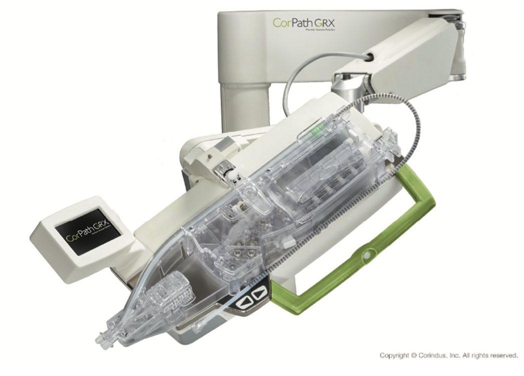
 The CorPath GRX robotic heart catheterization system (Corindus Vascular Robotics) is the first FDA-approved robotic platform designed to assist interventional cardiologists with percutaneous coronary intervention. It is designed to decrease radiation exposure to the cardiologist and also features enhanced control of catheter advancement and stent placement with sub-1 mm intraoperative measurements (Figure 1).1
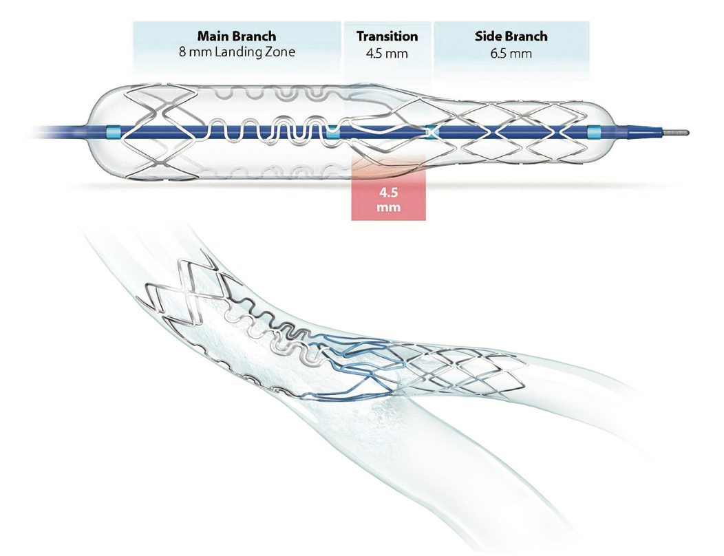
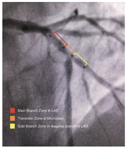
The Tryton side branch stent (Cordis, A Cardinal Health company) is the first FDA-approved stent designed specifically for bifurcation lesions. The specialized design of the Tryton stent has three distinct zones demarcated with radiopaque markers for proper placement at the side branch, which allows for minimal metal-to-artery ratio in the proximal segment and ease of integration of the traditional drug-eluting stent (DES) in the main vessel (Figure 2).2 In this report, we describe a novel utilization of the CorPath GRX robotic heart catheterization system for precise placement of a Tryton side branch stent.
Case Report
A 58-year-old woman with progressive anginal symptoms over several weeks presented to the outpatient clinic for further evaluation. She had a history of dyslipidemia and family history of premature coronary artery disease. Physical exam revealed normal heart sounds and rhythm, without jugular venous distention. The rest of the physical exam was unremarkable. Electrocardiogram showed normal sinus rhythm with no significant ST segment changes. Duke treadmill stress testing showed diffuse, 3 mm ST depression two and a half minutes after the start of exercise. Precordial pain was improved with sublingual nitroglycerin and the patient was optimized on beta blockers, aspirin, and statin. Transthoracic echocardiogram showed normal left ventricular function without hemodynamically significant valvular or pericardial disease.
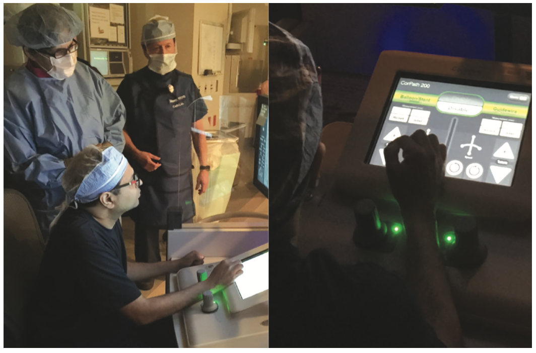
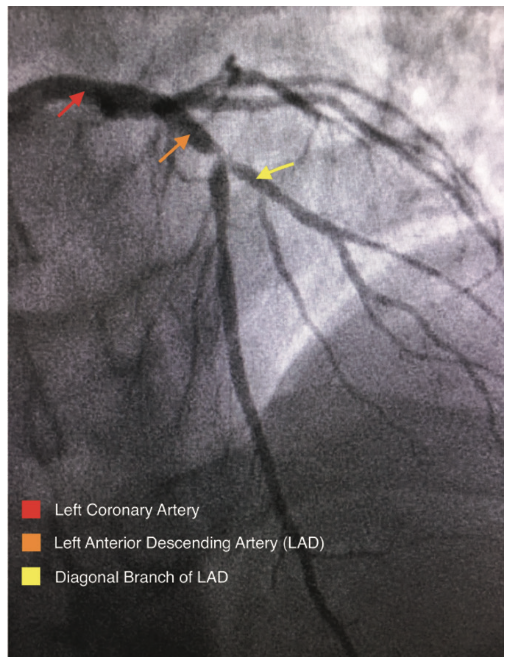
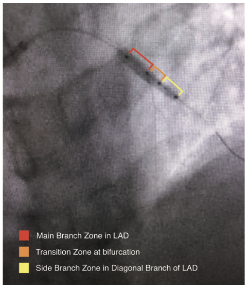
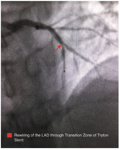
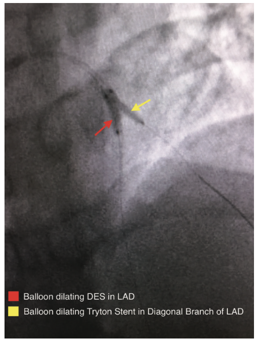
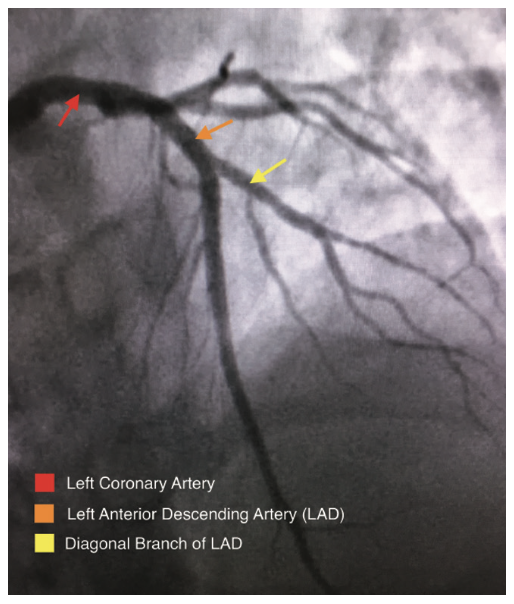
A coronary angiogram was performed, revealing a critical bifurcation stenosis of the mid left anterior descending artery (LAD) and a large diagonal branch vessel which was felt to be the culprit vessel causing her symptoms (Figure 3). Using the CorPath from a 6 French radial approach, a guidewire was placed across the lesion into the diagonal branch of the LAD and a second guidewire was placed across the lesion into the mid LAD (Figure 4). After pre-dilating the lesion, the CorPath was used to measure the longitudinal length of the diagonal branch at the ostium. An appropriately sized Tryton stent was placed over the diagonal branch guidewire and advanced to the site of the lesion. The stent was then carefully advanced by utilizing the CorPath such that the mid-markers precisely straddled the ostium of the diagonal branch to within 1 mm (Figure 5). The Tryton stent delivery system balloon was inflated and the stent was successfully deployed in an appropriate position (Figure 6). The Tryton stent balloon was withdrawn and the diagonal branch guidewire was rewired through the transition zone of the Tryton stent into the main vessel distal to the ostium of the diagonal branch, taking care not to retract proximal to the Tryton stent. Once the guidewire was successfully placed in the LAD through the transition zone, the “trapped” original LAD guidewire was removed (Figure 7). The length needed for the LAD stent was measured from the proximal edge of the Tryton stent to beyond the distal edge of the LAD lesion, and a drug-eluting stent (DES) was advanced over the same guidewire into position within the main branch zone of the Tryton stent and extending into the LAD, and then deployed. After removal of the DES balloon, an additional guidewire was selected and advanced into the diagonal branch, taking care to ensure that it was advanced within the lumen of the LAD stent. Two appropriately sized balloons were selected and advanced into position in the diagonal branch and LAD along their respective guidewires. Simultaneous kissing balloon inflation was performed (Figure 8). Repeat angiography confirmed adequate stent expansion and demonstrated dramatic improvement in coronary perfusion (Figure 9).
Discussion
The treatment of bifurcation lesions with percutaneous intervention continues to present a unique challenge to interventional cardiologists. Due to the geometric limitations of traditional drug-eluting stents, the procedure often requires additional time and can be technically difficult. Treatment of bifurcations has been traditionally performed with various techniques, using traditional stents, but each strategy has its own advantages and limitations, and no technique is suitable for every lesion. Several creative methods have been employed, including provisional stenting, V-stenting, simultaneous kissing stent double barrel technique, the crush technique, T-stenting, and the culotte technique, each with its specific advantages and limitations. The common practice of provisional stenting is often susceptible to complications such as restenosis of the side branch. Other stenting techniques using traditional stents can create problems with complete lesion coverage and increased metal-to-artery ratio in the proximal segment.3 As interventionalists search for more definitive treatment of these common lesions, there have also been reports of modifications of the above techniques from various approaches, such as a recently described modified crush technique from a radial approach.4 The Tryton side branch stent, which was also placed using a radial approach in this case, incorporates a Tri-Zone technology design with a specially sized side branch zone, a transition zone for full coverage of the ostium with proper placement, and a main branch zone with minimal metal-to-artery ratio for easier rewiring of the main vessel in order to integrate a DES. These zones are demarcated with radiopaque markers to aid in precise placement at the side branch prior to rewiring the main branch. Accurate placement of the Tryton stent is paramount for full vessel coverage of the side branch with the side branch zone, while keeping the ostium of the main branch open for efficient rewiring and placement of traditional DES.5
The CorPath GRX, while originally designed for reduction in radiation exposure, features sub-millimeter measurement capabilities and enhanced wire advancement control. It has been suggested that this unique capability may increase the accuracy of stent length selection and placement, and decrease the incidence of longitudinal geographic miss.6 In the case of Tryton stent placement, longitudinal accuracy is especially important; it is possible that accurate deployment of the Tryton stent may be more efficiently and consistently obtained with the use of the Corindus robotic system, potentially leading to improved patient outcomes. The placement of the Tryton stent with the Corindus robotic system proved successful in this particular case and further prospective study of this technology combination in comparison to manual stent placement, both with Tryton side branch stents and traditional drug-eluting stents, is warranted to determine differences in procedure efficiency and efficacy.
Conclusion
The Tryton side branch stent is a specially designed stand-alone device that has been shown to effectively stent bifurcation lesions. The CorPath GRX robotic heart catheterization system was developed for reducing radiation exposure to the interventionalist, but also features precise intraoperative measurement capability and wire control. We describe a novel use of this measurement capability, which may enhance precise delivery of the Tryton stent and allow for facilitated reentry with a traditional DES in the main vessel.
References
- Maor E, Eleid MF, Gulati R, et al. Current and future use of robotic devices to perform percutaneous coronary interventions: a review. J Am Heart Assoc. 2017 Jul 24; 6(7). pii: e006239. doi: 10.1161/JAHA.117.006239.
- Tryton Medical Receives FDA Approval for Tryton Side Branch Stent to Treat Significant Coronary Bifurcation Lesions [Press release]. Available online at https://www.trytonmedical.com/tryton-medical-receives-fda-approval-tryton-side-branch-stent-treat-significant-coronary-bifurcation-lesions/. Accessed February 2, 2018.
- Finch W, Lee MS. Percutaneous coronary intervention for coronary bifurcation lesions. Rev Cardiovasc Med. 2017; 18(2): 59-66.
- Marrero O, Tai Z. Modified crush via 6 French radial access. Cath Lab Digest. 2017 Nov; 25(11): 46-48. Available online at https://www.cathlabdigest.com/article/Modified-Crush-6-French-Radial-Access. Accessed February 2, 2018.
- Grundeken MJ, Winter RJ, Wykrzykowska JJ. Safety and efficacy of the Tryton Side Branch Stent™ for the treatment of coronary bifurcation lesions: an update. Expert Rev Med Devices. 2017 Jul; 14(7): 545-555. doi: 10.1080/17434440.2017.1338135.
- Carrozza JP. Robotic-assisted PCI – a new approach to the transcatheter treatment of coronary artery disease. Cath Lab Digest. 2012 Oct; 20(10). Available online at https://www.cathlabdigest.com/articles/Robotic-Assisted-PCI-%E2%80%93-New-Approach-Transcatheter-Treatment-Coronary-Artery-Disease. Accessed February 2, 2018.
1University of Texas Health Science Center - Texas College of Osteopathic Medicine; 2Adjunct Clinical Assistant Professor at the University of North Texas Health Science Center, Interventional Cardiologist, Heart Center of North Texas; 3Medical Director of Interventional Cardiology, Baylor Jack and Jane Hamilton Heart and Vascular Hospital; Fort Worth, Texas.
Disclosure: The authors report no conflicts of interest regarding the content herein.
The authors can be contacted via Michael Byers, MS-III, at michael.byers@my.unthsc.edu.


