Bringing Cardiology and Neurointervention Closer Together: A Conversation With Camilo R. Gomez, MD, MBA
A Conversation With Camilo R. Gomez, MD, MBA
A Conversation With Camilo R. Gomez, MD, MBA
© 2026 HMP Global. All Rights Reserved.
Any views and opinions expressed are those of the author(s) and/or participants and do not necessarily reflect the views, policy, or position of Cath Lab Digest or HMP Global, their employees, and affiliates.
Camilo R. Gomez, MD, MBA
Camilo R. Gomez, MD, MBA, can be contacted at crgomez@health.missouri.edu.
Editor's Note: A PDF of this article is available by clicking the red PDF icon to the right of the article title.
Camilo R. Gomez, MD, MBA, is a vascular, critical care, and interventional neurologist whose work helped establish interventional neurology as a field in the United States; he was also among the first physicians certified in the subspecialty after its formal recognition. Dr. Gomez popularized a new sense of urgency in stroke care through concepts such as “time is brain” and “Code Stroke,” bringing a systems-based approach familiar to interventional cardiologists into acute neurovascular treatment. His career has spanned stroke program leadership, carotid stenting, and neuroendovascular rescue, and he is a Professor of Neurology and continues clinical practice at the University of Missouri.
In this conversation, Dr. Gomez speaks about the procedural and physiologic overlap between cardiology and neurointervention, making the case that the two specialties have much to learn from each other as endovascular care moves into new vascular territories.
You came up with the phrase “time is brain.”1 Can you talk about that journey?
 Yes. I actually plagiarized it from a cardiology friend, Frank Aguirre. Frank was a resident when I was an intern, a year ahead of me, and we have known each other for many years. He is phenomenal, one of the best cardiologists I have ever known. Frank and I were lecturing in a program for the Army, and he spoke before me. He was talking about “time is muscle.” I heard that and thought, “That could be used by us.” So I wrote the editorial, got it published,1 and I kid you not, I received a lot of hate mail for that editorial from my own vascular neurology peers.
Their response was, essentially, “How dare you write this when we have no treatment for stroke?” Most people do not know that part of the story. And yet, here we are.
Then I wrote another editorial,2 showing mathematically that “time is brain” is not exactly correct, because I figured that if somebody was going to be my detractor, it might as well be me.
Time is really relative. I called it the stroke theory of relativity. Five hours of ischemia do not mean the same thing in two different patients, because of several factors, such as collaterals. Just as two lesions can have different fractional flow reserve (FFR) even though they look alike, two patients do not necessarily have the same hemodynamic circumstances. So time has a relative meaning. It is still true that faster is better, but “faster” may mean something different for Mr. Jones than it does for Mr. Smith.
What would you like the interventional cardiology field to know about acute stroke intervention?
The treatment of acute stroke has followed in the footsteps of the treatment of acute myocardial infarction. If you consider the dichotomy of ST-elevation myocardial infarction (STEMI) versus non-STEMI, or transmural versus subendocardial infarction (and I am dating myself because I go back to the older nomenclature), in the same way, in stroke, there is large arterial occlusion and small arterial occlusion. For large arterial occlusions, endovascular treatment is essentially the best way to treat these patients. For smaller arterial occlusions, I think intravenous thrombolysis does a very good job. Those are the two extremes.
The question is where they meet in the middle. Now we are treating what we call not-so-large arterial occlusions. We are moving into middle-sized vessels, particularly when the volume of tissue at risk of infarction is large enough to predict disability.3 As long as we have the means, and there is constantly new technology arriving, I think these patients can be treated. We have catheters now that simply did not exist when I started in this field in the 1990s. When I started working with Mort Kern, all we had was intra-arterial thrombolysis. Then we began moving into clot removal with snares and other tools that were not really designed for that purpose. It was painful to do, but we recognized that clot removal was the way forward.
Interestingly, you are now seeing that in the coronaries as well. Many people are doing clot removal because it is increasingly recognized that not every case of acute coronary occlusion is due to in situ plaque occlusion. The clot may come from somewhere else.
That is the bread and butter, so to speak. But from the perspective of interventional neurology, we do many other things. After I worked with Mort Kern, I worked with Gary Roubin for a number of years, and we helped bring carotid stenting to where it is today. Carotid stenting has essentially replaced carotid endarterectomy. Frankly, I do not know why anyone would have a carotid endarterectomy in 2026, especially with the latest results of the CREST-2 trial, which showed a much more significant benefit from stenting. All I could say was, that was shocking. We told them so back in 1994.
We also perform stenting in the vertebrobasilar system. We do intracranial stenting. I did an intracranial stent earlier this week. And believe it or not, to this day, I still use coronary stents inside the brain because they are the best stents around.
I published the first case of coronary stenting of the middle cerebral artery,4 so we have been doing this for a long time. There is a significant overlap between our two fields. Of course, I am biased, and I freely admit that, because I came into this through cardiology.
I will tell you an anecdote. I had recognized around 1992 or 1993 that neurologists needed to be involved in this field. At the time, interventional neuroradiology was dominated by radiologists, and because of turf battles, they would not allow neurologists to become involved. We had gone through an entire political process at Saint Louis University, where we proposed this to radiology, and radiology turned us down.
One day, I was standing in front of the elevators in the hospital and bumped into Mort Kern, whom I knew. He said, “You ought to take a look at some of the things we are doing,” because this was the beginning of balloon angioplasty and stenting. I said, “As a matter of fact, Mort, I am interested. Why don’t I just come down and start working with you and learn a few things?” He said, “Come on down.”
It was life-changing. I have written and published about this. We had a phenomenal program beginning to develop, a joint collaboration, but for political reasons, the dean essentially shut us down.5 I resigned my tenured position, left the university, and went to the University of Alabama, where I worked with Gary Roubin for a while. The rest is history.
Today, we do balloon angioplasty and stenting. We treat aneurysms with intracranial stenting, intravascular devices, and coil embolization. We do liquid and solid embolization for tumors and subdural hematomas. The newest area, and probably one of the best things we do, is embolizing patients who have had a subdural hematoma to prevent recurrence. It is not necessarily a sophisticated procedure, it is actually simple, but it is one of the most successful procedures we do, and it takes literally about 15 or 20 minutes.
Can you talk more about the use of aspiration thrombectomy and where you see this technology headed?
For us, aspiration thrombectomy has become the primary way to treat acute stroke. Back in the 1990s, we came from the era of intra-arterial thrombolysis. The problem with thrombolytic agents is that it is like owning a rattlesnake as a pet: it is not a matter of if you are going to get bitten, but when. Sooner or later, it is going to bite you.
The problem we used to face was that you would place the thrombolytic agent, and it would clear some of the vessel, but then some of it would remain there and clear the branches six hours later. By that time, the tissue is more infarcted, and it is going to bleed. It becomes a mess.
We started thinking very early about how to mechanically remove clots without having to use thrombolytic agents. We published on this, including a chapter in Critical Care Clinics on what we called neuroendovascular rescue.6,7 The idea was: how do we remove clots mechanically?
In the 1990s, we started using what we had available, which were snares. But if you can imagine trying to capture a three-dimensional structure with a snare, it is a labor of love. It could be done, but it was not reliable, replicable, or fast.
Fast-forward to the development of devices such as stent retrievers. Around 2010 to 2015, stent retrievers became the main technology. You deploy the device, which is essentially a stent that remains tethered, and then you pull the clot out. That works, up to a point.
But the more elegant and faster approach is with the newer aspiration catheters. We now have aspiration catheters that are .035 inches, which I can place into branches, including cortical branches, all the way to the cortex, and remove clots. I have images of small clots coming out and the vessel reopening. If that is an eloquent area of the brain, it may save the patient from aphasia, which is a major disability.
So, to me, aspiration is the first, second, and third step. I do not use a retriever anymore unless I have to, because we have better catheters now. Frankly, most of the time, we do not even have to hook them up to a pump. We can use a $2 VacLok syringe (Merit Medical) to aspirate the clot. I think aspiration has been very, very good for the treatment of acute stroke.
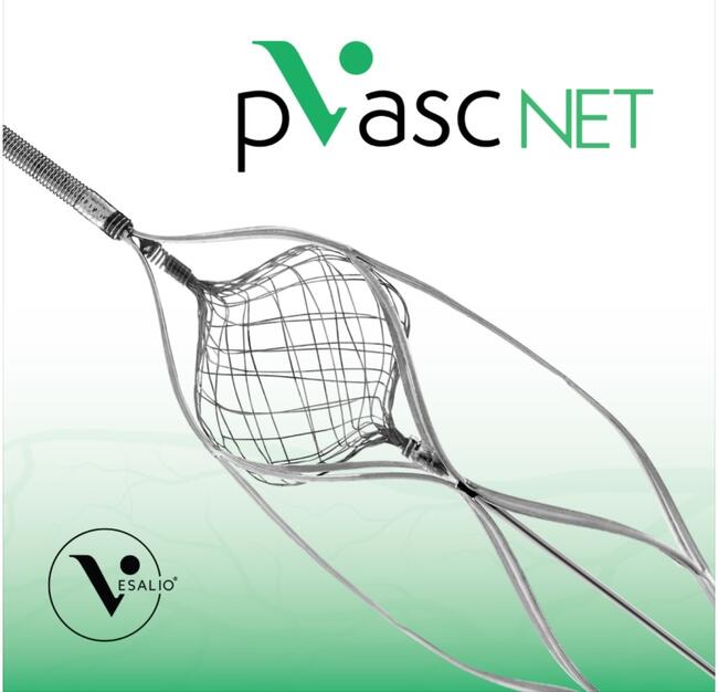
The other dimension is its use as an adjunct to other procedures. For example, there was a vertebral occlusion I treated earlier this week. The patient presented with a stroke and a vertebral occlusion right at the origin, behind the subclavian. When we finally opened a channel, we could see an intraluminal thrombus on the other side. Because it was a smaller vessel, we placed a distal protection device, which is like a little basket. We pulled the basket back, it captured the clot, we closed the basket, and we have pictures of the clot after it was removed. Then we were able to place a stent and fix the vessel without the same risk.
How do intravascular ultrasound (IVUS), FFR, and physiologic assessment fit into neurointerventional decision-making?
Let me back up to the standard of care. One of the good things for everyone is that noninvasive vascular imaging has changed and become very sophisticated. A lot of the answers we could not get noninvasively before can now be obtained through computed tomography (CT) angiography, magnetic resonance (MR) angiography, or even ultrasound. But there is still a niche of patients who require invasive diagnosis and invasive intervention.
The pathway is different. One of my partners is Adnan Qureshi, and he and I are among the most senior interventional neurologists in the United States. I met him when I first started getting involved in intervention in the 1990s, when he was a resident at Emory and I went there to lecture. That helped launch his career into neurocritical care and neurointervention, which is what he does. He and I have been thinking outside the box for many, many years. We are probably one of the only groups that systematically uses IVUS, FFR, and similar tools, which many people in the field do not even know exist.
One thing I think escapes many people is that angiography by itself is really a form of luminography. All you can see is the lumen of the vessel. The physiology, which Mort has been emphasizing for many years, and I think he is right on target, is critically important. In fact, that is one of the things that first got me interested in working with him: the physiologic measurements.
The question is whether the lesion is causing a hemodynamic effect that we can correct by stenting it or fixing the vessel. If the answer is no, then you are just wasting a good stent. You may create what I call cosmetic angiography, but you are not helping the patient. I think that distinction is very important.
So we go beyond angiography. We use IVUS, FFR, and other physiologic tools. We have actually placed IVUS in the venous sinuses of the brain for stenting in patients who have sinus stenosis with increased intracranial pressure, what used to be called idiopathic intracranial hypertension and is now called chronic cerebral venous hypertension syndrome. On the venous side, we can do that.
We start with noninvasive testing, and then for whatever remains unanswered, particularly if there is a potential intervention, we move to invasive evaluation.
For carotid stenting, particularly, I would imagine IVUS and FFR would be especially valuable.
Yes, absolutely, particularly in cases where the degree of stenosis is not critical. In most of those patients, the cat is already out of the bag. If you have someone with a perfusion scan showing decreased perfusion in the hemisphere, and an angiogram showing a near occlusion or a ruptured plaque, you can spend some time on it, but chances are you are not going to find anything surprising.
The real question, which Mort Kern and I discussed in an essay we wrote in 1997,8 is what to do with a patient who has a moderate degree of angiographic narrowing, but you still do not know whether the fractional flow reserve is sufficient. Some of those lesions will be significant, and some will not. That is where these non-angiographic diagnostic tests become useful.
The other important point is that we are probably one of the few groups systematically treating subacute carotid occlusions. For example, a patient presents with a carotid occlusion in the cervical segment. The artery is occluded, but the patient does not have a major clinical syndrome because they have collaterals. In the past, those patients might have been bypassed, but bypasses do not work very well. They often close up, and you are not truly bypassing the carotid; you are bypassing to a branch of a branch of a branch. The literature supports that, chronically, patients with these occlusions do not do well and continue to have problems.
What we do now, when we think the occlusion is very recent — for example, if it happened that morning — is keep the patient on anticoagulants for several days and let some of the clot settle down. Then we go in and cross the occlusion. I use coronary chronic total occlusion (CTO) wires.
Once we have a landing zone, the beauty of IVUS is that it can tell us what is on the other side of the occlusion. Since we cannot see through the occluded segment angiographically, IVUS helps us determine whether there is intraluminal thrombus and how to deal with it.
Sometimes the occlusion is there, but there is no clot. In that case, we can use a balloon, place a stent, and be done. Other times, the entire segment is full of clot. In those cases, you have to aspirate some of that clot. Otherwise, even with protection devices, you risk losing fragments, and then it becomes a fishing expedition, which you do not want.
IVUS is very useful for that. We now do those cases systematically. Dr. Qureshi and I have been very interested in this area and are accumulating a whole series of these cases.
What are the concerns about reaching the brain or the carotids from radial access?
The only real concern is access. Sometimes the anatomy is not friendly enough. What we have learned is not to do radial for the sake of doing radial.
I am convinced that, overall, the radial approach is better than the femoral approach for the patient. It is more comfortable, more convenient, less risky, and so on. Nobody has to convince me of that. But when I moved here from Chicago in 2019, I had not done a single radial case.
I told Dr. Qureshi and my other partners, “If we do not move into radial, we are falling behind, because the world is moving to radial. We need to start.” We started our own program here, and it has proved to be the right thing to do.
What we have learned is not to go radial simply for the sake of going radial. We make sure that, based on the procedure we are planning and the patient’s anatomy, we choose the route that is most suitable for what we are trying to do. That is particularly important when going from the right radial artery to the left carotid system. Making that turn in the arch can be difficult if the vessels are too separated, and we simply do not yet have the best technology for doing that.
That is improving. It is conceivable that later this year, or within the next year or two, we will have technology that allows us to do this regardless of anatomy. But right now, when I lecture about endovascular rescue, where time is critical, I talk about radial access for stroke. We wanted to do stroke cases through the radial approach, and we still do.
But now we let the anatomy and the clinical situation dictate the approach. We do not force the decision, because the femoral approach is still straightforward for us.
Physically in the body, are there areas where neurointervention did not typically go, but where you are starting to see opportunities to provide benefit — new vascular territory, let’s say?
I think probably the more distal vessels. That is where the field is moving.
Proximally, I have done subclavian and innominate interventions. I actually did a subclavian yesterday that went very well. It was essentially an occluded subclavian. Proximally, we have the technology. Carotids and proximal vertebrals are a done deal. Intracranially, the large arteries are very much a done deal. As I said, I did one earlier this week. But when you start getting into cortical vessels, then you start looking at thrombectomy for smaller vessels, not-so-large arterial occlusions, or smaller, more distal aneurysms. In the past, you could reach them with a microcatheter, but you might not have had the coils or the technology to fix them.
Even flow-diversion devices are becoming smaller and smaller. I predicted in the 1990s that coiling of aneurysms was going to disappear, that it was passé, and people criticized me for that because they were thinking about the here and now, as opposed to the future.
But if you look at where we are going, we are moving toward technology that goes beyond coiling. In a procedure to treat an aneurysm, entering the aneurysm is the single riskiest point of the entire procedure. If you can enter the aneurysm once and place one device, as opposed to entering repeatedly to place four or five coils, you reduce risk. Every time you place a new coil, you are expanding the volume inside the aneurysm. That is the trade-off, and I think that is where we are gaining.
For some reason, I do not think we move in the neurointerventional world at the same speed that cardiologists do. Cardiologists are bolder and, I think, savvier about moving forward. We have a lot to learn from them.
We did not move into radial intervention until much later than cardiology. Cardiologists paved the way for that. In fairness, it is much easier for them to access the heart from the radial artery than it is for us to access the brain, with one exception, which is the right vertebral artery. For them, it is a straight shot to the heart. For us, we have to go down and then come back up, and that creates a series of difficulties sometimes.
That said, I did a carotid stent yesterday through the radial artery, and the distal protection time, from the moment I deployed the distal protection device to the moment I removed it, was essentially seven minutes. So we can move quickly, even via radial access.
You mentioned above that one of the newest procedures you do is embolizing patients who have had a subdural hematoma to prevent recurrence. Can you share more?
One of the major problems with subdural hematomas over the years has been that they can become chronic and reaccumulate. You operate on them, drill another hole, evacuate the hematoma, and then they reaccumulate.
There has been a growing body of literature showing that the reason they reaccumulate is inflammatory leakage of blood through the vessels that supply the subdural space, particularly branches of the middle meningeal artery. We go into the middle meningeal artery with a microcatheter and inject a liquid embolic agent. It takes about 10 minutes, and it shuts down the middle meningeal artery. The hematomas do not reaccumulate; they regress and the patients get better.
In fact, the FDA recently approved TRUFILL, which is the J&J MedTech n-BCA glue liquid embolic agent, for use in the middle meningeal artery.
There are numerous endovascular procedures that we do. The challenge we face is that, unlike cardiology, we have not had several generations of devices ahead of us. For example, because of my background, I still use a lot of coronary wires and coronary equipment. Around here, I am one of the few who does that, and I am also the only one who works with a manifold. The other interventionists have not been brought up in that world, and I do not know why anyone would not work with a manifold. It baffles me, but it is what it is.
When I saw Mort’s wonderful essay on his departure from intervention,9 I could feel his pain. Even though I have been reading Cath Lab Digest for many years, it occurred to me that maybe it is time for us to start bringing the two specialties a little closer together, because I think there is a lot to be learned from cardiology.
Frankly, every once in a while, I have a cardiologist come talk to me and ask, “What kind of coils can I use if I have a coronary perforation?” or something similar. We have helped them with cases like that because we use all of that material.
How is your interventional program organized, and what do you think neurointervention can learn from the cardiac catheterization lab model?
We have our own support team, but let me give you a little more detail. I think it is a real problem that we do not have more sharing with cardiology. This is the first place in my career where I have not worked together with cardiology colleagues.
That was not my choice. When I came here, that was simply the way it was set up. It is organized through radiology, and in my opinion, that is a real problem because the mindset is very different. I am used to a clinical mindset, whereas in radiology, the role is often more ancillary.
At Loyola in Chicago, I think we did something very well. One of the successes of my three years there was that we created a multidisciplinary interventional center. Within the same resources, we had interventional cardiology, interventional neurology, interventional neurosurgery, vascular interventionists, vascular surgeons, endovascular surgeons, and radiology, all working together.
That model made many things easier. Inventory, for example, becomes much easier. If you spend money on a balloon, there are multiple people who can use that balloon. Here, if I have to do a case where I need coronary equipment, I have to go two flights up and “shop,” as I call it, like going to Walmart, to bring all that equipment back. Then it has to be marked and tracked according to whether I used it. It is as clumsy as it can be.
I miss working within a cardiology environment. I wish cardiac catheterization laboratories would give masterclasses to other groups on how to manage these kinds of programs, because I think the philosophy and the practicum are very different.
Camilo R. Gomez, MD, MBA, can be contacted at crgomez@health.missouri.edu.
References
1. Gomez CR. Editorial: time is brain! J Stroke Cerebrovasc Dis. 1993;3(1):1-2. doi:10.1016/S1052-3057(10)80125-9
2. Gomez CR. Time is brain: the stroke theory of relativity. J Stroke Cerebrovasc Dis. 2018;27(8):2214-2227. doi:10.1016/j.jstrokecerebrovasdis.2018.04.001
3. Gomez CR, French BR, Siddiq F, Qureshi AI. Triage of stroke patients for urgent intervention. In: Edgell RC, Christopher KM, eds. Neurointervention in the Medical Specialties. Springer; 2022:73-91. doi:10.1007/978-3-030-87428-5_6
4. Gomez CR, Misra VK, Campbell MS, Soto RD. Elective stenting of symptomatic middle cerebral artery stenosis. AJNR Am J Neuroradiol. 2000 May;21(5):971-3
5. Gomez CR. My way: negotiation and quality, not debate. In: Strategic Conversations: Winning Communication Strategies for Life’s Most Important Interactions. Successbooks; 2025:203-212.
6. Gomez CR, Wadlington VR, Terry JB, Tulyapronchote R. Neuroendovascular rescue: nonthrombolytic approach to acute brain ischemia. Crit Care Clin. 1999;15(4):755-776. doi:10.1016/S0749-0704(05)70086-5
7. Gomez CR, Orr SC, Soto RD. Neuroendovascular rescue: interventional treatment of acute ischemic stroke. Curr Treat Options Cardiovasc Med. 2002 Oct;4(5):405-419. doi: 10.1007/s11936-002-0020-6
8. Gomez CR, Kern MJ. Cerebral catheterization: back to the future. J Stroke Cerebrovasc Dis. 1997 Jul-Aug; 6(5): 308-312. doi:10.1016/s1052-3057(97)80211-x
9. Kern M. My last day in the cath lab: a personal reflection. Cath Lab Digest. 2025; 33(12): 5-7. Accessed June 8, 2026. https://www.hmpgloballearningnetwork.com/site/cathlab/clinical-editors-corner/my-last-day-cath-lab-personal-reflection