Cardiac Hemangioma Opacification During Cardiac Catheterization: “Now You See Me, Now You Don’t”
© 2026 HMP Global. All Rights Reserved.
Any views and opinions expressed are those of the author(s) and/or participants and do not necessarily reflect the views, policy, or position of Cath Lab Digest or HMP Global, their employees, and affiliates.
Richard Casazza, MAS1; Armando Seitllari, MD1; Khaled Abaza, MD2; Arsalan Hashmi, MD1
1Department of Cardiology, Maimonides Medical Center, Brooklyn, New York
2Hartford HealthCare Heart & Vascular Institute, Middletown, Connecticut
Disclosure: The authors report no conflicts of interest regarding the content herein.
The authors can be contacted via Richard Casazza, MAS, at rcasazza@maimo.org.
Abstract
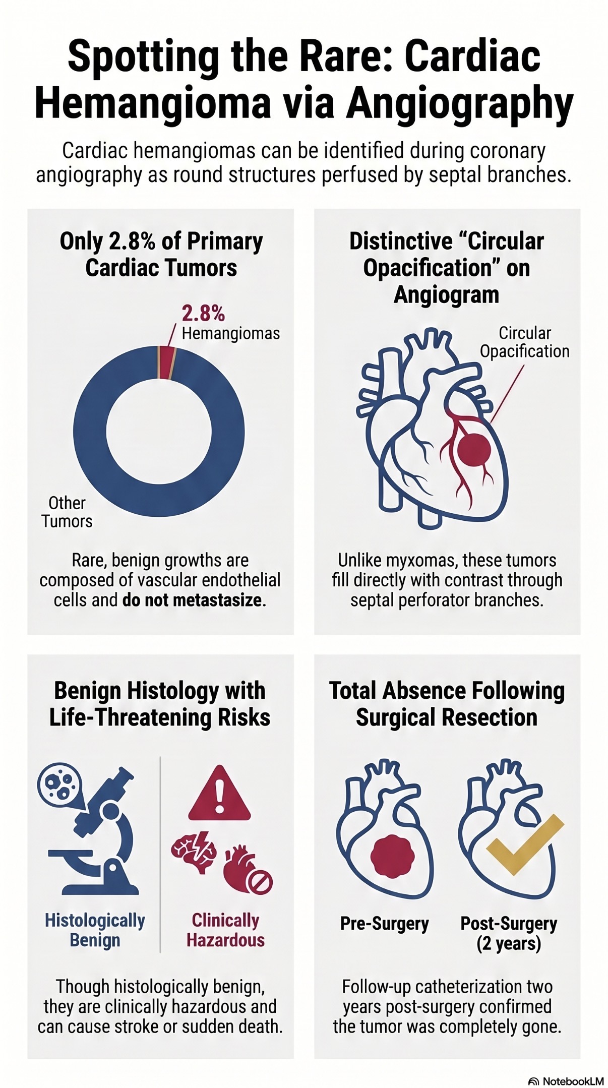
Cardiac hemangiomas are a rare cardiac tumor. We present a 53-year-old male who underwent cardiac catheterization before and after surgical resection of a cardiac hemangioma. We demonstrate the opacification of a cardiac hemangioma during cardiac catheterization appearing as a round, circular structure being perfused by septal perforator branches. On follow-up catheterization, post-surgical resection, the structure is completely absent. In conclusion, cardiac hemangiomas can be visualized during cardiac catheterization and this case demonstrates what they can appear as angiographically.
Cardiac hemangiomas are rare primary tumors of the heart and occur in 2.8% of primary cardiac tumors.1 They are typically comprised of benign proliferative endothelial cells lining blood vessels with increased vascularization.2 Cardiac hemangiomas typically get larger over a longer course of time and don’t have the ability to metastasize. The vascular channels are lined by endothelial cells with moderately pleomorphic, sometimes atypical, nuclei and focal tuft formation. Mitoses rarely occur. Histopathologic features of cardiac hemangiomas are identical to those elsewhere in the body. Based on the predominant type of the proliferating vessels, hemangiomas are classified into cavernous, capillary, and arteriovenous types.2 Moreover, they are well circumscribed by an outer surface. However, case reports of hemangiomas invading the conductive tissue of the heart suggest that they are unlike the usual benign tumors. Therefore, despite histopathologic benignity, they are viewed as clinically hazardous, attributable to life-threatening complications such as stroke and death. Owing to its rarity, to our knowledge this disorder is derived from case reports, and no comprehensive review or clinical guidelines are available.3
Case Presentation
A 53-year-old male was referred to the cardiology outpatient clinic for a routine cardiac evaluation by his primary care provider due to a strong family history of cardiac disease and sudden cardiac death. He had no prior history of cardiac conditions and had never been evaluated by a cardiologist. The patient denied chest pain, shortness of breath, fatigue, palpitations, syncope, dizziness, fever, chills, or weight loss. He reported adequate exercise tolerance and well-controlled chronic conditions.
During the initial assessment, a transthoracic echocardiogram revealed a cardiac mass. To further characterize the lesion — specifically to differentiate between thrombus and tumor — cardiac magnetic resonance imaging (MRI) was performed, showing a mobile, enhancing, pedunculated lesion in the anterior septal apical region of the left ventricle, consistent with a cardiac myxoma. The patient subsequently underwent successful surgical excision of the left ventricular mass. Pathology from the excised tissue suggested either a hemangioma or an organizing thrombus, with no evidence of malignancy.
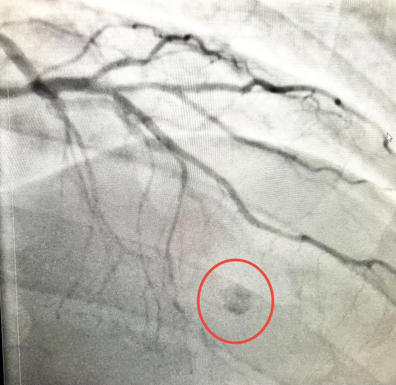
Additionally, the patient had a cardiac catheterization prior to his surgical resection of the tumor demonstrating non-obstructive coronary artery disease. A circular opacification (Video 1) was noted adjacent to several septal branches in the right anterior oblique (RAO)/cranial view (Figure 1).
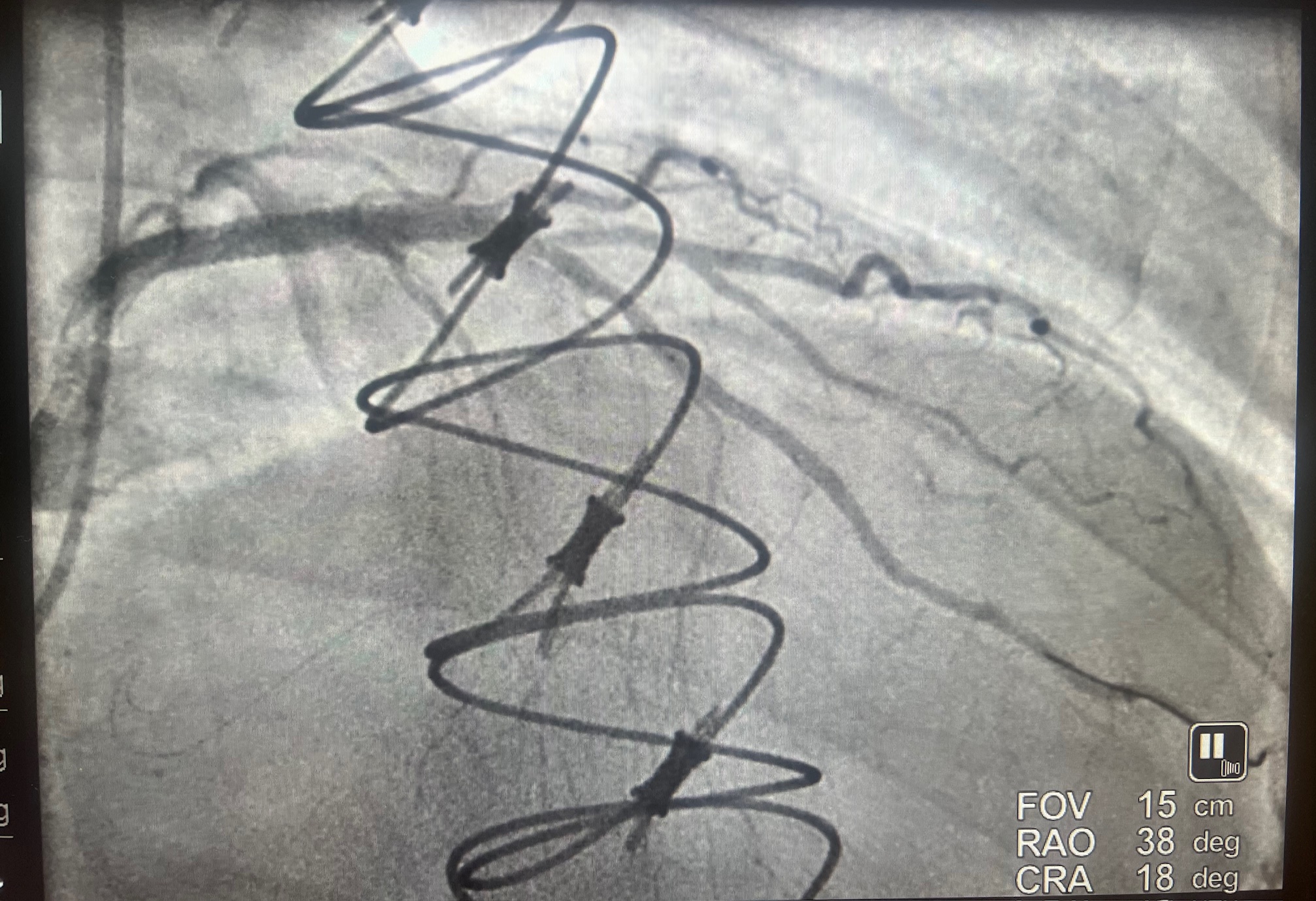
Subsequently, two years later, the patient underwent another coronary angiogram for angina and shortness of breath. Upon catheterization, an 80% lesion was seen and treated with percutaneous coronary angioplasty and stenting. Additionally, the round opacification was absent, as that was seen on the previous angiogram (Figure 2).
Case Details
History and Physical Exam
Past Medical History:
• Hypertension
• Hyperlipidemia
• MTHFR gene mutation
• Gout
Past Surgical History:
• None
Medications:
• Amlodipine 10 mg daily
• Lisinopril 10 mg daily
• Atorvastatin 20 mg daily
• Ezetimibe 10 mg daily
• Aspirin 81 mg daily
Allergies:
• No known drug allergies
Physical Examination and Vital Signs:
• Height: 182.8 cm
• Weight: 107 kg
• Temperature: 36.6°C
• Heart Rate: 73 bpm
• Blood Pressure: 125/80 mmHg
• Oxygen Saturation: 96% on room air
General: Well-developed, in no acute distress
Psychiatric: Normal mood and affect
Neurological: Alert and oriented to person, place, and time; no focal deficits; strength and sensation intact
Skin: Warm and dry
Respiratory: Clear breath sounds bilaterally
Cardiovascular: Normal S1 and S2, with a systolic murmur heard at the apex
Abdomen: Soft, non-tender, non-distended; normal bowel sounds
Musculoskeletal/Extremities: Full range of motion in all extremities without weakness
Edema: +1 bilateral lower extremity edema
Varicose Veins: Present bilaterally in lower extremities
Investigations and Treatment
Electrocardiogram (EKG):
• Normal sinus rhythm
• No ST-T wave abnormalities
• Normal QT interval
Transthoracic Echocardiogram (May 2023):
• Hyper-echogenic 0.9 cm mass attached to the mid-anteroseptal wall of the left ventricle; Differential: old thrombus vs cardiac tumor
• Normal left and right ventricular function
• Trivial pericardial effusion
• No significant valvular abnormalities
Cardiac MRI (June 2023):
• 9 x 4 mm mobile, enhancing pedunculated lesion in the anterior septal wall near the apex of the left ventricle
• Findings most consistent with cardiac myxoma
Surgical Summary (June 2023)
• Under general anesthesia, median sternotomy and cardiopulmonary bypass were performed. The heart was arrested with cardioplegia. Access to the mass was gained via a transseptal approach through the mitral valve. The mass was excised with its stalk and sent to pathology. The heart was irrigated and rewarmed. The atrial septum and right atrium were closed. After de-airing, the heart resumed sinus rhythm and pacing wires were placed. The patient was weaned off bypass without complications. A chest tube was placed, and the sternum and incision were closed in layers. The patient was transferred to recovery in stable condition.
Left Ventricular Mass Biopsy (Post Surgical):
• Pathology suggested hemangioma vs organizing thrombus
• No malignant cells identified
Initial Cardiac Catheterization (June 2023):
• Normal left main coronary artery
• 30% stenosis in the proximal left anterior descending (LAD) artery
• Normal circumflex artery
• Small, non-dominant right coronary artery with luminal irregularities
Repeat Coronary Angiogram (2 years later):
• 80% stenosis in the proximal LAD
• Successfully treated with drug-eluting stent placement
• Opacification of the circular structure previously was absent on the subsequent coronary angiogram
Discussion
In this case report, we demonstrate the opacification of a cardiac hemangioma during cardiac catheterization. Cardiac tumors are generally diagnosed by echocardiography. However, this rare case demonstrates angiographic signs that can assist in diagnosis. Angiographically, this tumor is different in appearance than a cardiac myxoma, which is also seen infrequently during angiography. In particular, this hemangioma filled slowly by septal branches. Moreover, this “slow” perfusion may be attributable to why it was a slow-growing tumor. Myxoma typically don’t opacify the same way as in our case. They are usually seen as a globular structure (typically during left ventricular angiograms) that is “outlined” by contrast, as opposed to, as in our case, a structure that is directly opacified by contrast. On follow-up angiogram, post resection, the opacification is clearly absent. This doesn’t preclude the possibility of recurrence; however, as cardiac hemangiomas don’t metastasize, the possibility of future tumor growth is remote.
Conclusion
Cardiac hemangiomas are rare tumors that may be seen angiographically during cardiac catheterization and appear as a circular opacified structure.
Highlights
• Cardiac hemangiomas are rare and account for 2.8% of primary cardiac tumors.1
• During cardiac catheterization, hemangiomas can appear as a circular or ovoid structure on coronary angiography.
References
1. McAllister HA Jr, Fenoglio JJ Jr. Tumors of the Cardiovascular System. Atlas of Tumor Pathology, 2nd series, fascicle 15. Washington, DC: Armed Forces Institute of Pathology; 1978.
2. Hara T, Ushijima T, Takemura H, et al. A case report of cavernous hemangioma of the left atrial epicardium. Kyobu Geka. 1998; 51(12): 1027-1029.
3. Berdica L, Kola E, Nakuci D, et al. Cardiac hemangioma presenting as a primary cardiac tumor. Cardiooncology. 2023; 9(1): 3. doi:10.1186/s40959-023-00154-5