








When Slow Reflow Turns Dangerous: Balloon Embolization as a Bailout Strategy for Wire-Induced Coronary Perforation During Primary PCI
JULY 2026
© 2026 HMP Global. All Rights Reserved.
Any views and opinions expressed are those of the author(s) and/or participants and do not necessarily reflect the views, policy, or position of Cath Lab Digest or HMP Global, their employees, and affiliates.
Muhammad Imran Hanif, MBBS, FCPS (Cardiology), FRCP (Edin), FSCAI1; Ahmad Usama Wali, MBBS2; Murad Ali Khan, MBBS2
1Professor of Cardiology, Department of Cardiology, Gulab Devi Teaching Hospital Lahore, Pakistan
2Postgraduate Resident, Cardiology
Department of Cardiology, Gulab Devi Teaching Hospital Lahore, Pakistan
The authors can be contacted via Ahmad Usama Wali, MBBS, at ahmadusama49@gmail.com.
Editor's Note: A PDF of this article is available by clicking the red PDF icon to the right of the article title.
Patient Presentation & Evaluation
A 75-year-old man with 30 pack-year smoking history presented with chest pain for 6 hours associated with palpitations and two episodes of syncope occurring during the same symptomatic period prior to hospital presentation. He also reported anorexia with unintentional weight loss for 6 months. On presentation, heart rate was 40 beats/min and blood pressure was 100/70 mmHg. Electrocardiogram demonstrated inferior wall myocardial infarction with complete heart block. Echocardiography revealed left ventricular systolic dysfunction with ejection fraction of 35%. A temporary pacemaker was inserted, and the patient was shifted to the cardiac catheterization laboratory for primary percutaneous coronary intervention (PCI).
Procedure
Coronary angiography showed dominant right coronary artery (RCA) with distal occlusion. An anomalous left circumflex artery arising separately from the RCA was also noted (Figure 1, Video 1).
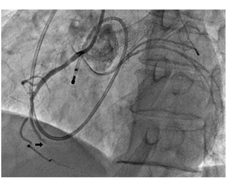
Video 1. RCA total occlusion, DES deployment, and distal coronary perforation.
The culprit RCA lesion was crossed using a Pilot 50 guidewire (Abbott Vascular). Following predilatation and deployment of two drug-eluting stents, check angiography demonstrated slow reflow (Figure 2). Slow reflow is a recognized complication of primary PCI and is commonly attributed to distal thrombus embolization, microvascular dysfunction, vasospasm, and reperfusion injury. In the presence of slow reflow, reduced antegrade contrast flow may obscure distal contrast extravasation, rendering a wire-induced perforation angiographically inapparent.
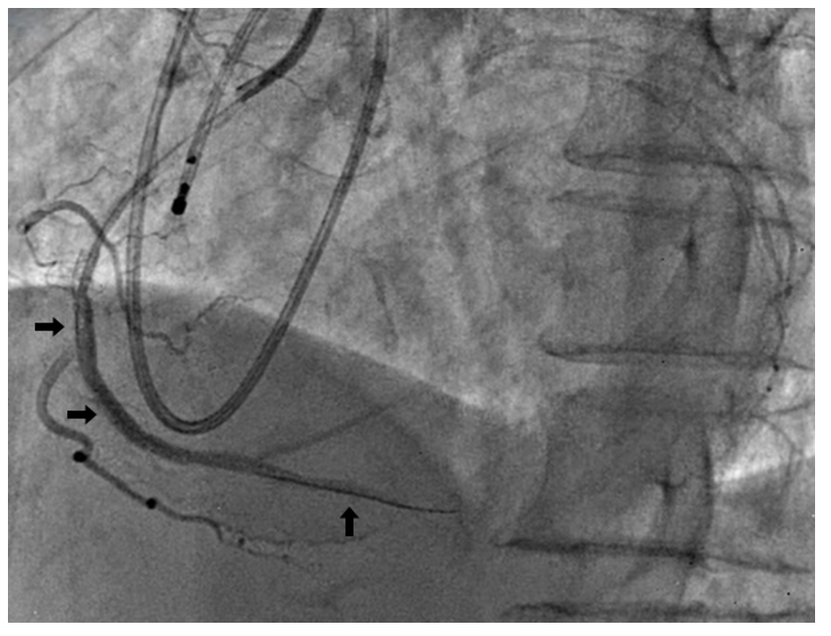
In our case, intracoronary nitroglycerin was administered in 200 μg boluses (total dose 1000 μg) for treatment of slow reflow. Following improvement in distal coronary flow, repeat angiography revealed contrast extravasation from a perforated side branch, consistent with an Ellis type III cavity-spilling perforation. This sequence is clinically important because delayed recognition of perforation may occur after administration of anticoagulation and dual antiplatelet therapy, increasing the risk of hemorrhagic complications (Figure 3, Video 1 above).
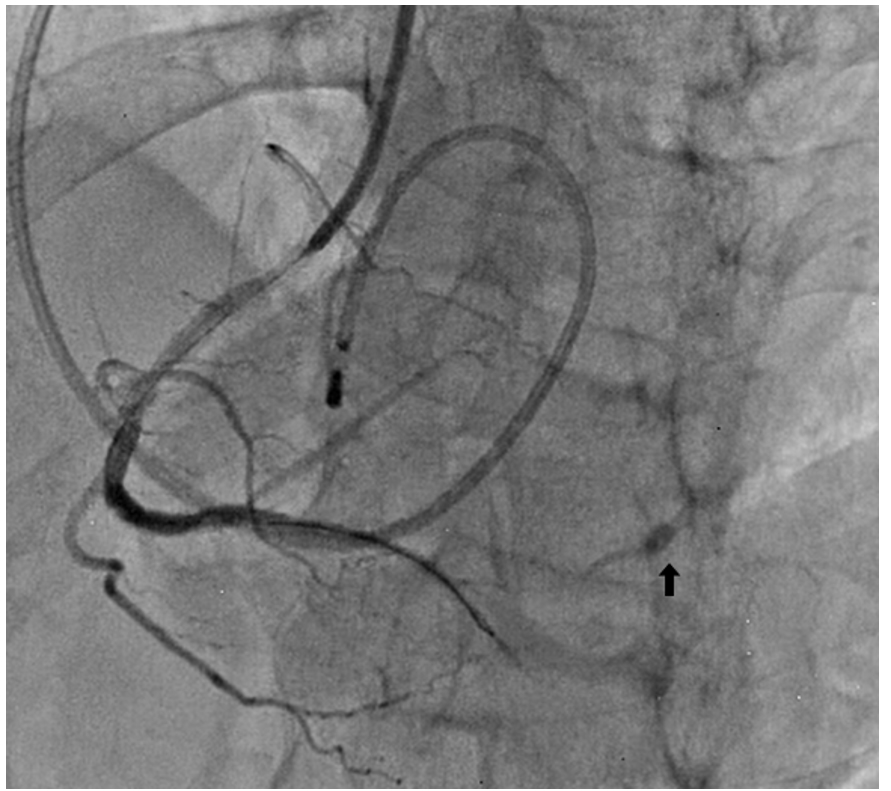
The patient remained hemodynamically stable with no pericardial effusion. However, persistent contrast extravasation was present in a patient who had received anticoagulation and dual antiplatelet therapy during primary PCI. Considering the distal location and small caliber of the perforated side branch, covered stent implantation was not technically feasible. Although coil embolization is a recognized treatment option for distal coronary perforations, coils were not readily available. Therefore, balloon embolization was selected as a readily available bailout strategy to achieve prompt vessel sealing.
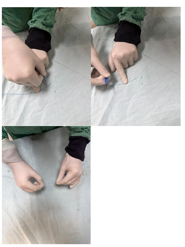
A 2.0 × 15 mm semi-compliant VexPander balloon (TT Medical, Inc.) was cut transversely using a sterile surgical blade. The sequential steps of the cut balloon technique are illustrated in Figure 4.
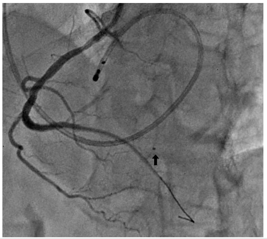
The distal balloon fragment was retained on the guidewire and advanced to the site of perforation. A second balloon segment was subsequently advanced over the same guidewire and used as a pusher to deliver the embolizing fragment distally (Figure 5). The pusher segment was not inflated and served solely as a mechanical support to facilitate distal delivery of the embolizing fragment. No additional delivery devices or microcatheter were required. Prolonged balloon tamponade was then performed proximal to the perforation using a 2.0 mm × 14 mm balloon inflated for 10 minutes. The guidewire was subsequently withdrawn, leaving the balloon fragment permanently implanted within the vessel.
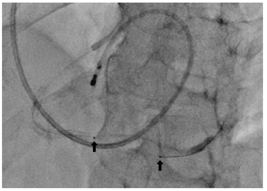
Repeat angiography demonstrated complete cessation of contrast extravasation (Figure 5, Video 2 below). Final angiography demonstrated complete resolution of contrast extravasation with restoration of distal flow (Figure 6, Video 2).
Video 2. Balloon embolization; sealing of perforation.
Discussion
Slow reflow during primary PCI can mask distal coronary perforation. Typical management principles include stopping further distal wire manipulation, instituting prolonged balloon inflation/tamponade, assessing for pericardial effusion, maintaining hemodynamic monitoring, considering anticoagulation reversal when appropriate, and selecting covered stent implantation or distal embolization based on perforation location. Balloon embolization may be considered a simple, readily available, and cost-effective bailout strategy for selected distal small-vessel perforations when standard embolization materials are unavailable. However, it should be performed with careful attention to the risks of distal vessel occlusion, embolic migration, thrombosis, and loss of side-branch territory.
Muhammad Imran Hanif, MBBS, FCPS (Cardiology), FRCP (Edin), FSCAI1; Ahmad Usama Wali, MBBS2; Murad Ali Khan, MBBS2
1Professor of Cardiology, Department of Cardiology, Gulab Devi Teaching Hospital Lahore, Pakistan; 2Postgraduate Resident, Cardiology; Department of Cardiology, Gulab Devi Teaching Hospital Lahore, Pakistan
The authors can be contacted via Ahmad Usama Wali, MBBS, at ahmadusama49@gmail.com.





