








Finalists From CRT’s 2026 Nurses and Technologists Interesting Cases Competition
The Cardiovascular Research Technologies (CRT) meeting was held March 7-10 in Washington, D.C.
The Cardiovascular Research Technologies (CRT) meeting was held March 7-10 in Washington, D.C.
© 2025 HMP Global. All Rights Reserved.
Any views and opinions expressed are those of the author(s) and/or participants and do not necessarily reflect the views, policy, or position of Cath Lab Digest or HMP Global, their employees, and affiliates.
Editor's Note: To view the article PDF, click the red PDF icon to the right of the article title.
Plan to submit your case for 2027! Click here to learn more about submitting CRT Interesting Cases.
In 2026, CRT continued a new tradition that began in 2024 — a competition focused on cases submitted by nurses and technologists, with three finalists asked to present their cases during CRT’s Nurses and Technologists session.
Cath Lab Digest is proud to share the featured CRT cases in this issue:
• Bridging the Gap: Unmasking Hidden Mechanisms in ANOCA – Falon Harper, BSN, RN
• Transcatheter Pulmonary Valve Replacement Post Ross – Brandy Smith, BS, RCIS, LMRT
• Beyond the Repair: Unmasking In-Stent Restenosis – Stephen M. Trice, RN, BSN
 Case #1. Bridging the Gap: Unmasking Hidden Mechanisms in ANOCA
Falon Harper, BSN, RN, MedStar Southern Maryland Hospital Center, Clinton, Maryland
Falon Harper, BSN, RN, can be contacted at falonharper@icloud.com.
This case highlights the importance of comprehensive coronary functional testing in a patient with persistent chest pain despite robotic coronary unroofing for a symptomatic myocardial bridge.
Initial Presentation
A 59-year-old female with a history of celiac disease initially presented with progressive exertional chest pain radiating to her left arm, fatigue, and reduced exercise tolerance. Despite a normal stress test, coronary angiography identified a symptomatic myocardial bridge involving the left anterior descending (LAD) artery. As her symptoms progressed, she underwent robotic coronary unroofing on January 8, 2025.
Persistent Symptoms
A few weeks after surgery, the patient developed a different type of chest pain. Her symptoms worsened with deep breathing and lying flat and were accompanied by shortness of breath. Light exercise also triggered her symptoms, while sitting upright provided relief. A computed tomography (CT) scan demonstrated no pericardial effusion; however, inflammatory markers were elevated, leading to a diagnosis of post-sternotomy pericarditis. She was treated with colchicine and NSAIDs, followed by corticosteroids due to persistent symptoms.
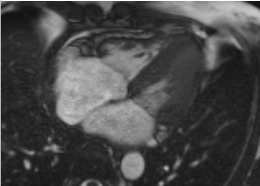
The patient was subsequently referred to a pericardial specialty clinic. Cardiac magnetic resonance imaging (MRI) confirmed active pericardial inflammation with evidence of pericardial thickening and edema (Figure 1). Treatment was escalated with the initiation of rilonacept and a planned long-term treatment course. Despite appropriate therapy and improvement in inflammation, the patient continued to experience chest discomfort, suggesting another mechanism was contributing to her symptoms.
What made this case particularly interesting was a new presentation of chest pain that differed from her prior symptoms. Activities such as carrying groceries, lifting bags, and bending over triggered episodes characterized by throat discomfort, neck tightness, chest pain, and a “heart-dropping” sensation. These symptoms were distinctly different from both her original myocardial bridge symptoms and her pericarditis-related pain.
The key question became: why was she still having chest pain?
Functional Testing & Diagnosis
Given her persistent symptoms and history of myocardial bridging, she was referred for repeat coronary angiography with comprehensive coronary functional testing. Coronary angiography demonstrated no significant obstructive coronary artery disease. The right coronary artery and circumflex artery were normal, and the previously unroofed mid-LAD myocardial bridge was not physiologically significant.
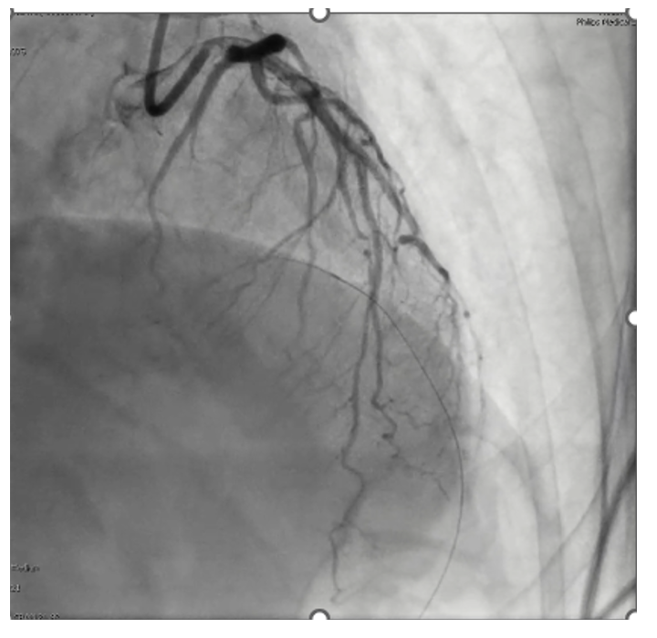
Coronary functional testing was performed to evaluate both endothelial-dependent and endothelial-independent pathways. During acetylcholine provocative testing, the patient remained asymptomatic at 2 mcg. At 20 mcg, mild angiographic spasm was observed without symptoms or electrocardiogram (EKG) changes. At 100 mcg, significant mid-LAD vasospasm occurred (Figure 2), accompanied by chest pain and ischemic EKG changes, meeting diagnostic criteria for epicardial coronary vasospasm.
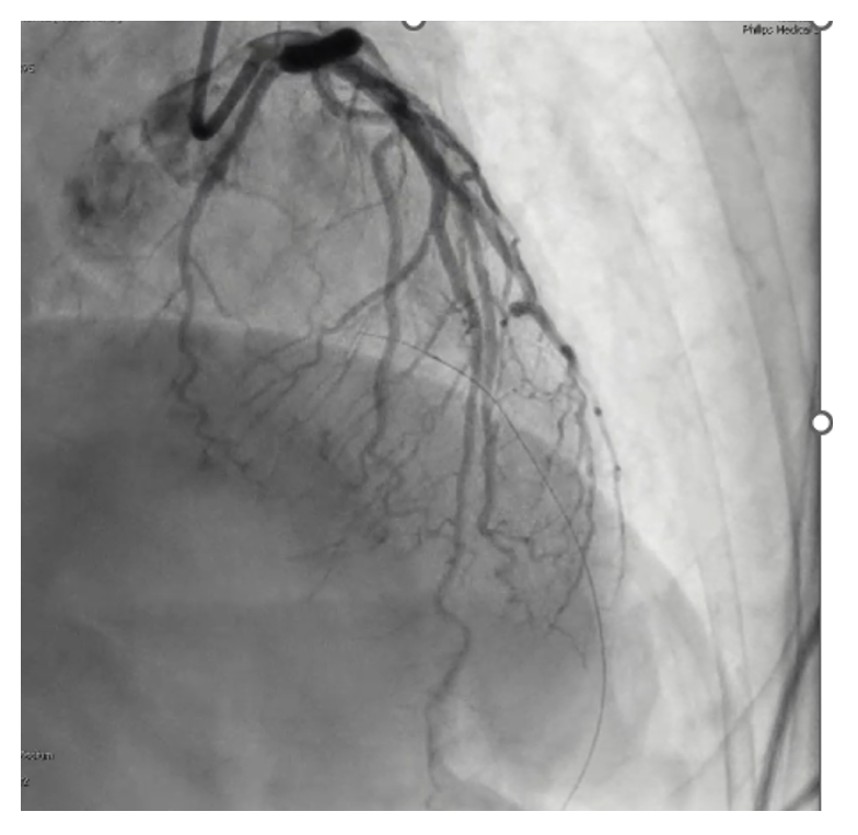
Following administration of intracoronary nitroglycerin, normal vessel caliber was restored (Figure 3). Coronary microvascular dysfunction assessment was then performed using thermodilution techniques. Physiologic testing of the epicardial coronary arteries was normal, and measurements of coronary flow and microvascular resistance were within normal limits, effectively ruling out coronary microvascular dysfunction as the cause of the patient’s symptoms.
The final diagnosis was insignificant coronary artery disease, a previously unroofed mid-LAD myocardial bridge, and epicardial coronary vasospasm confirmed by positive acetylcholine provocative testing. Medical therapy was tailored accordingly with initiation of calcium channel blocker therapy and statin treatment. The patient has remained asymptomatic since treatment was initiated.
Discussion & Conclusion
One might wonder whether coronary vasospasm contributed to the patient’s symptoms long before surgical unroofing was performed. Regardless, comprehensive coronary functional testing identified the underlying etiology and allowed therapy to be tailored to the patient’s specific endotype. The beautiful thing about performing comprehensive coronary functional testing is that it identifies the underlying etiology of ANOCA (angina with non-obstructive coronary arteries) and allows medical therapy to be tailored accordingly.
Myocardial bridges are complex, and surgical unroofing does not always resolve symptoms. In patients with ANOCA, persistent chest pain may occur in the absence of obstructive coronary disease, making comprehensive coronary functional testing essential. Ultimately, identifying the correct mechanism of angina is critical before pursuing intervention. In the cath lab, anatomy guides us, but physiology defines the diagnosis.
Falon Harper, BSN, RN, can be contacted at falonharper@icloud.com.
 Case #2. Transcatheter Pulmonary Valve Replacement Post Ross
Brandy Smith, BS, RCIS, LMRT Cath Lab Technologist, Baylor Scott and White The Heart Hospital; Adjunct Faculty at The Cardiovascular Institute, Plano, Texas
Brandy Smith, BS, RCIS, LMRT, can be contacted at brandy.smith2@bswhealth.org.
Significant advancements in structural heart interventions have revolutionized the management of complex valvular disease. These innovations have expanded treatment options, particularly for patients who are at higher risk of complications. This case study is a prime example of innovation leading to improved outcomes and shorter recovery times, particularly for patients with complex disease processes.
Patient Presentation and Evaluation
Our patient was a 73-year-old male who had a Ross procedure by Dr. Ross in 1990 that was performed when the patient was 38 years old. The Ross procedure is an innovative cardiac surgical procedure that involves replacing the diseased aortic valve with the patient’s own pulmonary valve (autograft), while the pulmonary valve and a portion of the pulmonary artery are replaced with a cryopreserved pulmonary homograft from a cadaver donor. Placing the pulmonary valve in the aortic position works because it is structurally similar in size and shape to the aortic valve, yet as a living tissue autograft, it can adapt to high-pressure systemic circulation. The Ross procedure is particularly favored for younger, active patients due to its advantages that include no lifelong anticoagulation requirement, minimal activity restrictions, and excellent long-term survival. This is particularly important for women of childbearing age who desire pregnancy without the complications and risks associated with anticoagulation. However, gradual degeneration of both the autograft and homograft valves occurs over time, often necessitating re-intervention.
Our patient, 3 decades post Ross procedure, was experiencing a significant reduction in physical capacity. After leading a very active lifestyle, including regular biking, walking, and skiing, he was experiencing New York Heart Association (NYHA) class II symptoms, including fatigue and shortness of breath with activity. Additionally, his medical history was notable for hypertension, hyperlipidemia, type 2 diabetes, chronic kidney disease stage IV (with a baseline creatinine of 2.8), and hyperparathyroidism.
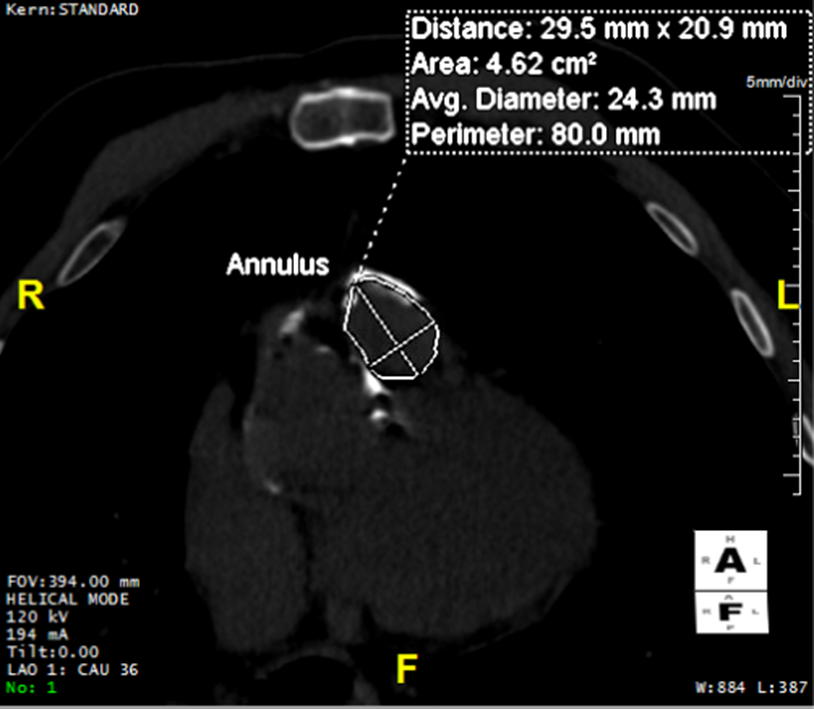
To protect the patient’s kidney function, contrast was avoided during the pre-procedural computed tomography (CT) scan (Figure 1). These images included annular area and right ventricular outflow tract (RVOT) measurements. Transesophageal echocardiography (TEE) revealed moderate pulmonic stenosis, severe pulmonic regurgitation, a preserved left ventricular ejection fraction (LVEF 55-60%), a dilated right ventricle with normal systolic function, and a mean transvalvular gradient of 27 mmHg. These findings indicated significant right-sided valve dysfunction.
Video: Pre pulmonary valve TEE
A collaborative approach to decision-making was utilized. The patient and the structural team, including cardiothoracic surgeons, interventional cardiologists, and imaging cardiologists, opted for a transcatheter pulmonary valve replacement (TPVR) as a minimally invasive alternative to surgical valve replacement.
Procedure
Vascular access was achieved via the right common femoral vein and artery using ultrasound and micropuncture techniques. The right common femoral artery was accessed with an 8 French (F) sheath, and an 8F Judkins left (JL4) catheter was placed in the left coronary system. The right common femoral vein was pre-closed with two Perclose devices (Abbott). An 8F sheath was accessed with a pulmonary wedge catheter. A Lunderquist wire (Cook Medical) was inserted into the pulmonary wedge catheter, and the sheath was exchanged to a 26F x 65 cm Gore DrySeal sheath (Gore Medical) following serial dilation.
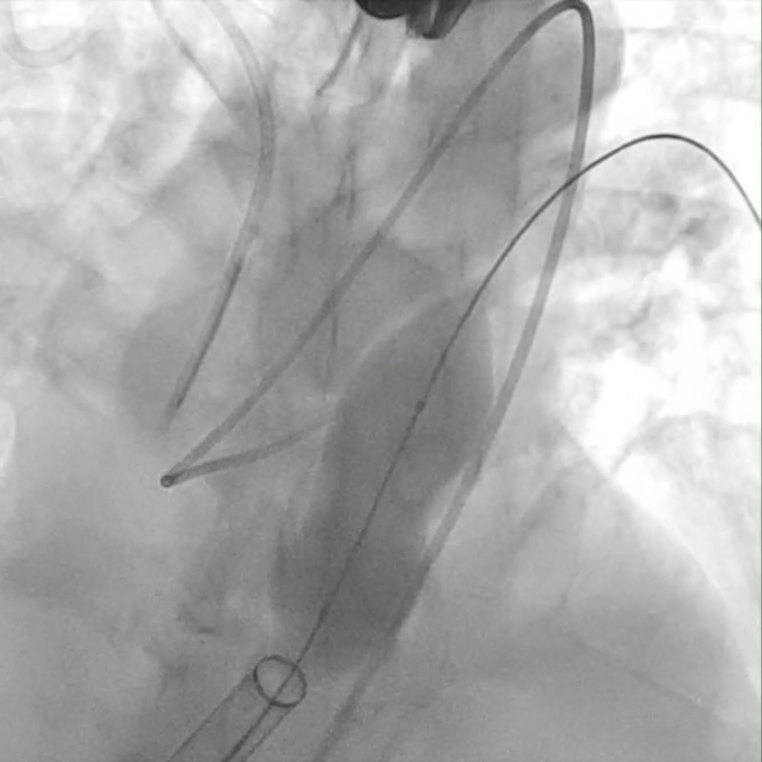
Even with careful pre-procedural testing, it was imperative to ensure that the left coronary system, particularly the left circumflex, would not be compressed by the new valve. The best way to visualize compression is to inflate a large balloon in the RVOT and pulmonary valve while simultaneously imaging the left coronary system. If the blood flow stops or is reduced, it is a hard stop for the procedure. The team elected to start with a 25 mm Z-Med balloon (B. Braun Interventional Systems). The imaging showed no compression, but the balloon was not large enough to get an accurate result.
Video: Initial 25 mm balloon too small.
The balloon was exchanged for a 28 mm Z-Med balloon that quickly showed adequate inflation by dog-boning at the pulmonary valve (Figure 2). This demonstrated that there was no compression in the left coronary system, and the procedure could continue.
Video: 28 mm balloon dog bone.
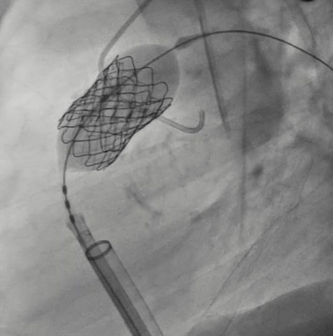
The structural team custom-built the new valve, which consisted of a Sapien 3 Ultra Commander 26 mm valve (Edwards) at the center, a Covered CP stent (4.5 mm x 28 mm) (B. Braun) placed over the valve, and a Palmaz 4010 Biliary stent (10 mm x 39 mm) (Cordis) on top of the valve and covered stent (Figure 3).
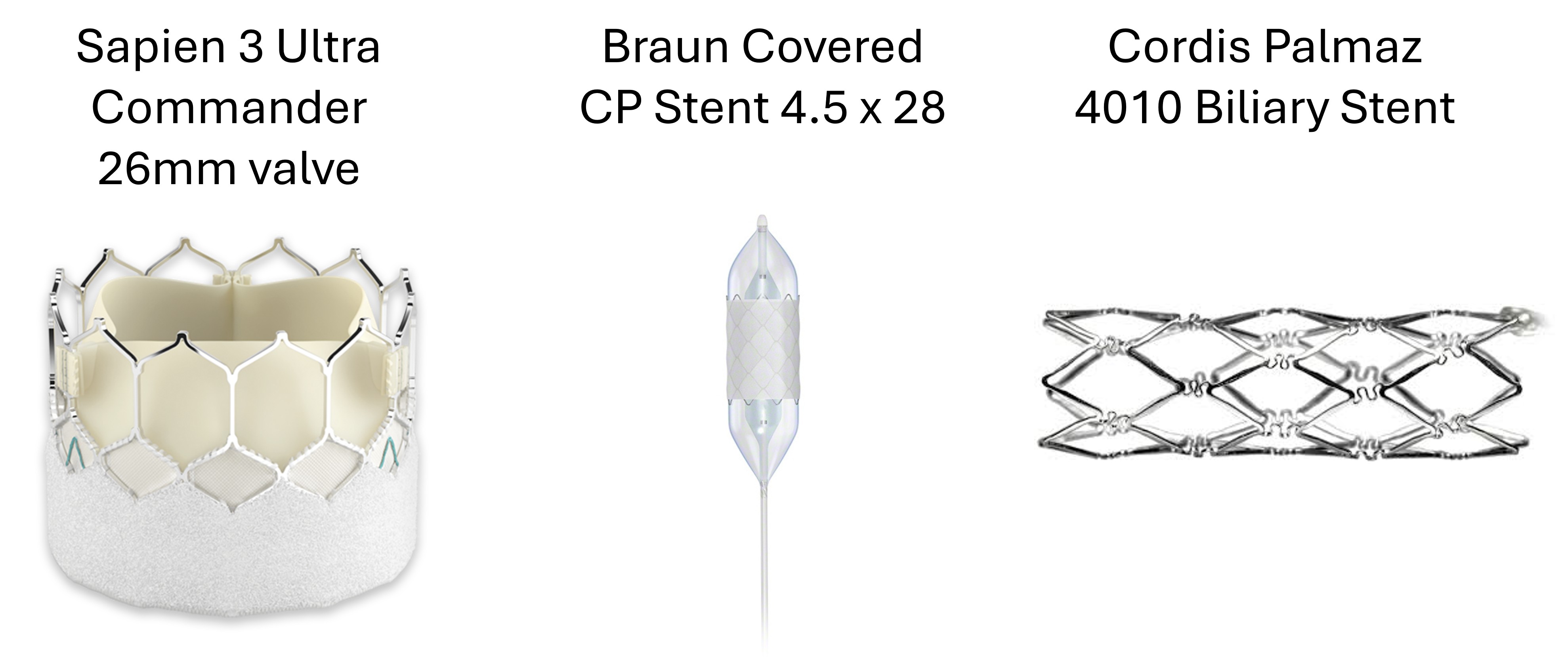
The covered stent prevents paravalvular leaks and the biliary stent provides structural support to the system. The valve and stents are all deployed simultaneously, unfolding together with the Sapien valve balloon inflation (Figure 4).
Video: Valve Check Pre Insertion
Video: Valve deployment
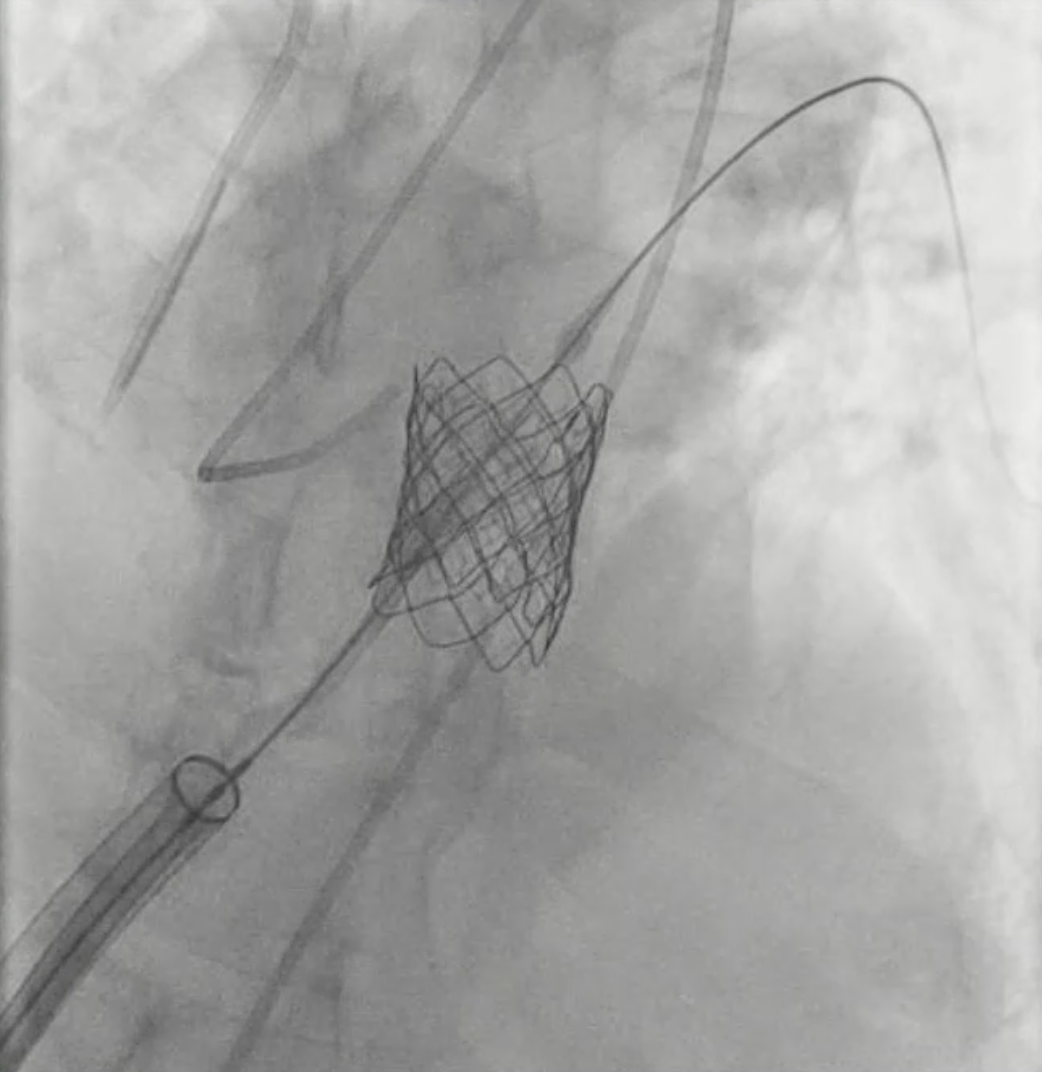
Angiography post deployment (Figure 5) confirmed that the valve was well-positioned with no evidence of compression. TEE demonstrated a well-functioning pulmonic valve with no evidence of regurgitation or stenosis. The mean gradient dropped by 22 mmHg to a healthy 5 mmHg. Hemostasis was achieved by completing the Perclose process and using an 8F Angio-Seal (Terumo) on the arterial access.
The patient remained in the hospital for 1 day. He resumed normal activities, including cardiac rehab, one week post procedure, without feeling short of breath or fatigue.
Video: Post procedure angiogram
Video: Post procedure TEE
Discussion and Conclusion
Transcatheter pulmonary valve replacement allows for larger implants, results in lower mortality and reduced 30-day readmissions, and is associated with fewer major cardiac events than surgical pulmonary valve replacement. TPVR is also a minimally invasive solution for high surgical risk patients. Recovery time is quick, and patients are typically able to resume normal activities one week post procedure, as our patient did.
Advancements in structural heart technologies and interventions are paving the way for less invasive procedures. Advanced imaging practitioners working with surgeons and interventional cardiologists can better identify suitable candidates for TPVR using advanced imaging modalities. Patients, even those with comorbidities, have increased options for improving health.
Brandy Smith, BS, RCIS, LMRT, can be contacted at brandy.smith2@bswhealth.org.
 Case #3. Beyond the Repair: Unmasking In-Stent Restenosis
Stephen M. Trice, RN, BSN Medstar Southern Maryland Hospital Center, Clinton, Maryland
Stephen Trice, RN, BSN, can be contacted at stephen.m.trice@medstar.net.
Initial Presentation
This case highlights the complex clinical course of a 43-year-old male with a history of hypertension, hyperlipidemia, and tobacco use, who initially presented with an acute inferior ST-elevation myocardial infarction (STEMI). Emergency medical services identified ST elevations on electrocardiogram (ECG), prompting rapid activation of a cardiac catheterization protocol. Coronary angiography revealed a culprit lesion requiring percutaneous coronary intervention (PCI) with drug-eluting stent placement in the right coronary artery (RCA). The procedure was complicated by complete heart block, necessitating temporary pacing.
Persistent Symptoms
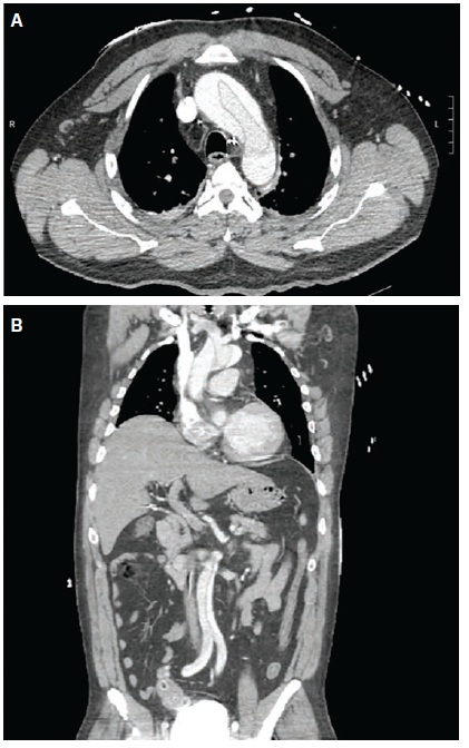
Despite technically successful intervention and stabilization, the patient continued to experience severe chest pain post procedure. Although repeat ECG demonstrated resolution of ST elevations, further imaging revealed concerning findings, including mediastinal widening. Subsequent computed tomography (CT) angiography confirmed an acute Type A aortic dissection extending from the ascending aorta into the RCA, explaining both the initial presentation and persistent symptoms (Figure 1A-B). The patient underwent emergent surgical repair involving ascending aortic replacement, hemiarch reconstruction, and sinotubular junction repair. Postoperatively, cardiac function remained preserved with normal ejection fraction.
Repeat Evaluation
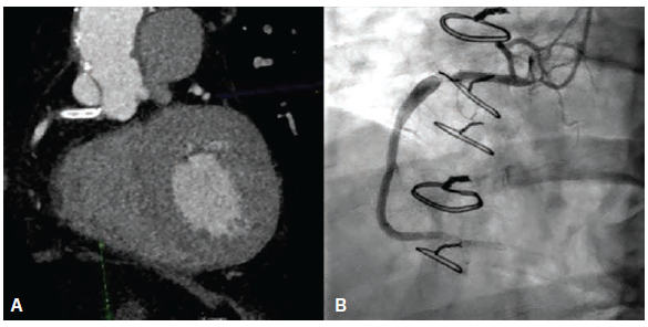
Five years later, the patient returned with recurrent chest pain, described as similar but less severe than his prior myocardial infarction. Laboratory evaluation showed elevated troponins with a rising trend, though ECG findings were non-specific. Given his history of aortic dissection, a cautious diagnostic approach was taken using coronary CT angiography (cCTA), which revealed a previously placed RCA stent with areas of uncertain patency (Figure 2A).
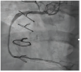
Further invasive evaluation with intravascular imaging identified two critical issues: severe in-stent restenosis in the distal RCA and significant extrinsic compression of the ostial RCA stent due to chronic changes related to the prior aortic dissection (Figure 2B). These findings explained the patient’s recurrent symptoms and ischemia. The patient underwent a successful high-definition intravascular ultrasound (IVUS)-guided PCI. The procedure included serial balloon dilations and placement of a new drug-eluting stent across the affected segments, achieving optimal blood flow restoration (Figure 3).
Discussion and Conclusion
This case underscores several important clinical lessons. First, aortic dissection can mimic or coexist with acute myocardial infarction, potentially delaying recognition if symptoms persist, despite apparent treatment success. Persistent chest pain after PCI, even with ECG normalization, should prompt further investigation for alternative or concurrent diagnoses. Second, coronary CT angiography can be a valuable noninvasive tool in high-risk patients, particularly when prior surgical history complicates decision-making. Finally, advanced intravascular imaging techniques such as IVUS play a crucial role in diagnosing and managing complex in-stent restenosis, enabling precise intervention and improved outcomes.
Overall, this case demonstrates the importance of maintaining a broad differential diagnosis in acute cardiac presentations and highlights evolving strategies in the management of complex coronary artery disease.
Stephen Trice, RN, BSN, can be contacted at stephen.m.trice@medstar.net.





