








Change Is the Only Constant in Life and in the Cath Lab: A Comment on the AIR-STEMI Trial
© 2026 HMP Global. All Rights Reserved.
Any views and opinions expressed are those of the author(s) and/or participants and do not necessarily reflect the views, policy, or position of Cath Lab Digest or HMP Global, their employees, and affiliates.
Morton J. Kern, MD, MSCAI, FACC, FAHA
Clinical Editor; Interventional Cardiologist, Long Beach VA Medical Center, Long Beach, California; Professor of Medicine, University of California, Irvine Medical Center, Orange, California
Disclosures: Dr. Morton Kern reports he is a consultant for Abiomed, Abbott Vascular, Philips, ACIST Medical, and Opsens Inc.
Dr. Kern can be contacted at mortonkern2007@gmail.com
On X @MortonKern
"The only constant in life is change"
— Heraclitus, the ancient Greek philosopher, around 500 B.C.
 The ever-changing nature of the universe reminds us of the ancient saying that “…there is nothing permanent except change,” signifying that all things are in a state of flux and nothing remains static over time. This adage applies to life in general, medicine as a field, interventional cardiology as a practice, and the cath lab as our work-world.
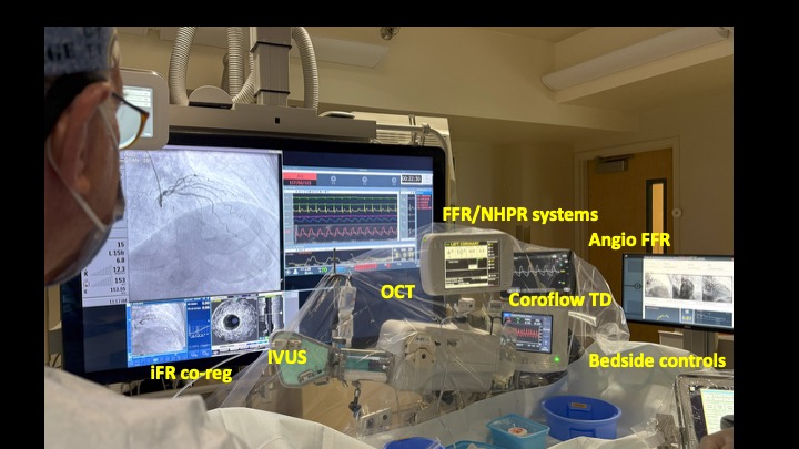
Thinking about change, I recall the equipment in the cath lab past and present. Today, everything is different from 2 decades ago — different x-ray systems and techniques, digital imaging replacing cinefilm, and no more dark rooms or developer chemicals. Cine projectors and older archiving systems are now fondly forgotten. The cath procedure itself transformed from a predominantly femoral-access procedure to one increasingly using radial access, along with different catheters, sheaths, and ultrasound guidance. The details of structural heart interventions change annually, if not more quickly, bringing new valves, wires, and delivery sheaths, along with a procedural language unique to structural heart teams and operators. The list of changes goes on. Change is the only constant. We all have changed, too (not just gotten older).
One of the biggest changes for me personally was when I could take call from home using the iPhone to see the ST-elevation myocardial infarction (STEMI) electrocardiogram (ECG) and make the decision about getting dressed in the middle of the night. In our cath lab conversations, we frequently talked about how we treated STEMI in years past compared to how we do so currently. Recall the treatment pendulum swinging between strategies — perform target vessel only versus complete revascularization at one setting, then staged settings, treating all vessels based on angiography alone, then treating all vessels guided by wire fractional flow reserve (FFR) physiology. STEMI studies have supported one strategy or another for how we should achieve complete revascularization, either all in one setting or staged, and with or without assessment of nonculprit bystander stenoses in adjacent vessels. This issue comes up frequently. The latest STEMI study, AIR-STEMI,1 prompted my introspection into the change that will evolve in future STEMI practice.
The AIR-STEMI trial,1 recently published in the New England Journal of Medicine, showed that physiologically guided complete revascularization using functional coronary angiography was better than conventional angiography-guided PCI in STEMI patients with multivessel disease. Functional coronary angiography derives FFR from the coronary angiogram, using 3D reconstruction of coronary images to estimate blood flow and the functional significance of a stenosis (eg, FFRangio or quantitative flow ratio [QFR]), rather than relying solely on visual assessment of percent diameter stenosis. The AIR-STEMI trial1 results favored functional angiography-guided complete revascularization over angiography-guided percutaneous coronary intervention (PCI), with a significantly lower incidence of the composite primary outcome. Only 51% of nonculprit lesions were physiologically significant, yet 95% were treated with PCI in the angiography-guided arm, where all lesions with ≥50% diameter stenosis were candidates for PCI. This suggests that functional guidance can substantially reduce unnecessary PCI, with less contrast-induced nephropathy and bleeding.
The approach to complete revascularization was in keeping with an earlier study, the COMPLETE trial,2 which established clear benefits for complete revascularization over culprit-only PCI in STEMI patients.
The COMPLETE trial,2 along with several other trials, appropriately resulted in an important change in guidelines and practice for STEMI patients in the cath lab.
The AIR-STEMI trial differs from prior studies in ways that may help explain the discordant results. Both the FLOWER-MI and FRAME-AMI studies used wire-based technology to physiologically assess nonculprit lesions, but the timing and application of physiology differed (see below).3,4
A quick look at FLOWER-MI. This trial randomized 1171 primary PCI STEMI patients with a nonculprit lesion to FFR-guided revascularization (n=590) versus angiography-guided revascularization (n=581). At 12 months, the primary outcome of death, MI, or urgent revascularization occurred in 5.5% of the FFR-guided group compared with 4.2% of the angiography-guided group (p=0.31). There was no difference in nonfatal MI (3.1 vs 1.7%, p=ns) or urgent revascularization (2.6 vs 1.9%, p=ns) between strategies. One can interpret the results to mean FFR-guided revascularization was not superior to angiography-guided revascularization in the STEMI patients in this study.
However, while some advocates of physiologically-guided acute coronary syndrome (ACS) interventions will be disappointed by FLOWER-MI study, I agreed with Dr. Nils Johnson from Houston, Texas, that one strong message from the trial was that FFR identified one-third of nonculprit lesions that could safely be deferred without major adverse cardiac events (MACE). A high FFR supports deferring unnecessary interventions, a finding repeatedly produced in FAME, FAME 2, DEFER, and other similar studies over the last 2 decades. As in FAME I, there is potential cost saving in FLOWER-MI, applying FFR to defer stenting in the nonculprit vessel.
FLOWER and FRAME-AMI utilized wire-based FFR assessments. In AIR-STEMI, functional coronary angiography (angiography-derived physiology) was systematically applied during both the index procedure (providing information beyond the determination of whether to treat a specific lesion), and also to procedural planning, through a predefined physiological treatment strategy for the nonculprit vessels. This approach overcomes key limitations of wire-based physiological assessment by removing the need to separate diagnostic assessment from procedural decision-making and potentially avoiding staged procedures. For this study, however, functional angiographic FFR was determined by a core laboratory rather than at the individual sites, which could limit its applicability to everyday practice.
Of note, AIR-STEMI largely assumes that physiologically significant nonculprit lesions are those that produce the majority of subsequent events. If subsequent events are instead related to disruption of thin-cap fibroatheroma, we should recognize that vessel anatomy requires intravascular imaging with a specialized catheter and is not available from functional angiography. Physiology tells us whether a lesion limits blood flow, but relatively little about its morphology. In the future, a pullback pressure gradient (PPG) with its determination of diffuse/focal disease distribution may suggest areas prone to plaque vulnerability. Currently, translesional physiology is used to defer non-flow-limiting lesions that nevertheless may carry high-risk morphological features. In the optical coherence tomography (OCT) sub-study of COMPLETE, approximately 47% of patients had at least one obstructive nonculprit lesion with thin-cap fibroatheroma (TCFA), suggesting that a substantial proportion of ACS patients had nonculprit lesions with morphology resembling the culprit lesions.5 This finding is particularly relevant because intracoronary imaging use in AIR-STEMI was low, only 3.1% in the physiology-guided and 5.1% in the angiography-guided group, which may also be relevant to stent-related outcomes.
The Bottom Line
My colleague Dr. Ziad Ali from St. Francis Hospital in New York reminded us about practice changing studies.6 He remarked that the AIR-STEMI study provided refined data on physiology-guided nonculprit lesion PCI, and, over time, “…medicine evolves with the totality of evidence rather than reacting to single trials.” As an example, he mentioned that it took a preponderance of evidence before intravascular imaging was elevated to its current routine use (my words). Furthermore, Dr. Ali emphasized that in STEMI management, we need to know how to best sequence and select revascularization strategies in patients with multivessel disease. It should be our goal to change or adapt our practice to provide best outcomes based on the latest available data. Let’s not be old dogs with old tricks.
References
1. Biscaglia S, Erriquez A, Colaiori I, et al; AIR-STEMI Trial Investigators. Complete revascularization guided by functional coronary angiography in STEMI. N Engl J Med. 2026 Sep 10;395(10):972-982. doi:10.1056/NEJMoa2605373
2. Mehta SR, Wood DA, Storey RF, et al; COMPLETE Trial Steering Committee and Investigators. Complete revascularization with multivessel PCI for myocardial infarction. N Engl J Med. 2019 Oct 10;381(15):1411-1421. doi:10.1056/NEJMoa1907775
3. Puymirat E, Cayla G, Simon T, et al; FLOWER-MI Study Investigators. Multivessel PCI guided by FFR or angiography for myocardial infarction. N Engl J Med. 2021 Jul 22;385(4):297-308. doi:10.1056/NEJMoa2104650
4. Lee JM, Kim HK, Park KH, et al; FRAME-AMI Investigators. Fractional flow reserve versus angiography-guided strategy in acute myocardial infarction with multivessel disease: a randomized trial. Eur Heart J. 2023 Feb 7;44(6):473-484. doi:10.1093/eurheartj/ehac763
5. Pinilla-Echeverri N, Mehta SR, Wang J, et al. Nonculprit lesion plaque morphology in patients with ST-segment-elevation myocardial infarction: results from the COMPLETE trial optical coherence tomography substudys. Circ Cardiovasc Interv. 2020 Jul;13(7):e008768. doi:10.1161/CIRCINTERVENTIONS.119.008768
6. Ali ZA. The opening gambit: thrombectomy to guide strategy in STEMI. J Am Coll Cardiol. 2026 Aug 29:S0735-1097(26)07584-4. doi:10.1016/j.jacc.2026.08.033




