EMS Patient Assessment: Cranial Nerves
As EMS clinicians we are always looking for tools to help us make better assessments, and to provide the best care and treatment for our patients. Our brains are our most important tool we have. They have served us since the first time another human being asked someone, “Take a look at this. What do you think is going on here?”
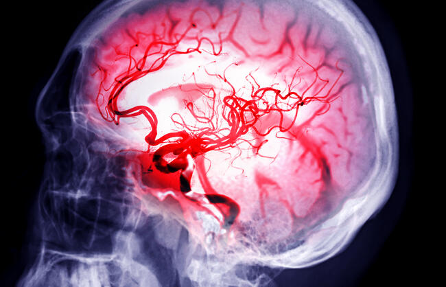
Cranial nerve (CN) assessment is often overlooked in the prehospital setting, yet it can provide some of the most powerful clues in diagnosing neurological emergencies. Those subtle differences in eye movement and pupillary response can help determine traumatic brain injury (TBI), identify rising intracranial pressure, and guide treatment and transport decisions for EMTs and paramedics. A focused CN exam doesn’t require extensive time or specialized tools, just keen observation and an understanding of what abnormal findings may mean.
In trauma this is especially true: Subtle neurologic changes speak more loudly than the injuries we see. CN assessment provides a rapid window into how trauma is affecting the nervous system. A few seconds spent assessing eye movement and pupil response can indicate the progression of intracranial bleeding or reveal early signs of herniation long before vital signs change.
Every EMS clinician has been in such chaotic situations. The patient condition may be evolving at an accelerated pace, so these rapid assessments can be the difference between missing a critical neurologic deficit and catching it early. In the field, where time and early recognition drive outcomes, a focused cranial nerve exam becomes more than an assessment tool; it’s a critical diagnostic GPS in determining priorities, destinations, and emergent interventions.
What Cranial Nerves Do
There are 12 pairs of CNs (I-XII) that emerge from the brain, controlling sensory, motor, or mixed functions for the head, neck, and torso.1-2 They are numbered by their attachment to the brain and have specific functions. They are:
- Olfactory (CN I): Smell.
- Optic (CN II): Vision.
- Oculomotor (CN III): Eye movement, pupil constriction.
- Trochlear (CN IV): Eye movement (superior oblique muscle).
- Trigeminal (CN V): Facial sensation, chewing.
- Abducens (CN VI): Eye movement (lateral rectus muscle).
- Facial (CN VII): Facial expressions, taste.
- Vestibulocochlear (CN VIII): Hearing and balance.
- Glossopharyngeal (CN IX): Taste, swallowing, salivary glands.
- Vagus (CN X): Heart rate, digestion, voice (widely distributed).
- Accessory (CN XI): Shoulder and neck movement.
- Hypoglossal (CN XII): Tongue movement.
We have all used mnemonics for memorizing everything in medicine and EMS. There are some very colorful ones for the CNs. One this author has used: “Ooh, Ooh, Ooh, to touch and feel very good velvet. Such heaven!”
Assess and Reassess
Regarding trauma, most of the bad TBI stuff presents about the same way:
- Headache
- Nausea/vomiting
- Altered mentation
- Neurologic deficits
You may have a baseball player who is struck with a line drive. He may be conscious, but confused, he may be complaining of nausea and vomiting, and he may be unsteady on his feet, with a poor gait. These signs and symptoms are indicative of concussion, but they could also signal an epidural hematoma. Monitoring and reassessment is crucial for this patient; both are serious injuries, but an epidural bleed may be fatal.3-6 This author is reminded of the death of actress Natasha Richardson, a famous example of a patient who succumbed to an epidural bleed following a skiing accident.7
During the secondary assessment, CNs may be assessed depending on the patient’s condition. Frequent reassessments, of not only vital signs, but of pupillary status and level of consciousness (LOC), is essential. Keep in mind that patients do not suddenly crash; we suddenly notice.
The Eyes Have It
It would be impractical to assess all 12 pairs of CNs in the chaotic, time-critical prehospital environment. As EMS clinicians instead we focus on rapid cranial nerve checks that affect vision, pupils, and neurologic status. They say that the eyes are the mirrors to the soul—they are also a reflection of the brain. The most important CNs are responsible for the evaluation of eye dysfunctions: CN II, III, IV, and VI.3-6
Some of these assessments will be dependent on the patient’s ability to follow commands. These three nerves control eye movement: CN III, IV, VI.1-6 In EMS, we test them together, not separately.3-6 When assessing eye movement, you may need to determine the extraocular movement (EOM). To do this, say to your patient: “Follow my finger with your eyes—only your eyes, don’t move your head.” Then move your finger in a large “H” or “X” pattern.
- The inability to move one or both eyes indicates a neurologic deficit (CN III, IV, VI).
- Paralysis of the lateral gaze is an early sign of rising intercranial pressure (ICP) in TBI. Because the abducens nerve, CN VI, is highly susceptible to stretch and compression, especially with increased ICP, your patient may have impaired lateral gaze as one of the earliest focal neurological deficits in TBI. Rising ICP causes downward/medial brainstem displacement and stretches/compresses the nerve. Because this is a physical impairment of the nerve, the deficit exists whether or not the patient can follow commands. The challenge is detecting it without voluntary eye movement. How do you do this?
You can detect impaired lateral gaze through spontaneous eye position. For example, if the eyes are deviated inward, one or both eyes are turned toward the nose instead of looking straight ahead; the eye(s) look crossed. This happens even when the patient is relaxed or unconscious. If the patient is conscious, they may or may not complain of vision problems. Another example is asymmetric resting eye positions—when the patient is not trying to look at anything, the eyes are not lined up together. One eye may be looking straight ahead. The other eye may be turned inward, outward, upward, or downward. Simply said, the patient’s eyes are not pointing in the same direction when they are relaxed. Again, the patient may be unconscious.
- Paralysis of the upward gaze may indicate an orbital floor fracture and EOM muscle entrapment. This is a mechanical issue and not neurological.
- If a patient reports double vision (diplopia) cover one eye; if symptoms improve, it’s likely a cranial nerve issue.
- Drooping eyelid (ptosis) CN III
- A patient who cannot move both eyes simultaneously in a specific direction, even when trying, is called midline gaze palsy, a major stroke indicator.
When you are asking your patient to move their eyes and follow an “H” or “X” pattern, you are also assessing the patient’s conjugate gaze. If the eyes do not move in unison, this is a problem that may indicate a neurologic lesion. Such a lesion may be caused by:3-6
- Stroke (ischemic blockage or hemorrhage)
- TBI with bleeding
- Brainstem or cranial nerve dysfunction
If you see nystagmus (rapid involuntary movement of the eyes), this could be indicative of intoxication, seizure, TBI, or stroke.
You can check CN II and CN III by performing consensual pupil check. This is another check that does not require a patient to be conscious. If an EMS clinician shines their penlight into one eye, and both pupils constrict, even when the opposite eye is blocked from the light, that is the consensual light reflex and indicates that both CN II and CN III are intact.
Regarding pupils, every EMS clinician has learned the term PERRLA, meaning
- Pupils
- Equal
- Round
- Reactive to Light and Accommodation
Equal pupils and reaction to light are the most basic tests to assess CN II and III. Reactive to light checks CN II by confirming that the eye can detect light and send that signal to the brain, and CN III controls pupillary constriction.
- CN III compression leads to unequal or unreactive unequal pupils with altered LOC. Shine your light into the patient’s eyes; check size, equality, reaction. When we see unequal or sluggish pupils in patients with altered LOC or who are unconscious, think rising intracranial pressure from a bleed in the brain or possible stroke. This local compression will often precede possible brain herniation, and this is our warning signal.
- If you see pupils that are bilaterally dilated and unresponsive, this is usually a late sign and may indicate catastrophic brainstem failure and herniation.
Other Signs of Brain Injury
Don’t forget our other physical manifestations of brain injury that we may see in conjunction with LOC and pupillary response:3-6
- Abnormal posturing, either flexion (indicating rising pressure above the brainstem) or extension (a much worse sign indicating a serious injury and rising pressure at the level of the brain stem)
- Seizures, which may be manifestation of acute brain injury, rising intracranial pressure, or cerebral hypoxia
Remember that hypoxia and hypotension are our two enemies when we treat any patient with TBI. Maintain blood pressure and keep your patient oxygenated and treat them immediately if they begin to fall to reduce mortality and morbidity.3-6
Keep it simple. If your patient is conscious and can follow commands, a quick test any EMT or paramedic can perform is: “See the Light, Follow the Finger.”3-6
- See the Light: CN II & III (pupils)
- Follow the Finger: CN III, IV, VI (eye movements)
Cranial nerve assessment is not about memorizing anatomy. It's about recognizing change. In EMS, a few seconds spent checking pupils and eye movement can reveal evolving brain injury long before vital signs deteriorate. These simple, repeatable observations help EMS clinicians identify life-threatening neurologic conditions, prioritize rapid transport, and communicate critical findings to receiving facilities.
When used consistently, focused cranial nerve checks become one of the most powerful tools EMS clinicians carry into the field.
References
- Drake R, Vogl AW, & Mitchell, AW. (2009). Gray's anatomy for students E-book. Elsevier Health Sciences.
- Cohen, BJ, & Hull, KL. (2020). Memmler's the human body in health and disease. Jones & Bartlett Learning.
- Chapleau W, Burba A, Pons, PM, Page D. (2011). The Paramedic Updated Edition (1st ed.). McGraw-Hill Education
- PHTLS Course Manual. National Association of Emergency Medical Technicians (US). 10th edition. Burlington, MA: Jones & Bartlett Learning, (2024)
- Pinto VL, Adeyinka A. Increased Intracranial Pressure. [Updated 2025 Sep 14]. In: StatPearls [Internet]. Treasure Island (FL): StatPearls Publishing; 2025 Jan. Available from: https://www.ncbi.nlm.nih.gov/books/NBK482119/
- Lulla A, Lumba-Brown A, Totten AM, et al. Prehospital Guidelines for the Management of Traumatic Brain Injury, 3rd Edition. Prehosp Emerg Care. 2023;27(5):507-538. doi: 10.1080/10903127.2023.2187905. Epub 2023 Apr 20. PMID: 37079803.
- ABC News. (2009, March 17). Natasha Richardson died of epidural hematoma after skiing accident. ABC News. https://abcnews.go.com/Entertainment/Movies/story?id=7119825&page=1