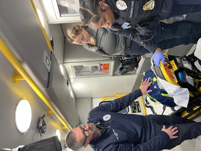
Studies Have Shown…Or Have They?
It seems like more studies are being published than ever, either completed or preliminary. An often misleading phrase that gets frequently mentioned in EMS is “studies have shown.” My immediate thought on reading those few words is, “maybe not.” Do they actually show what they claim, and is it significant? More importantly, is it representative of your data or merely an average of what everyone else does?
Epinephrine: Seven Decades of Guessing
An area of widespread interest in recent years is the use of epinephrine in cardiac arrest. The “standard” 1 mg adult dose for cardiac arrest was formulated based on early 1960s studies using 10 kg dogs who were deliberately asphyxiated. The study is old enough now that those dogs would be eligible for Medicare.
Amazingly, that same dose used on those dogs whose weight was roughly equivalent to a one-year old child is also used on a 400 lb patient, often regardless of arrest etiology or downtime. Despite many different variations that have been tried, the correct dose or method of administration is still not well known. It’s way past time to realize that one size rarely fits all in emergency medicine.
Limitations
A small but important part of any study is to note its limitations, which are usually listed near the end, way beyond the more commonly read abstract. A recent study examined the use of IM epinephrine for anaphylaxis and concluded that an initial dose of 0.5 mg might be more effective than the more common 0.3 mg.
A significant shortcoming was that the injection site often wasn’t recorded. What if a majority of the lower doses were administered in the more commonly used deltoid site? A larger muscle would likely have more rapid absorption and lead to better first injection results. Of note, 30 years ago I worked under guidelines that gave us the discretion to use anywhere from 0.3 mg to 0.5 mg based upon patient and presentation.
Is the Comparison Valid?
Other recent studies have been based on alternative placement of defibrillation pads, whether for first or secondary shocks. An overall advantage of other locations is often cited, based upon aggregate data. One drawback is a presumption that the initial placement is correct. Chests and breasts also vary in size, shape, and density. The most effective positioning for a deep, thick chest may vary from a wide, thin one. Averages can be misleading.
Look through much of what has been published on the matter and you will often see pictures, illustrations, or videos showing more of an anterior-anterior placement rather than true anterior-lateral. Even major guideline updates from the end of 2025 give very specific locations for pads. If the initial placement is incorrect then simple correct repositioning represents vector change and might alone be more effective.
Is the Conclusion Valid?
Other studies purport to show the lack of efficacy for transcutaneous pacing (TCP) in the field, including variable electrical capture. Most importantly, while electrical capture may be an objective, the goal of pacing is to improve perfusion. Perhaps the best surrogate for this is the use of end-tidal carbon dioxide (ETCO2) monitoring. While not an exact correlation, a patient who requires pacing will likely have a low reading that will rise with true mechanical capture.
These studies often don’t note how a pulse was confirmed, and I strongly believe that pulse confirmation was frequently performed using the carotid artery. If you use a high enough energy level to get capture, what seems to be a pulse is often due to muscle contractions from the electricity. Lacking end-tidal monitoring, a more distal pulse such as the radial is more accurate. While again not an exact correlation to blood pressure, someone who has no radial pulse pre-pacing but gets it back as a result has improved perfusion (a good general guide for any patient, pacing or not).
Mental Status
An even more basic method of determining the efficacy of treatment is mental status. A patient who is altered or unresponsive prior to treatment then becomes alert after care is rendered likely has improved perfusion. Besides pacing, this pertains to almost anything from simple patient positioning to whole blood administration. Fancy monitors aren’t needed to determine this but paying attention to your patient is.
A recent presentation on TCP led me to conclude that verification of true mechanical capture should include at least two of the following parameters: improved mental status, a radial pulse rate that matches that of the pacer, increased ETCO2, a consistent SpO2 reading and increased blood pressure. Any one parameter alone is likely subject to error or misinterpretation but the presence of at least two should increase confidence of efficacy.
Death by Association
Another important aspect is that studies often show correlation rather than cause and effect. To demonstrate the difference, sudden cardiac arrest is often a cause of death. On the other hand, properly performed CPR should not cause death but approximately 90% of patients who receive CPR die. As a result, CPR is associated with death but clearly does not cause it unless major retraining is indicated.
Evaluate Carefully
Years ago, Ernest Hemingway noted that a good writer should have a well-developed crap detector (edited for mixed company). Apparently, this also is handy for an enlightened reader as many of the studies that I have seen lately prompt me to think, “that’s crap.” Ensure that any studies you undertake or digest are Carefully Researched And Planned and do not merely show Concluded Results As Proposed.