


Use of High-Frequency Electrical Stimulation in Gastrocutaneous Fistula Closure: A Case Report
© 2024 HMP Global. All Rights Reserved.
Any views and opinions expressed are those of the author(s) and/or participants and do not necessarily reflect the views, policy, or position of Wounds or HMP Global, their employees, and affiliates.
Abstract
Background. Gastrocutaneous fistula is a rare complication following Roux-en-Y gastric bypass, a commonly performed bariatric surgery. While most ECFs respond to conservative management, some do not close despite adequate nutritional support, infection source control, and drainage management. As such, the chronicity of these difficult-to-treat wounds can be physically and economically costly to patients. Case Report. A 53-year-old female with a history of Roux-en-Y gastric bypass developed a gastrocutaneous fistula secondary to a perforated gastrojejunal ulcer, requiring immediate surgical intervention. After being discharged from the hospital, 37 days of conservative management and NPWT did not reduce the size of the fistula tract. To help control the patient’s chronic abdominal pain and increase the rate of wound healing, the patient underwent treatment with HFES (20 kHz) delivered using a handheld transcutaneous electrical nerve stimulator. This electrotherapy was found to reduce the majority of the patient’s pain within the first treatment session. The patient's fistula also began to decrease in size within 1 week of initiating treatment. Conclusion. This case report details the successful closure of a gastrocutaneous fistula after administration of HFES 3 times a week over the course of 25 days. The mechanism of action of HFES and its role in the wound healing process are also discussed.
Abbreviations
Abbreviations: BMI, body mass index; CT A/P, computed tomography of the abdomen and pelvis; ECF, enterocutaneous fistula; EF, electric field; GE, gastroesophageal; HFES, high-frequency electrical stimulation; NPWT, negative pressure wound therapy; OR, operating room; TEP, transepithelial potential.
Introduction
Electrical stimulation has been studied as an adjunct therapy to normative treatments for ECFs, with many studies focusing on microcurrent therapy aimed at replicating the endogenous EF present in wounded skin.1-3 Compared with microcurrent therapy, the benefits of HFES in wound care have yet to be largely explored.
This case report describes the successful closure of 1 patient's abdominal wound and gastrocutaneous fistula with HFES delivered by the TrueRelief device (TrueRelief, LLC; hereafter transdermal electrical nerve stimulator). This device, which was cleared by the US Food and Drug Administration in 20214 for the management of chronic pain and postoperative acute pain, emits a 20-kHz transcutaneous current (herein referred to as HFES) via 2 steel-tipped probes.
Case Report
In 2002, a 53-year-old female underwent Roux-en-Y gastric bypass surgery as a treatment for morbid obesity. The patient reported weighing over 400 lb prior to surgery (BMI > 65), along with a history of hypertension, type 2 diabetes, and chronic kidney disease. Her comorbid conditions all resolved or improved following gastric bypass surgery, and the patient achieved a normal BMI. In 2021, following a medically uneventful period of 19 years after the Roux-en-Y gastric bypass, this patient presented with a perforated gastrojejunal anastomotic ulcer (marginal ulcer) secondary to nonsteroidal anti-inflammatory drug usage. The course of her hospital stay as well as wound care with HFES after hospital discharge are described.
Hospital stay: day 1
The patient's course of illness began when she awoke with sudden-onset abdominal pain radiating to the left shoulder. The pain progressively worsened throughout the morning, and it was exacerbated by deep breathing. The patient presented to a local hospital for pain and underwent CT A/P, which showed free air near the GE junction.
Later that afternoon, the patient was transferred by air to a larger medical center under the working diagnosis of a perforation at the GE junction. The CT A/P was reviewed, and the diagnosis was changed to that of a perforated marginal ulcer. The patient was hypotensive on arrival and was immediately admitted and taken to the OR for diagnostic laparoscopy. No identifiable perforation was discovered, so a peritoneal lavage was performed and drains were placed. Shortly after that, the patient experienced hemodynamic instability and was admitted to the intensive care unit.
Hospital stay: days 2 to 6
The patient's condition improved on days 2 to 5, but she soon developed left upper quadrant pain and abdominal distension. On day 6, another CT A/P was performed, showing extravasation of contrast material. The patient was returned to the OR for exploratory laparotomy that afternoon, which revealed an obvious perforated marginal ulcer. A Graham patch was placed at that time to treat the ulcer. The fascia was left open, and an NPWT dressing was placed.
Hospital stay: days 9 to 15
On day 9, the patient was returned to the OR. The fascial border of the wound was necrotic, and it was debrided. The abdomen was irrigated and left open, and a new NPWT dressing was placed.
At days 10 to 13, the wound drainage became bilious, and the patient became febrile.
The patient was returned to the OR on day 14, but no reperforation was evident, despite bile staining around the GE junction. A washout was performed, and the abdomen was left open. The patient remained septic.
On day 15, the patient was returned to the OR for placement of an endoscopic stent across the perforation. Her condition immediately improved.
Hospital stay: days 18 and 31
On day 18, the patient was returned to the OR once again for abdominal closure. The fascia was noted to be friable, and the abdomen was closed by buttressing the fascia superficially with Phasix mesh (BD) and using an inlay XenMatrix AB biologic mesh (BD). An NPWT dressing was placed.
The patient improved sufficiently to be discharged on day 31 to a skilled nursing facility with continuing wound care.
Hospital stay: day 35
The patient was returned to the hospital with concerns of leakage through the NPWT dressing and excess purulent drainage. Further radiographic evaluation revealed that the stent had migrated distally. The stent was removed, and an NPWT dressing was replaced over the open wound. During the dressing change, bile was noted to be freely flowing into the abdominal cavity. NPWT controlled the drainage. An upper endoscopy was performed, and a large ECF was noted at the gastrojejunostomy. Using the endoscope as a guide, a Dobhoff tube was placed distal to the anastomosis. Attention then was turned to local wound care and nutritional support. The patient was ultimately able to tolerate full-strength tube feedings via a Dobhoff tube.
The patient was discharged home in stable condition with NPWT controlling the drainage. The NPWT device was applied with exclusive dressing around the wound and black foam over the fistula and wound (75 mm Hg continuous pressure). This increased granulation tissue development; however, it also kept the fistula open by sucking fluid from the stomach.
Wound care course after hospital discharge: days 36 to 71
Despite adequate nutritional support and serial dressing changes, the fistula continued to leak bilious fluid during every NPWT dressing change, and there was no change in fistula size. The patient also reported chronic abdominal pain.
Wound care course after hospital discharge: day 72
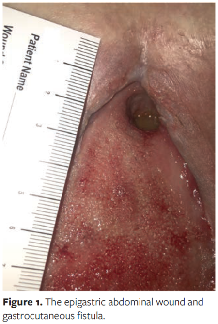
In an effort to help control the patient's chronic abdominal pain, HFES therapy was started. At the onset of treatment, the patient's wound measured 18.5 cm × 4.5 cm × 0.3 cm, centered epigastrically. The fistula measured 12 mm in diameter and was located at the superior-most aspect of the wound (Figure 1).
Treatment with the transdermal electrical nerve stimulator consisted of 10-minute applications of 20-kHz current across the wound. One probe was placed on each side of the wound, and the position of the probes was altered every 30 seconds to 1 minute, in order to fully treat the entire wound. The fistula tract was treated separately with the same constant current for 5 minutes. At the end of treatment, the patient's NPWT dressing was replaced. This treatment was administered 3 times a week over a 25-day period. The wound was debrided as necessary immediately prior to these treatments.
Prior to the first treatment, the patient reported abdominal pain of 2 out of 10 (0 = no pain, 10 = worst imaginable pain). Immediately after the first treatment, the patient reported that her pain had decreased to 0.
Wound care course after hospital discharge: days 76 to 97
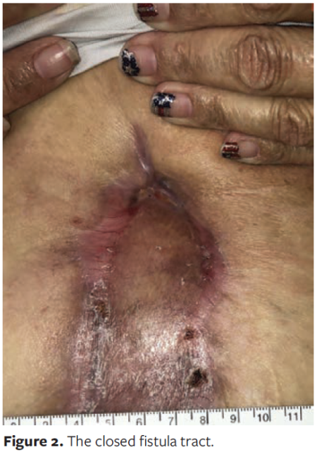
On day 76, less bilious drainage was noted originating from the wound during the NPWT dressing change. By day 78, the fistula had decreased in size to 8 mm, and less gastric content was being produced. By day 85, the fistula had decreased in size to 6 mm, and there was minimal drainage from the fistula site. The fistula tract was completely closed on day 97, with no evidence of further drainage (Figure 2).
Wound care course after hospital discharge: day 112 and beyond
The patient underwent split-thickness skin graft placement on day 112. This was performed without complication and resulted in 90% graft take.
Over the following weeks the skin graft healed completely, and at the time of this writing there had been no further drainage from the fistula site. The patient was able to resume a regular diet without supplemental support.
Discussion
The hallmark of ECF treatment consists of nutritional support, limiting oral intake, controlling drainage, and protecting the skin.5 Despite this, many ECFs do not heal entirely. A retrospective study of 23 patients with postoperative ECF revealed a 69.6% closure rate, with 5 high-output fistulas (21.7%) resulting in death due to sepsis, severe malnutrition, and organ failure.6 A 2022 meta-analysis by Gefen et al7 of 3078 patients with ECF revealed a healing rate of 89%, with a recurrence rate of 11.1% and a mortality rate of 8.5%. No standardized management exists for gastrocutaneous fistula, a subtype of ECF with an incidence rate of 1.7% to 4.0% after bariatric surgery.8 Although the rate of incidence of gastrocutaneous fistula is small, it presents a very difficult problem, because treatment options are limited, often are customized, and have variable success rates that are largely dependent on the individual patient's condition and ability to heal.9
Many different approaches to the management of gastrocutaneous fistula have been explored, including surgical and endoscopic options such as stenting, clipping, and placement of T-tubes or cardiac septal occluders.8,10-12 Since the discovery of biological TEPs in skin, electrical stimulation for wound healing has become another an item of interest as an adjunct therapy for chronic wounds.
In the 1800s, German physiologist Emil Du Bois-Reymond discovered that the skin conducts electricity and that, as reviewed recently by Luo et al,13 the flow of current in injured tissue (due to the directional transport of ions by epithelial cells) forms a TEP. Based on these discoveries, theories about the so-called skin battery were developed, based on the work of Barker et al. In 2 foundational studies by Barker et al (1982) and Chiang et al (1992) on glabrous human skin and bovine cornea, respectively, it was further shown that wound sites develop an endogenous EF.13 Poorly healing, chronic wounds have been found to exhibit suppressed endogenous EFs.13 There has been much interest in applying an exogenous EF to mimic the biological response and restore the electrical component of electric factors in wound healing.
In their systematic review, Luo et al13 analyzed the effect of exogenous electrical stimulation on inflammation, blood flow, cell proliferation and migration, and wound scarring. They noted different types of current (ie, direct current, alternating current, pulsating current) and their effects on the 4 main stages of wound healing: coagulation phase, inflammation phase, proliferation phase, and remodeling phase.
The HFES used in the current case report is a pulsed direct current; according to Luo et al,13 such current recruits immunocytes, shortens the inflammation phase by decreasing the expression of pro-inflammatory cytokines, increases tissue blood flow, and promotes the migration of epithelial cells, among other things. A few recent studies, including a study by an author of the current case report and a study delineating the mechanism of action of HFES, have shown similar antibacterial and anti-inflammatory results.
In 2019, Barki et al1 reported that a wireless electroceutical dressing prevented and disrupted bacterial biofilms in wounds in a porcine model. Biofilms promote infection by sheltering bacteria and inhibit skin barrier function by silencing E-cadherin, an essential component of epidermal tight junctions.1 Similarly, in 2019, Yu et al2 created a microcurrent dressing that achieved a statistically significant increase in wound healing rates in rats. Those authors attributed the increased wound healing rates to reduced inflammation duration and increased growth factor expression. Compared with NPWT, microcurrent electrical stimulation was found to be at least as effective in reducing wound surface area according to a 2019 study of patients with burn wounds.3 However, whereas HFES was used the current case report, the aforementioned studies used microcurrent electrical stimulation or silver-zinc microcell batteries.
While HFES has not been as extensively studied as either microcurrent electrical stimulation or silver-zinc wireless electroceutical dressings, Hatch and Lavor14 successfully used HFES to reduce pain, improve microvascular blood flow, and reduce bacterial load in 9 patients with chronic wounds. Five (56%) of the 9 patients demonstrated an increase in perfusion of periwound tissue, and the average percent wound closure increased from 23.77% to 64.58% after the addition of HFES to the wound care regimen. At the time, the authors acknowledged that "the exact mechanism specific to the present device has yet to be elucidated."14 In 2022, however, Yang et al15 demonstrated in a murine model that HFES delivered via transdermal electrical nerve stimulator resulted in downregulation of the HMGB1 gene and reduction in the secretion of inflammatory modulators in sensory neurons.
Given the prolonged course of fistula treatments and the significant cost and morbidity to the patient, additional modalities that facilitate fistula closure are of great value. HFES therapy has the potential to further accelerate wound healing if it is used as part of routine dressing changes. The efficacy of HFES in accelerated wound healing may be further studied by comparing wound resolution in wounds with expected closure times.
Patients can also benefit from the pain reduction effects of HFES, which are attributed to the disruption of sensory nerve secretory activity.15 In the current case, the patient's pain improved immediately after the first treatment, from 2 out of 10 to 0.
Limitations
The primary limitation of this study is the sample size, which is limited to a single patient. Further studies are needed to investigate the efficacy of HFES in gastrocutaneous fistula closure. For example, a double-masked, randomized, sham-controlled trial could be conducted to assess the effect of HFES on ECF closure. ECFs involving the intestinal tract are more common than gastrocutaneous fistulas, so it would be easier to obtain participants for such a study. The primary end points in such a study would be the rate of fistula closure (fistula diameter over time in mm/day) before and after initiation of HFES, and the percentage of fistula closure, that is, change in fistula area over time, calculated as follows:
πr2 initial
within X days after initiation of transdermal electrical nerve stimulator treatment, where r = fistula radius.
Conclusion
This case report presents a post-gastric bypass patient who developed a gastrocutaneous fistula secondary to a perforated marginal ulcer. The lengthy postoperative course resulted in a high-output fistula that was controllable with NPWT and nutritional support but did not improve after 37 days of treatment. HFES therapy was added to the treatment regimen, after which the fistula decreased in size within 1 week and closed completely within 25 days. The patient ultimately underwent split-thickness skin grafting and achieved complete wound closure shortly thereafter. Although this case does not confirm the ability of transdermal electrical nerve stimulator to reduce pain or facilitate wound healing and fistula closure, it does suggest that future investigation is warranted. Treatment modalities that reduce wound care costs and accelerate wound healing should be studied in the interest of improving outcomes of patients with chronic wounds.
Acknowledgments
Authors: Diego Silva-Mendoza, BS1,2; Dylan Joule, BS1,3; Michael Lavor, MD4; and Matthew J. Weiner, MD5
Affiliations: 1Saguaro Surgical, Tucson, AZ; 2Saint Louis University School of Medicine, St. Louis, MO; 3The University of Arizona College of Medicine - Tucson, Tucson, AZ; 4Wound Care, Saguaro Wound Care Clinic, Tucson, AZ; 5Bariatric Surgery, Tucson Medical Center, Tucson, AZ
ORCID: Joule, 0009-0005-5885-3158; Silva-Mendoza, 0009-0007-4915-3744
Disclosure: The authors disclose no financial or other conflicts of interest.
Correspondence: Matthew J. Weiner, MD; Medical Director of Bariatric Surgery, Tucson Medical Center, 5301 E. Grant Road, Tucson, AZ 85712; drweiner@tucsonwls.com
Manuscript Accepted: December 6, 2023
References
1. Barki KG, Das A, Dixith S, et al. Electric field based dressing disrupts mixed-species bacterial biofilm infection and restores functional wound healing. Ann Surg. 2019;269(4):756-766. doi:10.1097/SLA.0000000000002504
2. Yu C, Xu Z-X, Hao Y-H, et al. A novel microcurrent dressing for wound healing in a rat skin defect model. Mil Med Res. 2019;6(1):22. doi:10.1186/s40779-019-0213-x
3. Ibrahim ZM, Waked IS, Ibrahim O. Negative pressure wound therapy versus microcurrent electrical stimulation in wound healing in burns. J Wound Care. 2019;28(4):214-219. doi:10.12968/jowc.2019.28.4.214
4. March 2021 510(k) clearances. U.S. Food and Drug Administration. Accessed January 24, 2024. https://www.fda.gov/medical-devices/510k-clearances/march-2021-510k-clearances.
5. Ghimire P. Management of enterocutaneous fistula: a review. JNMA J Nepal Med Assoc. 2022;60(245). doi:10.31729/jnma.5780
6. Noori IF. Postoperative enterocutaneous fistulas: management outcomes in 23 consecutive patients. Ann Med Surg (Lond). 2021;66:102413. doi:10.1016/j.amsu.2021.102413
7. Gefen R, Garoufalia Z, Zhou P, Watson K, Emile SH, Wexner SD. Treatment of enterocutaneous fistula: a systematic review and meta-analysis. Tech Coloproctol. 2022;26(11):863-874. doi:10.1007/s10151-022-02656-3
8. Liagre A, Queralto M, Levy J, et al. Treatment of persistent large gastrocutaneous fistulas after bariatric surgery: preliminary experience with endoscopic Kehr's T-tube placement. Obes Surg. 2022;32(4):1377-1384. doi:10.1007/s11695-022-05935-y
9. Strong AT, Guerrón AD. Revisional bariatric surgery for chronic complications necessitates custom surgical solutions. Mini-invasive Surg. 2022;6:37. doi:10.20517/2574-1225.2021.137
10. Negm S, Mousa B, Shafiq A, et al. Endoscopic management of refractory leak and gastro-cutaneous fistula after laparoscopic sleeve gastrectomy: a randomized controlled trial. Surg Endosc. 2022;37(3):2173-2181. doi:10.1007/s00464-022-09748-z
11. Goparaju A, Cherasard P, Kella V, Levine J, Brathwaite C. A231 laparoscopic repair of chronic gastro-cutaneous fistula from the excluded stomach 19 years after gastric bypass. Surg Obesity Related Dis. 2019;15(10):S84-S85. doi:10.1016/j.soard.2019.08.176
12. Kumaira Fonseca M, Coelho NH, Manica JL, Ramblo RR, Spier IE, Seabra AP. Endoscopic management of a chronic gastrocutaneous fistula after bariatric revisional surgery using a novel cardiac septal occluder. GE Port J Gastroenterol. 2022:30(Suppl 1):52-56. doi:10.1159/000526507
13. Luo R, Dai J, Zhang J, Li Z. Accelerated skin wound healing by electrical stimulation. Adv Healthc Mater. 2021;10(16):e2100557. doi:10.1002/adhm.202100557
14. Hatch DC, Lavor M. Decreasing pain and increasing the rate of chronic wound closure with the use of a noninvasive bioelectronic medical device: a case series. Wounds. 2021;33(5):119-126. doi:10.25270/wnds/2021.119126
15. Yang H, Datta-Chaudhuri T, George SJ, et al. High-frequency electrical stimulation attenuates neuronal release of inflammatory mediators and ameliorates neuropathic pain. Bioelectron Med. 2022;8(1):16. doi:10.1186/s42234-022-00098-8


