Advantages of a Radial Approach for Endovascular Interventions in “Global” Renal and Mesenteric Ischemia
© 2026 HMP Global. All Rights Reserved.
Any views and opinions expressed are those of the author(s) and/or participants and do not necessarily reflect the views, policy, or position of Cath Lab Digest or HMP Global, their employees, and affiliates.
Robert L. Minor, Jr, MD
Interventional Cardiologist and Endovascular Specialist, Billings Clinic Heart and Vascular at Community Medical Center
Missoula, Montana
Disclosure: Dr. Minor reports serving as a consultant for Medtronic.
Robert Minor, MD, can be contacted at rlm816@aol.com.
Introduction
In patients with renovascular hypertension and ischemic nephropathy due to bilateral renal artery stenosis, and cases of acute renal infarction, treatment now favors endovascular therapy. Chronic mesenteric ischemia, and life-threatening cases of acute mesenteric infarction without peritonitis also warrant consideration of endovascular therapy over traditional surgery. However, endovascular therapy for atherosclerotic renal and mesenteric artery disease can be extremely challenging when using femoral access, given the frequent need to traverse tortuous and often calcified aorto-iliac anatomy in elderly patients. In addition, guide catheter backup may often be suboptimal in the setting of steep downward takeoffs of these visceral arteries or widened abdominal aortas. Use of the radial artery approach reduces technical challenges by avoiding aortoiliac anatomy, allowing easier visceral artery cannulation, and providing coaxial guide catheter support. Two complex cases involving “global” renal and mesenteric ischemia are presented where the radial approach greatly facilitated endovascular therapy that included the use of filter embolic protection.
Case #1: Renal Interventions
This is a 65-year-old male lifelong smoker, with known chronic left subclavian artery occlusion, stable leg claudication, and poorly controlled hypertension on 4-drug therapy, who presented with right flank pain and hematuria. A renal artery duplex ultrasound demonstrated atrophy of the right kidney to 7.1 cm, with a normal left kidney size of 10.8 cm. Right renal artery flow could not be detected, with left renal artery to aortic velocity ratio markedly increased to 4.8. Computed tomography angiography (CTA) demonstrated probable total occlusion of the right renal artery and severe stenosis of the left renal artery, 100% left iliac artery and 70% stenosis of the right iliac artery, with fusiform enlargement of the abdominal aorta to 4.6 cm. Upon presentation, blood pressures in the right arm were repeatedly >200/100 mmHg, and serum creatinine was 1.59 mg/dl with glomerular filtration rate (GFR) = 45 mL/min.
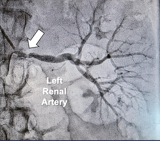
Using right radial artery access, a 6 French (Fr) Launcher MB2 110 cm long guiding catheter (Medtronic) was advanced over an .035-inch Wholey guidewire (Medtronic) to confirm 80% proximal left renal artery stenosis (Figure 1).
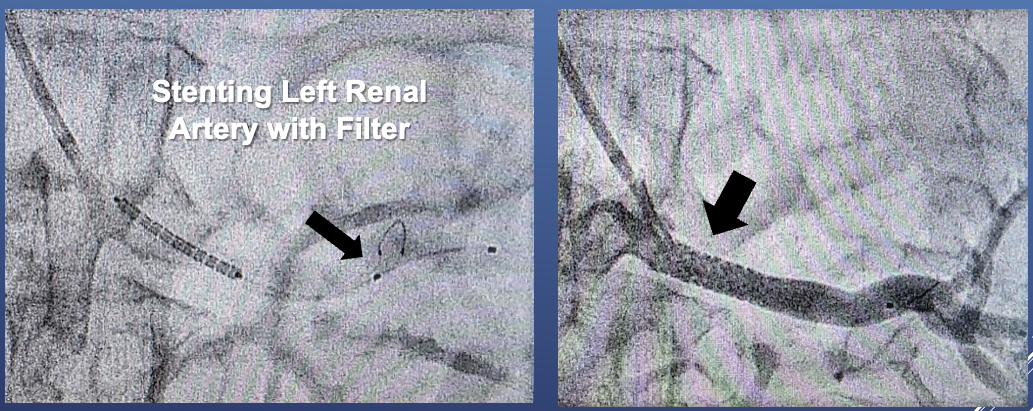
Given concerns about embolization risks from interventions in a possible solitary functioning left kidney, a 180 cm .014-inch Grand Slam coronary guidewire (Asahi Intecc) was used to cross the disease, and a 320 cm .014-inch SpiderFX 6 mm capture filterwire (Medtronic) was deployed in the distal main left renal artery. Pre-dilatation was performed with an .014-inch Viatrac 14 Plus Rx 6 x 15 mm balloon (Abbott), followed by an .014-inch Herculink Elite balloon-expandable 7 x 18 mm stent (Abbott) (Figures 2A-B). Upon filter retrieval, inspection revealed successful capture of embolic debris.
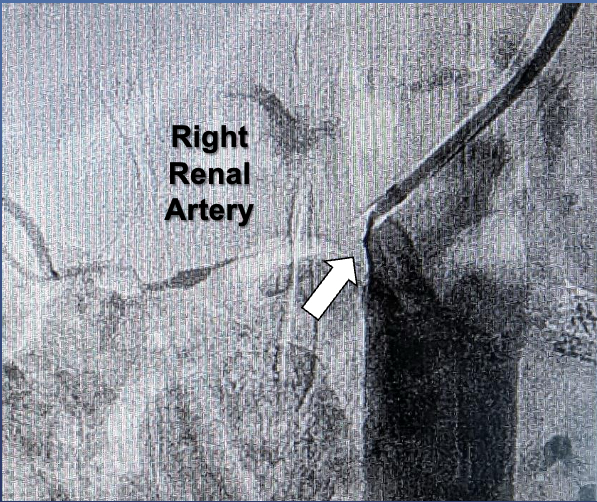
The MB2 guide catheter was then advanced and rotated over the .035-inch guidewire to the opposite aortic wall to attempt to identify the right renal artery. Digital subtraction angiography (DSA) revealed a long 99% stenosis of the artery with slow filling into the intrarenal branches (Figure 3).
Using the .014-inch guidewire to traverse the disease, an .014-inch NC Euphora 2.5 x 15 mm coronary balloon (Medtronic) was used to predilate the disease, followed by intravascular ultrasound study (IVUS, OptiCross 6 HD 60 MHz, Boston Scientific). IVUS demonstrated diffuse atherosclerotic disease extending beyond the first secondary branches of the right renal artery (Figures 4A-B).
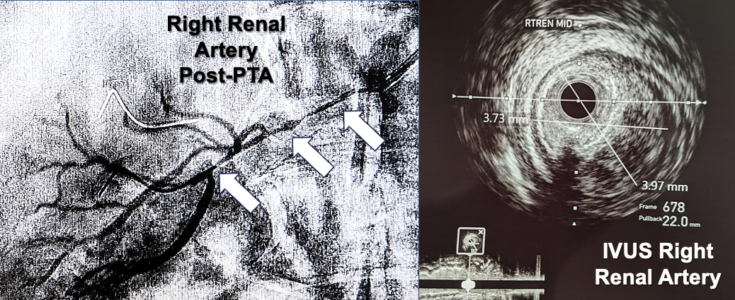
The renal artery media-to-media diameters in the intrarenal segment were 2.7 mm, and in the main right renal artery were 3.2 mm in the distal segment and 3.9 mm in the mid segment. A 3 x 38 mm Onyx coronary stent (Medtronic) was deployed to cover the right renal artery ostium, and post-dilated with a NC Euphora 4.0 x 20 mm balloon. A second overlapping 2.5 x 15 mm Onyx coronary stent was deployed to cover the disease extending into the intrarenal segment, with restoration of normal arterial flow (Figures 5A-B).
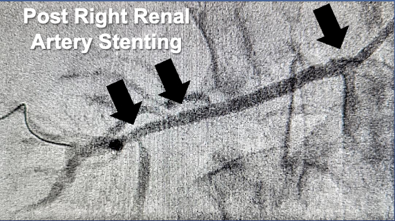
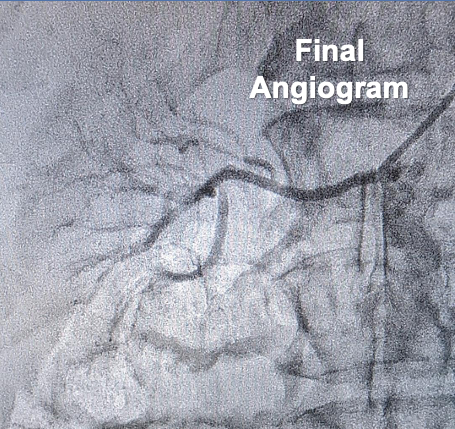
Subsequently, serum creatinine fell to 1.22 mg/dl with normalization of GFR>60 mL/min, and blood pressure remained well-controlled on 2-drug therapy.
Case #2: Mesenteric Interventions
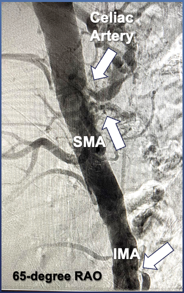
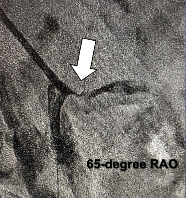
This is an 83-year-old male with severe peripheral arterial disease, right below-the-knee amputation and iliac artery stenting, who presented with severe, recurrent postprandial abdominal pain and bloody stools. He was referred for immediate catheter-based angiography with suspected threatened mesenteric infarction. Using right radial artery access, abdominal aortography using DSA performed in a 65-degree right anterior oblique (RAO) projection demonstrated 80% celiac artery ostial stenosis, 99% superior mesenteric artery (SMA) ostial stenosis, and 99% inferior mesenteric artery (IMA) ostial stenosis (Figure 6).
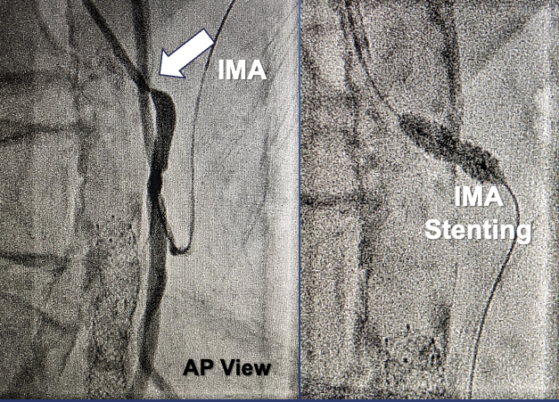
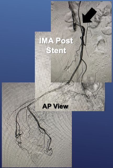
Using a 6 Fr MB1 110 cm long guiding catheter (Medtronic) advanced over an .035-inch Wholey guidewire (Medtronic), the IMA was cannulated in the anteroposterior (AP) projection, and a 180 cm .014-inch Grand Slam coronary guidewire (Asahi Intecc) was used to cross the lesion. The IMA was predilated with an .014-inch NC Euphora 5 x 12 mm balloon (Medtronic), followed by deployment of a .014-inch Herculink Elite balloon-expandable 5 x 12 mm stent (Abbott) (Figures 7A-B), with restoration of excellent flow into the vasculature of the descending and sigmoid colon (Figure 8).
Using the 65-degree RAO angle, the MB1 guide was rotated anteriorly over the .035-inch guidewire and slowly withdrawn to allow immediate cannulation of the SMA. Given the ulcerated appearance of the SMA stenosis with suspected intraluminal thrombus (Figure 9A), a 320 cm .014-inch SpiderFX 6 mm capture filter wire (Medtronic) was deployed over an .014-inch guidewire in the mid SMA for embolic protection.
The SMA lesion was predilated with an NC Euphora 5 x 12 mm balloon, followed by deployment of an .014-inch Herculink Elite balloon-expandable 5 x 18 mm stent. Final deployment was performed using an .014-inch Viatrac 14 Plus Rx 6 x 15 mm balloon (Figure 9B).
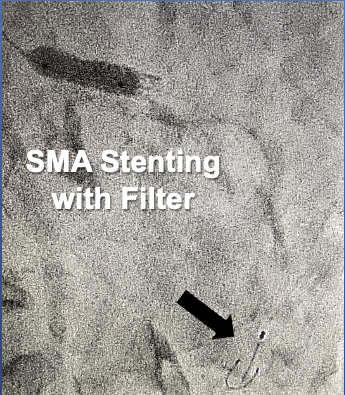
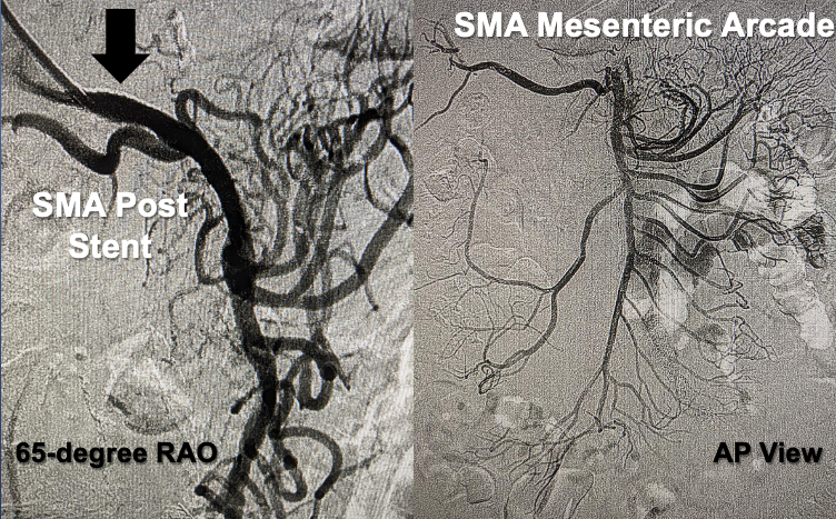
Filter retrieval revealed capture of embolic debris. DSA demonstrated excellent flow into all colic artery branches of the mesenteric arcade circulation with no evidence of embolization (Figures 10A-B).
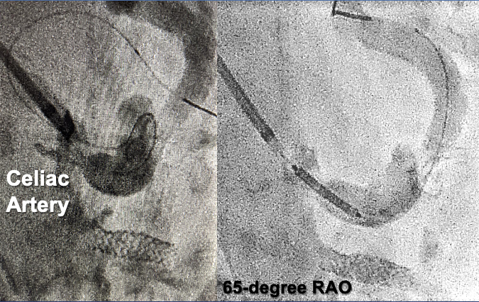
Maintaining the 65-degree RAO angle, the MB1 guide was again slowly withdrawn over the .035-inch guidewire with immediate cannulation of the celiac artery. The .014-inch guidewire was used to traverse the lesion with initial passage of the guidewire into the gastric artery, followed by repositioning and advancement into the main splenic artery to better support stent advancement (Figures 11A-B).
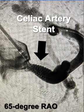
The celiac artery was predilated with the NC Euphora 5 x 12 mm balloon, followed by deployment of an .014-inch Herculink Elite balloon-expandable 7 x 15 mm stent (Figure 12A).
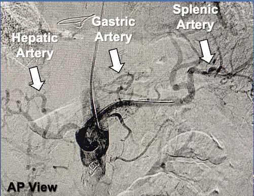
Final DSA in the AP view showed brisk filling of the gastric, splenic and hepatic arteries (Figure 12B). The patient had complete resolution of his abdominal pain, and during his hospitalization was able to resume a normal diet.
Tips and Tricks For the Radial Approach
Endovascular therapy for ostial renal and mesenteric artery stenosis is often challenging from femoral artery access when aortoiliac tortuosity and acute angulations of visceral artery takeoffs are encountered. From the femoral approach, curved renal double curve (RDC) and coronary Judkins Right (JR)4 and left internal mammary (LIMA) guiding catheters are commonly used. However, use of these guides to find the ostium of the diseased artery requires rotation and “dragging” of the guide tip along the diseased aortic arterial wall. This increases risks for embolization of aortic wall plaque fragments, and “snow-plowing” of plaque, which can occlude the target artery when critical ostial disease is present. In addition, these guide catheters often do not provide good backup support, especially when larger aortic diameters are present. When radial access is used, not only can the aortoiliac tortuosity be avoided, but coaxial cannulation of the origin of renal and mesenteric arteries can be easily achieved.
In the cases described using right radial access, the 6 Fr multipurpose MB1 and wider curved MB2 guide catheters were selected because of their steeper tip angulation, as their curve geometry provides better backup against the opposite aortic wall when compared to standard multipurpose guide catheters. These MB guide catheters are available in longer 110 cm lengths that extend their reach into the distal abdominal aorta and even the iliac arteries from radial artery access. After advancement of the MB guide catheters from right radial artery access over a supportive .035-inch guidewire to a position distal to the target artery, rotation of the guide tip to the wall of the aorta correlating with the target artery origin was greatly facilitated by keeping the .035-inch guidewire inside the guide catheter. Slow withdrawal of the guide catheter with small dye injections allowed identification and easy cannulation of the target arteries with less trauma to the aortic wall.
With coaxial guide catheter orientation and support, .014-inch guidewire passage and SpiderFX filter wire placement was easily achieved for the left renal artery and SMA interventions, with excellent platform support for balloon and stent passage. Following stent deployment, advancement of the guide tip over the deflating deployment balloon allowed the guide tip to enter a stable position in the mid segment of the stent lumen. This important technique facilitates easy passage of the retrieval catheter for filter removal, without catching or trapping the filter on stent struts.
Conclusion
The radial artery approach for renal and mesenteric endovascular interventions should be first considered over femoral access, as it can reduce the technical difficulty of these procedures, and lower the risk for access site complications (Table 1).
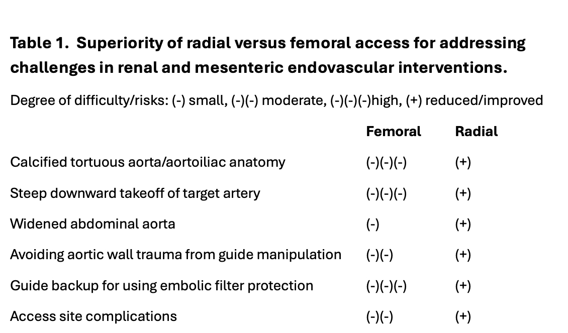
As demonstrated, the radial approach greatly facilitated bilateral renal and “triple” mesenteric endovascular interventions during index procedures for treatment of “global” organ ischemia. Only rare cases of extreme thoracic and suprarenal aortic tortuosity may still favor the femoral approach for these endovascular interventions.
Robert L. Minor, Jr, MD
Interventional Cardiologist and Endovascular Specialist
Billings Clinic Heart and Vascular at Community Medical Center
Missoula, Montana
Disclosure: Dr. Minor reports serving as a consultant for Medtronic.
Robert Minor, MD, can be contacted at rlm816@aol.com.