From Maps to Mechanisms: The Power of the OPAL HDx™ Mapping System in Identifying Critical Isthmus
© 2026 HMP Global. All Rights Reserved.
Any views and opinions expressed are those of the author(s) and/or participants and do not necessarily reflect the views, policy, or position of EP Lab Digest or HMP Global, their employees, and affiliates.
EP LAB DIGEST. 2026;26(8):6.
Usman A Bhatti, MD
Modern electrophysiology (EP) has entered an era in which information is abundant. High-density mapping systems routinely generate thousands of electrograms within minutes. As a result of these advances, the primary challenge facing electrophysiologists is rarely data acquisition but instead is the map interpretation.
The integration of the FARAWAVE™ NAV Pulsed Field Ablation (PFA) Catheter with the OPAL HDx™ Mapping System software 7.0 (Boston Scientific) offers an efficient and intuitive workflow. With the introduction of localized mapping, OPAL HDx system streamlines the transition from diagnosis to therapy while maintaining focus on mechanism identification.
Case Description
A patient presented for an EP study and catheter ablation. Surface electrocardiogram and intracardiac recordings suggested an organized left atrial (LA) flutter with a cycle length of approximately 316 ms. The FARAWAVE NAV catheter was used to create a detailed LA activation map within minutes.
OPAL HDx system software 7.0 allows for map collection using 2 bipole configurations: conventional and localized. The conventional configuration uses electrode 3 (E3) from each spline to create an interspline bipole (eg, A3-B3). The localized configuration uses E3 and electrode 1, 2, and 4 (E124) on each spline to create an intraspline bipole (eg, A3-A124).
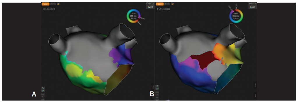
Initially, the activation map was created using the conventional E3-E3 bipole configuration (Figure 1A). While the activation sequence suggested a macroreentrant mechanism, propagation appeared relatively diffused, and the critical isthmus was not readily apparent.
The map was subsequently reprocessed to use the localized E3-E124 bipole configuration (Figure 1B). Reprocessing was completed quickly and did not require additional mapping. The resulting activation map revealed a discrete conduction corridor traversing the anterolateral LA, unmasking the critical isthmus sustaining the flutter circuit.
Electrogram analysis within this region demonstrated fractionated, low-amplitude signals consistent with slow conduction through a protected isthmus (Video). The improved signal selectivity provided by the localized bipole configuration rapidly focused attention on the physiologically relevant portion of the circuit responsible for maintaining the tachycardia.
Discussion
This case highlights how bipole configuration can fundamentally expand arrhythmia interpretation, even when analyzing the same mapping dataset. By improving near-field signal resolution and reducing far-field signal contribution, the localized configuration transformed a complex activation map into a clearly defined mechanism. The ability to map using the localized mapping on OPAL HDx system software 7.0 streamlined workflow while maintaining focus on the physiology sustaining the arrhythmia.
Conclusion
Ultimately, the value of mapping lies not in the volume of data collected but in the clarity with which it defines the underlying arrhythmia mechanism—and in how efficiently that understanding can be translated into effective treatment.
Video
Video. LA mitral annular flutter. Electrogram analysis within the diastolic corridor demonstrated fractionated low-amplitude signals consistent with slow conduction through the protected isthmus channel.
Author Bio
Usman A Bhatti, MD, is a cardiac electrophysiologist at AdventHealth Orlando with a focus on complex atrial and ventricular arrhythmia management. His professional interests include advanced electroanatomic mapping, zero-fluoroscopy electrophysiology, intracardiac echocardiography integration, and procedural workflow optimization. Dr Bhatti is an active educator, investigator, and national faculty member dedicated to advancing innovation and best practices in catheter ablation.
Disclosures: Dr Bhatti has completed and returned the ICMJE Form for Disclosure of Potential Conflicts of Interest. He reports support for the present manuscript and reports payment or honoraria for lectures, presentations, speakers’ bureaus, manuscript writing, or educational events.
This content was published with support from Boston Scientific.