




Advancing ICE-Guided LAAC: Improving Procedural Efficiency and the Patient Experience
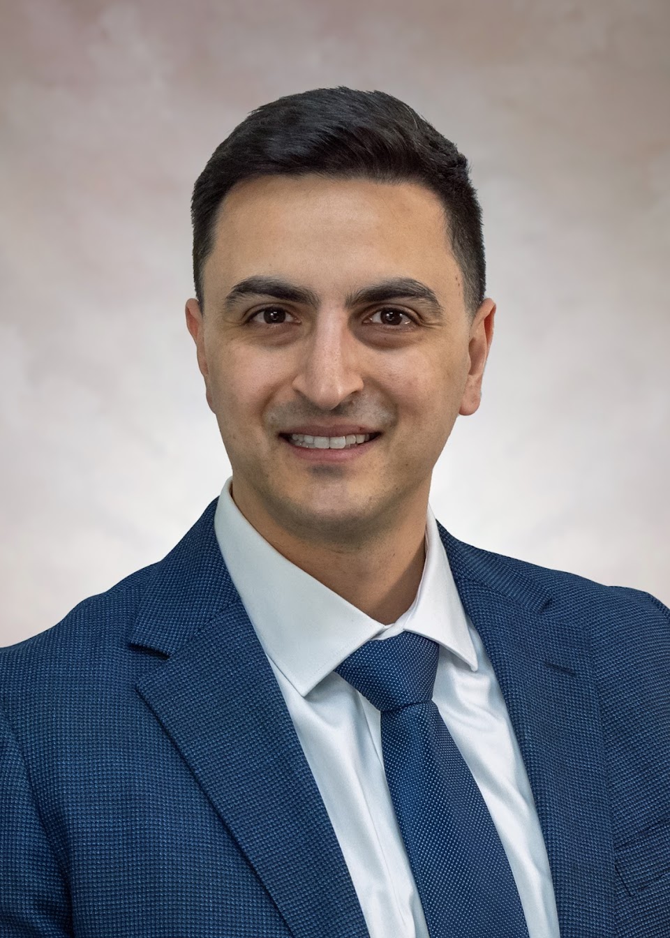
Interview With Adnan Alexander Kassier, MD, FACC, FSCAI
Interview With Adnan Alexander Kassier, MD, FACC, FSCAI
© 2026 HMP Global. All Rights Reserved.
Any views and opinions expressed are those of the author(s) and/or participants and do not necessarily reflect the views, policy, or position of EP Lab Digest or HMP Global, their employees, and affiliates.
EP LAB DIGEST. 2026;26(11):17-18.
Interview by Jodie Elrod
In this interview, we speak with Adnan Kassier, MD, about the intracardiac echocardiography (ICE)-guided left atrial appendage closure (LAAC) program at University of Michigan Health-Sparrow and the use of 3-dimensional imaging to support precise device placement and peri-device leak assessment. He also discusses how this approach enables conscious sedation, improves procedural efficiency and scheduling flexibility, and supports same-day discharge for approximately half of patients.
Can you tell us about your EP program at University of Michigan Health-Sparrow and the volume of LAAC procedures you perform?
 We have a robust program at University of Michigan Health-Sparrow, with multiple providers performing ICE-guided LAAC. The team initially consisted of 2 structural heart cardiologists and 1 electrophysiologist. We recently added another structural heart cardiologist and 3 electrophysiologists. We performed 125 LAAC procedures in 2025 and are on pace to surpass that volume in 2026. We expect our volume to continue to grow with the addition of these new providers.
How has incorporating 3D ICE changed the way your program approaches LAAC procedures, particularly compared with traditional transesophageal echocardiography (TEE) guidance?
The ICE-guided approach provides greater flexibility in procedural scheduling because the procedure can be performed under conscious sedation, without requiring general anesthesia or an additional cardiologist dedicated to imaging. Conscious sedation also facilitates treatment of patients who may be at higher risk for general anesthesia, including those with chronic respiratory failure. An additional advantage of having the same operator perform both the ICE imaging and device implantation is the ability to directly correlate imaging findings with catheter manipulation and procedural anatomy in real time. In my experience, this integration facilitates precise device positioning and may contribute to improved procedural outcomes and efficiency.
During LAAC, where do you find 3D ICE provides the greatest clinical value—from evaluating the LAA and transseptal puncture to device positioning and deployment?
My typical workflow includes evaluating the LAA with an ICE catheter positioned in the right heart chambers. After transseptal puncture, I place the ICE catheter in the LA and obtain more images of the appendage to better understand the anatomy. After device deployment, I utilize ICE to confirm appropriate sizing and adequate seal. I personally find that 3D ICE provides the greatest clinical value during the following stages:
- Transseptal puncture: I first attempt to align the transseptal puncture so that I can direct the guide toward the appendage and ensure coaxial alignment with appendage post transseptal. The ability to maneuver the ICE catheter myself during the transseptal puncture provides valuable guidance on how to move the catheter. For example, clocking the ICE catheter allows me to visualize the appendage, helping me determine how to clock the transseptal catheter to align it with the appendage.
- Before device deployment: 3D ICE allows me to evaluate the appendage, confirm sizing, and identify any small additional lobes that need to be covered by the device.
- After device deployment: 3D ICE allows me to evaluate the device for appropriate position and seal. I find 3D ICE particularly useful for evaluating peri-device leaks. I manipulate the catheter myself to evaluate every angle and use color flow with X-plane imaging to thoroughly assess the device in the aortic, mitral, and pulmonic views. In some cases, I also use the supramitral view for confirmation. Additionally, the ability to digital steer and obtain more imaging angles from the same ICE catheter location, without manipulation, is very valuable and, in my opinion, provides a thorough assessment of the device similar to that achieved with TEE.
As your experience with ICE-guided LAAC grows, what benefits are you seeing for procedural efficiency, anesthesia requirements, and the overall patient experience?
Greater experience and continued practice naturally lead to greater proficiency. In my own experience, I began performing ICE-guided LAAC approximately 4 years ago and have transitioned almost entirely to an ICE-guided approach over the past 3 years. During that time, I have significantly improved my proficiency and efficiency by continuing to refine the technique and optimize my procedural workflow. Currently, we generally perform 3 cases in a single day, with each procedure taking approximately 90 minutes. We typically reserve operational capacity and allocate buffer time to accommodate other procedures in cath lab.
Regarding anesthesia, our electrophysiology colleagues generally perform LAAC in conjunction with atrial fibrillation ablation as a concomitant procedure and therefore continue to utilize general anesthesia. In contrast, the structural heart team performs LAAC under conscious sedation. Both approaches have demonstrated excellent outcomes and low complication rates consistent with the national average. As discussed above, conscious sedation offers the additional advantage of allowing LAAC to be performed in patients who may be at higher risk for general anesthesia, while also providing greater flexibility in scheduling and procedural workflow.
Overall, ICE-guided LAAC has been associated with a favorable patient experience and an efficient procedural pathway. Approximately 50% of our patients are discharged on the same day. For those who are not discharged the same day, this is often related not to a procedural issue but to practical considerations, such as patients living a significant distance from the hospital or lacking adequate support at home.
Disclosures: Dr Kassier has completed and returned the ICMJE Form for Disclosure of Potential Conflicts of Interest. He reports receiving honoraria payment from Siemens Healthineers for participation in lectures, presentations, and manuscript writing related to the AcuNav 4D ICE catheter.