





From Visualization to Procedural Intelligence: The Next Era of Intracardiac Imaging in Electrophysiology
Interview With Christopher Jones, MD
Interview With Christopher Jones, MD
© 2026 HMP Global. All Rights Reserved.
Any views and opinions expressed are those of the author(s) and/or participants and do not necessarily reflect the views, policy, or position of EP Lab Digest or HMP Global, their employees, and affiliates.
EP LAB DIGEST. 2026;26(11):6-8.
Interview by Jodie Elrod
In this interview, Christopher Jones, MD, discusses how the automated imaging capabilities of SONATA (Johnson & Johnson MedTech), integrated with real-time 4D intracardiac echocardiography (ICE), can enhance anatomical clarity, procedural efficiency, and decision-making in complex electrophysiology (EP) procedures. He explores how SONATA may reduce cognitive workload, support the ICE learning curve, and help advance integrated imaging across EP and structural heart procedures.
EP procedures continue to evolve in complexity. What has been missing from conventional intracardiac imaging, and what challenges have remained unresolved in today’s workflow?
 ICE provides excellent real-time anatomy, but physicians and operators still need to mentally translate a 2D image into the 3D space. A limitation during training is understanding where the ICE catheter is moving. Without experienced ICE users to guide you, anatomy can become a limiting factor because no one wants to advance the catheter into the wrong space.
CARTOSOUND (Johnson & Johnson MedTech) and similar technologies have helped by incorporating ICE imaging into Fast Anatomical Mapping or electroanatomic mapping to translate it into 3D. But much of the value still depends on operator experience and manual interpretation.
SONATA closes that gap. It can clarify anatomic variations for experienced ICE users while supporting newer users, leveling the playing field and improving efficiency. It can assist with transseptal access and identifying pulmonary veins (PVs), including distinguishing a single left-sided PV from a difficult-to-visualize small inferior left PV. SONATA makes that anatomy clearer and the workflow efficient.
How does SONATA address those longstanding challenges, and why do you view it as a significant step forward from traditional CARTOSOUND workflows?
Traditional CARTOSOUND was valuable because it incorporated ICE imaging-derived anatomy directly into CARTO (Johnson & Johnson MedTech), bringing imaging and mapping together in one system. SONATA takes that further through artificial intelligence-based automation, automatically transforming ICE images into anatomy within the map and identifying and labeling structures.
It is not about identifying structures we do not know are there; it provides clarity and reduces the time needed to confirm a structure. Rather than relying solely on our ICE interpretation, SONATA helps confirm the anatomy.
This is valuable when images are not anatomically pristine. SONATA helps translate them into defined, anatomically typical anatomy we can operate with, which is often not the case.
Automation is a central part of SONATA. Which features have had the greatest impact on your workflow? And what are some of the features that really elevate intracardiac imaging that maybe a new user would appreciate?
Automatic chamber creation and anatomic reconstruction, with less manual contouring and manipulation, reduce the cognitive attention spent building anatomy. When I begin an atrial fibrillation (AF) ablation, I spend less time creating true-to-life 3D left atrial (LA) anatomy. After a few catheter turns, I can be ready to ablate or map, depending on the operator’s comfort level—whether proceeding with anatomy around the veins or evaluating the substrate for scar formation or burden.
That can reduce procedure time—not by removing important parts, but by reducing added steps. The 10 minutes it may take to create an LA chamber or determine the optimal location to cross for a transseptal become much easier. You can focus on entering the atrium, completing the ablation, and getting the patient off the table, with less LA chamber dwell time and less procedural risk.
That is what we want and have seen so far. I have used it heavily, and it reduces catheter time in the body.
A major benefit is staying focused on the procedure: catheter position, anatomy, treatment strategy, and the other things we need to consider, rather than clarifying surrounding structures. This matters in redo procedures, where small atretic veins can be difficult to identify. CARTOSOUND is excellent at bringing out that anatomy so we can integrate it and develop our strategy.
NUVISION NAV 4D ICE (Johnson & Johnson MedTech) acquires anatomic information while SONATA translates it into the CARTO environment. It takes an already excellent NUVISION NAV catheter and adds another level of benefit. This imaging enables us to perform these procedures using a minimally invasive, percutaneous approach.
SONATA integrates with both NUVISION NAV and SOUNDSTAR CRYSTAL (Johnson & Johnson MedTech). Regardless of which option you use in an ablation strategy, you get that benefit. SOUNDSTAR CRYSTAL is an ultrasound catheter, and SONATA lets you maintain those expectations for the procedure.
One of the biggest benefits of automation is that it gives me my attention back. It clarifies the anatomy so I can focus on the procedure and my role as an electrophysiologist. It allows less efficient users to operate at the level they want—getting patients in and out of the room and off the table quickly, with quicker ablations, less LA dwell time, and less catheter time in the body.
Beyond improving efficiency, how does the integration of real-time 4D ICE into the CARTO ecosystem enhance your understanding of cardiac anatomy, and why is that especially valuable during more complex procedures?
Two-dimensional ICE gives you a slice. It is useful but limited compared with today’s capabilities. With 4D ICE, you get broader spatial context—depth, superior and inferior orientation, and a larger view of the chamber instead of a single plane. Catheter manipulation becomes easier because you understand right and left, up and down, without continually turning the catheter to determine your surroundings. With every turn, we get more information, helping us move efficiently. That is the key: efficiency and clarity.
It makes relationships between structures more intuitive by relating what you see to the actual anatomy. We know the chamber is 3D, not flat. That spatial awareness helps you position the catheter and clarify where the valve is relative to the appendage, where the veins are relative to the appendage and posterior wall, how far the antrum extends, and what the internal anatomy looks like.
You can see what the catheter is interacting with in real time as you move around the pulmonary vein isolation (PVI) lines. The inside of the heart is not smooth—it is contracting, with muscle bundles and variable anatomy. Seeing that in real time helps you balance and know the catheter’s location.
Integrating that with CARTO connects what I see anatomically with what I do as an electrophysiologist. The differences may seem slight, but they matter.
How does real-time 4D ICE visualization of catheter–tissue contact influence pulsed field ablation (PFA) delivery and procedural decision-making, particularly when using VARIPULSE (Johnson & Johnson MedTech) or other PFA catheters?
I could talk all day about that. I am 100% for contact force and clear visualization of catheter-tissue contact. I use VARIPULSE frequently and am always looking to optimize contact, even though the variable-loop catheter already provides point clouds indicating contact.
With SONATA providing clear anatomy and 4D ICE, you can look down the barrel of a vein and, in real time, manipulate the variable loop to hug the wall before delivering PFA. That is especially important for newer PFA users because it reinforces maintaining catheter position during pulse delivery. You can see contact in real time, and without the catheter touching, we are not delivering what we hope to deliver.
One reason I use VARIPULSE over other PFA catheters is that contact force and everything else are integrated. That is key. With other PFA catheters, in right-sided workflows or without dedicated ultrasound or ultrasound-trained mapping staff, it can be difficult to know what you are contacting. The catheter may appear to be touching during PFA, but you do not have the same confirmation.
Combining high-quality imaging—whether viewing the fossa from the right side or from within the LA, similar to WATCHMAN (Boston Scientific) implantation—with visualization of catheter-tissue contact and point clouds on the integrated map creates a night-and-day difference.
Long term, I think we will see fewer return procedures because we are delivering PFA as designed: contacting the atrium, achieving good delivery, and monitoring it in real time. If I do not have good ICE images and confirmation of contact, I am not sure what delivery we can guarantee.
Can you share an example where the combination of automation and integrated 4D imaging changed how you approached a particularly challenging case?
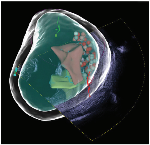
I recently performed a PVI where ultrasound initially suggested there was no left superior PV, but SONATA indicated one was present. It was a truncated, atretic vein that led nowhere but remained electrically active. Because of its unusual location and angle, it had been missed previously with 2D ICE, which essentially shows anatomy in slices.
SONATA quickly identified the vein, and I visualized it in 4D with the NUVISION NAV catheter. I crossed with the ICE catheter and confirmed it was a small, truncated PV. When I mapped it, it was electrically active. Even mapping alone was difficult to interpret unless you could actually see the catheter entering the vein.
In this redo case, identifying that vein let us complete the full PVI and get the patient out of AF. Without SONATA identifying the anatomy and NUVISION allowing me to visualize it, it would have been a long, difficult case—both to prove it was a vein and determine whether this atypically located structure should be incorporated into the isolation.
It was almost vertical, extending from the top of the LA. Initially, it looked like a pouch you would not normally address, but farther out, it was clear it was a vein.
In that complex case, what specific clinical insights did SONATA and integrated 4D imaging provide that you may not have had otherwise?
It clarified how to approach complex anatomy, plus the information needed to apply my knowledge and determine how to manage it. It allowed me to treat the patient efficiently and achieve the best outcome. Without it, the procedure would have been longer and difficult.
I think the combination of the 4D catheter and SONATA changed that patient’s future. She is no longer in AF, and we are not facing another invasive procedure because we incorporated anatomy that was not found or addressed during her previous ablation. The technology made a meaningful difference.
With SONATA, having that auto-labeling and telling you where you are within space, how does that impact the learning curve now for those who aren’t necessarily using ICE on a day-to-day basis or they’re just newly adopting it?
My first thought is teaching interventionalists how to perform transseptals—what the veins, appendage, and other structures look like on ICE. ICE is an acquired skill; you are not born knowing how to interpret ICE images, which differ from fluoroscopy. Being able to show someone, in real time, the fossa ovalis, superior vena cava, and PVs, and where to target on the fossa relative to those structures, makes a big difference in understanding anatomy.
Interpreting anatomy on the fly during a procedure is difficult, which is why we spend so much time learning it in fellowship. Outside of fellowship, that adds a layer of difficulty. Auto-labeling and spatial orientation can be valuable for fellows and new ICE users because they know where they are going and what they are looking at, which builds confidence.
Much of the hesitation with a transseptal comes from not knowing where you are going and not wanting the needle directed toward the wrong chamber or structure. Now you can see the veins, identify the left atrial appendage, and understand how far you are going. During ablation, you can see the PVs and valves.
As a teaching tool, whether in an academic setting or during onsite training, I think that clarity will help physicians overcome learning obstacles and more easily provide the care their patients need.
As more physicians adopt a concomitant treatment strategy, how does the integration of SONATA with CARTO and VARIPULSE help support those more comprehensive procedures?
CARTO becomes a common environment for mapping, imaging, and therapy. That is what everyone wants because all 3 are needed for great care. SONATA contributes integrated anatomical information, while VARIPULSE brings CARTO-integrated PFA therapy.
Combining AF treatment with another intervention increases imaging requirements. In concomitant procedures, for example, we use 4D ICE for precise WATCHMAN implantation. Now we can apply that visualization, movement, and deployment to AF ablation.
As EP and structural heart procedures increasingly converge, how could SONATA’s automation and integrated imaging capabilities extend beyond EP to support interventional cardiology, including mitral and tricuspid interventions?
It is just the tip of the iceberg. When you consider MitraClip or TriClip (Abbott), as well as structural heart procedures more broadly, everything is becoming increasingly minimally invasive. Visualization is key, particularly for physicians without extensive ICE training, such as interventional cardiologists moving into WATCHMAN and valvular procedures. Intuitive access to anatomy and spatial orientation can be valuable.
People may associate 4D ICE with EP and mapping, but this is also structural imaging. It shows the heart’s structure in real time—the contours of the chamber and walls, the small nooks, and valve movement—information fluoroscopy cannot provide.
It provides that information on demand, giving structural interventionalists, surgeons, and electrophysiologists the visualization needed for the procedure. I have yet to hear an interventionalist say, “I have too much imaging.” What I hear is, “This has worked out very well.”
As EP continues to advance, how do you see intelligent, automated imaging platforms like SONATA shaping the future of EP? What does the next era of intracardiac imaging look like?
The more visualization, the better. With SONATA, 4D imaging, intelligent image acquisition, automation, anatomical recognition, and mapping integration, everything is moving toward greater integration and visualization. Imaging will evolve from providing visualization to becoming part of procedural intelligence.
Procedures are evolving and better visualization is needed to improve efficiency and reduce procedure time. That will become important as we learn more about ablating different substrates, particularly in the ventricle. Ventricular tachycardia is an area where we are headed, using chamber reconstruction and assessing wall thickness in the LA and left ventricle, as well as understanding aneurysms and other electrically active ventricular areas—similar to how we evaluate and visualize the PVs in the atrium today.
More automation should allow physicians to spend less time manipulating technology and more time interpreting information for clinical decisions. SONATA is not just about automation or saving clicks. It is about integration, providing better anatomical information, and reducing cognitive workload so we can focus on patient care rather than building a map. It frees up that bandwidth while providing the imaging and information we need.
Looking ahead, as we think about structural procedures in the EP space, we have to go beyond WATCHMAN and consider where this technology can take us next.
I encourage physicians to use ICE as much as possible so they understand how to incorporate it into everyday procedures. Most people will find that it provides a benefit. The more you can see, the more informed you can be. The field is moving in that direction, so we need to start using it.
The transcripts were edited for clarity and length.
Disclosures: Dr Jones has completed and returned the ICMJE Form for Disclosure of Potential Conflicts of Interest, and has no conflicts of interest to report.
This content was published with support from Johnson & Johnson MedTech.
US_ELP_NAVI_422284