Infected Wound in a Lower Extremity Hematoma in a Patient With Thrombocytopenia After Renal Transplantation
© 2026 HMP Global. All Rights Reserved.
Any views and opinions expressed are those of the author(s) and/or participants and do not necessarily reflect the views, policy, or position of Wounds or HMP Global, their employees, and affiliates.
Abstract
Background. Renal transplantation is a highly effective treatment for end-stage renal failure. However, prolonged administration of immunosuppressive agents is necessary, which may elevate the risk of infection, particularly in cases associated with injury. These medications have the potential to induce thrombocytopenia, which can complicate wound management, because mechanical debridement is contraindicated in such cases. The paucity of literature on infected hematoma following renal transplantation is striking. Case Report. The case reported herein is that of a hematoma wound infection located on the skin of the lower extremity, which occurred as a result of a car accident that took place 20 years after renal transplantation. The patient had been experiencing thrombocytopenia due to prolonged immunosuppressant therapy for 2 months prior to this incident. A combination of ultrasonic debridement and moist wound dressings was used for effective management of the infected wound. Conclusion. The successful use of ultrasonic debridement in conjunction with autolytic debridement can not only prevent bleeding during debridement but also effectively remove necrotic tissue, thereby promoting favorable wound healing and enabling patients to resume normal daily life.
Kidney transplantation is a proven and effective treatment for end-stage renal failure, with the ability to restore renal function and markedly enhance patient quality of life, in addition to alleviating depression.1,2 However, as organ transplantation has advanced novel challenges have been noted, such as posttransplant rejection, infections due to bone marrow suppression resulting from immunosuppressive therapy, and thrombocytopenia.3,4
Management of wounds after injury is a complex process, beginning with hemostasis, the initial phase of physical wound healing. In the event of tissue damage and consequent bleeding, the body’s immediate response is to induce temporary vasoconstriction, thereby facilitating thrombus formation. Concurrently, platelets aggregate and activate coagulation factors, promoting clot formation and achieving hemostasis, which initiates the wound repair process.5 In the event of diminished thrombus formation and impaired coagulation, wounds are prone to delayed healing and the development of subcutaneous hematomas and infections. This, in turn, can lead to a deterioration in the overall condition of the wound. The risk of wound infection is increased following kidney transplantation due to the use of high-dose immunosuppressants, which suppress the patient’s immune function. The interplay of these factors contributes to the complexity of wound treatment.
This case report discusses management of a wound in a patient with coagulation disorders in whom a lower extremity skin hematoma infection occurred following renal transplantation.
Case Report
A 49-year-old female patient diagnosed with renal failure attributed to chronic glomerulonephritis underwent allogeneic kidney transplantation under general anesthesia 2 decades ago. Since then, she has been on a regimen of oral antirejection medications, including azathioprine and prednisone, a glucocorticoid.
Two months before presenting to the authors of this report, the patient sustained a contusion to the left lower limb in a road traffic accident. This injury was initially treated at a local hospital, but management there was suboptimal. The patient presented to Xinhua Hospital with deteriorated nutritional status, a hemoglobin level of 99 g/L, and a platelet count of 67 × 109/L. Vascular ultrasound Doppler imaging revealed clear deep veins in the lower limbs, with no evidence of thrombus formation or limb swelling.
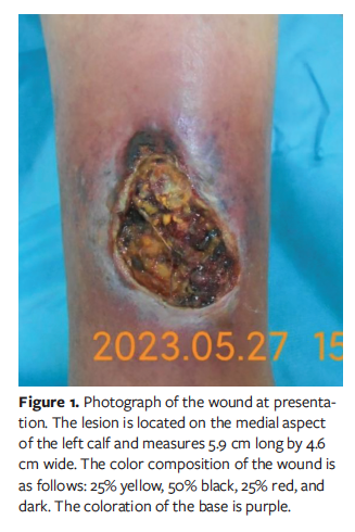
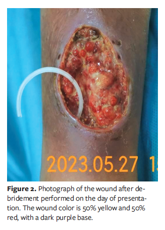
Physical examination of the wound was conducted. The wound was located on the medial aspect of the left calf and measured 5.9 cm × 4.6 cm (Figures 1 and 2). Wound coloration was characterized as 25% yellow, 50% black, and 25% red. After debridement, the wound was 50% yellow and 50% red, with a dark purple base and a sinus tract measuring 0.9 cm long at the 7-o’clock position.
The numerical rating scale (NRS) was used to assess pain (score range of 0-10, with 10 indicating the worst pain), with a resting pain score of 4 and a score of 6 during dressing changes. The exudate was characterized as dark yellow, watery, and moderate in quantity (classified as “medium” because it ranged from 5 mL-25 mL over 24 hours).5 The olfactory classification was assigned a grade of 4 on a 6-point scale, where grade 0 indicates a strong odor perceptible across a ward or consulting room, and grade 4 suggests an odor perceptible only to the patients themselves.
The patient presented with an infected wound characterized by necrotic tissue adhering to a blood clot, necessitating urgent debridement. However, she had a subnormal platelet count and exhibited coagulation dysfunction, which contraindicates mechanical debridement.5
Consequently, an alternative approach was selected. Ultrasonic debridement was performed at every dressing change during the first month, with each session lasting 15 minutes to 20 minutes. This was done in conjunction with use of a silver alginate foam dressing to facilitate autolytic debridement and eliminate necrotic tissue and congestion. The dressing was changed every 2 days to 3 days based on the amount of exudate.
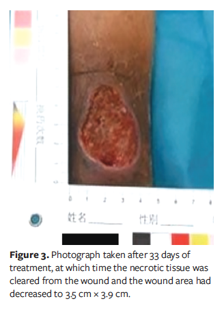
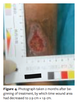
After 33 days, the necrotic tissue was cleared from the wound and the wound area had reduced to 3.5 cm × 3.9 cm (Figure 3). Two months after the beginning of treatment, the wound dimensions had significantly reduced to 2.9 cm × 1.9 cm (Figure 4). At that time the wound bed was completely red, indicating healthy granulation tissue, and the sinus tract was fully epithelialized, with no resting pain (NRS score of 0). Pain during dressing changes was minimal (NRS score of 2). The exudate was yellowish, watery, and minimal (classified as “small”). The infection was identified as a chronic bacterial biofilm. There were no signs of erythema on the periwound skin, which had a normal temperature and normal dorsalis pedis artery pulsation.
The main treatment principles include continued biofilm removal and promotion of epithelial tissue growth, with close monitoring of the patient’s condition for signs of rejection following a renal transplant, or changes in wound status. Given the presence of a bacterial biofilm at the wound site, plantar gel (applied every 2 days) was chosen as the primary dressing, with a foam dressing as the secondary dressing. The secondary foam dressing was changed every 2 days, coinciding with the gel application, to maintain a moist wound environment. This combination was intended to promote epithelial growth and maintain the moisture balance essential for optimal wound healing.
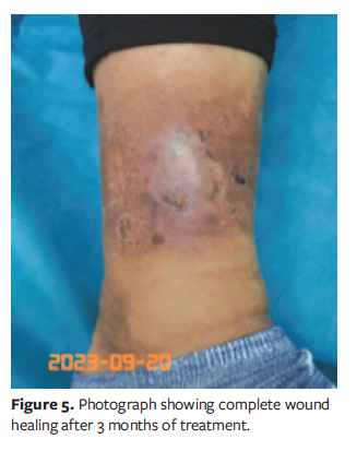
As shown in Figure 5, wound healing was complete within 3 months. There were no complications throughout this period, including no signs of rejection of the transplanted kidney. However, the patient’s urologist, L.C., recommended regular monitoring of key blood parameters. This included monitoring platelet count, hemoglobin levels, and renal function, as well as routine urine analysis, in addition to periodic ultrasound examination of the urinary system. The patient provided written informed consent to share details and photographs of her treatment.
In terms of dietary management, it is important that the patient adhere to a diet that is low in salt, fat, and sugar. It is also important that the food they consume is fresh and hygienic. The diet should be balanced, with an equal intake of meat and vegetables, to ensure a nutritious and moderate diet and to avoid overeating. This approach supports overall health and helps to manage the patient’s renal condition after transplant.
Discussion
Thrombocytopenia is a common complication following kidney transplantation. It is often associated with the use of immunosuppressive therapy6-7 and can significantly delay wound healing. In patients with thrombocytopenia, even minor trauma can result in severe bleeding. Studies have demonstrated that the rapid recruitment of platelets to the wound site and the subsequent release of growth factors are crucial initiators of tissue repair.8 Although platelets can influence wound inflammation, they do not significantly affect the proliferative phases of repair, such as wound closure, angiogenesis, and collagen synthesis. In the current case, it was imperative to remove the early hematoma while ensuring that no bleeding was induced.
Although necessary for posttransplant care, immunosuppressants can increase the risk of wound infection due to their suppression of the body’s immune function. Infection is one of the most common complications following renal transplantation and is a major cause of transplant failure. Pseudomonas aeruginosa is the predominant pathogen in these cases, which aligns with findings from related studies.9
Although the use of immunosuppressants and glucocorticoids can disrupt wound healing, these medications are indispensable for preventing transplant rejection in the patient discussed in the current report. The potential adverse effects of these medications are overshadowed by the serious consequences of kidney rejection. In this case, the risk of transplant rejection outweighs the risk of delayed wound healing. Thus, when devising nursing strategies, the overarching principle must be to prioritize the patient’s overall well-being in accordance with the principle of “First, do no harm.” In collaboration with the urology department, judicious use of antibiotics can mitigate the risk of wound infection, and silver ion dressings can provide local anti-infection treatment.
To address the challenge of decontamination without exacerbating bleeding, the authors of this case study used a combination of ultrasonic and autolytic debridement using a novel dressing. Ultrasonic debridement was performed at a frequency of 2 to 3 times per week (at each dressing change) during the initial 4-week period. The duration of each session ranged from 15 minutes to 20 minutes, depending on the wound area and the amount of necrotic tissue. This regimen effectively removed necrotic tissue and bacterial biofilm without inducing bleeding, which was of particular concern given the patient’s thrombocytopenia.
Ultrasonic debridement promotes wound healing through the following mechanisms.10,11 First, low-frequency ultrasound can create a so-called cavitation effect within the irrigation stream. This atomizes water droplets and, under pressures of up to 1000 atm, propels these droplets into the abscess cavity, resulting in a more thorough removal of bacteria and fungi from both the superficial and deep layers of the wound. This method offers the benefits of reduced pain, improved patient compliance and comfort, and decreased mechanical contact. Second, given that necrotic tissues have a lower tensile strength than healthy tissues, ultrasound debridement can be tailored to remove necrotic tissues with minimal or no damage to normal and fresh granulation tissues. Third, ultrasonic debridement can also utilize the microjet effect resulting from the disintegration of cavitation bubbles to increase the local transcutaneous partial pressure of oxygen in the wound, thereby protecting fresh granulation tissue and accelerating wound healing.
Limitations
This case report has limitations. Because it describes a single case, the findings and specific treatment success (ultrasonic and autolytic debridement) cannot be generalized to other patients with similar conditions; individual responses can vary significantly. This report does not include a direct comparison with conventional treatment methods or other alternative approaches to managing infected wounds in transplant patients with thrombocytopenia. This makes it difficult to definitively claim that the approach described herein reduces healing time more effectively than other methods. Success was observed in a specific type and size of wound (a lower extremity hematoma infection measuring ~27 cm² initially) in a stable, long-term transplant recipient. The applicability of this approach to larger wounds, different locations, more severe coagulopathies, or patients with active rejection or infection is unknown.
Conclusion
For patients with thrombocytopenia and coagulation disorders after renal transplantation, the successful application of ultrasonic debridement combined with autolytic debridement can not only prevent bleeding during debridement but also effectively remove necrotic tissue, thereby promoting favorable wound healing and enabling patients to resume normal daily life.
Author and Public Information
Authors: Hongyu Wang, MSN1; Lin Chen, MD2; Qian Gao, BSN1; Guirong Shi, MSN1; and LiPing Jiang, DNP1
Affiliations: 1Wound Center, Xinhua Hospital, Shanghai Jiao Tong University School of Medicine, Shanghai, China; 2Department of Urology, South Campus, Shanghai Sixth People’s Hospital, Shanghai Jiao Tong University, Shanghai, China
Author Contributions: Hongyu Wang and Dr Lin Chen contributed equally to this work.
Disclosure: The authors disclose no financial or other conflicts of interest.
Ethics Statement: The patient provided written informed consent to publish details and photographs of her case.
Correspondence: Lin Chen; Department of Urology, South Campus, Shanghai Sixth People’s Hospital, Shanghai Jiao Tong University, 6600 NanFeng Road, FengXian District, Shanghai, China, 201499
Manuscript Accepted: April 21, 2026
References
1. Huang L. Clinical comparison of quality of life between kidney transplant patients and hemodialysis patients. Journal of Qiqihar Medical College. 2016;37(12):1620-1621.
2. Li H, Li S, Li G, et al. Study on quality of life of patients with kidney transplantation and hemodialysis and related influencing factors. Chinese Journal of General Medicine. 2013;15(35):4152-4155.
3. Du C, Miao Y, Zhou H, et al. Long-term outcomes of kidney transplantation in 2332 patients under cyclosporine A. Guangdong Medical Journal. 2016;37(6):904-908. (Accession Number: edscoj.gdyx201606036)
4. Morath C, Zeier M, Süsal C. Increased risk of infection-associated death with incompatible kidney transplantations. Transpl Int. 2017;30(12):1209-1211. doi:10.1111/tri.12995
5. Wang L, Hu A, eds. 奄口造口失禁牡科湯理 [Wound, Ostomy, and Continence Specialty Nursing]. Beijing, China: People's Medical Publishing House; 2018.
6. Malyszko J, Basak G, Batko K, et al. Haematological disorders following kidney transplantation. Nephrol Dial Transplant. 2022;37(3):409-420. doi:10.1093/ndt/gfaa219
7. Gierczak V, Jouve T, Malvezzi P, et al. Impact of immunosuppressive strategies on post-kidney transplantation thrombocytopenia. Transplant Proc. 2021;53(3):941-949. doi:10.1016/j.transproceed.2020.09.014
8. Szpaderska AM, Egozi EI, Gamelli RL, DiPietro LA. The effect of thrombocytopenia on dermal wound healing. J Invest Dermatol. 2003;120(6):1130-1137.
9. Sinha S, Shailja S, Kumari N, Kumar S. . Phenotypic detection of multidrug resistance and metallo-β-lactamase production among clinically relevant non-fermenters at a medical centre in India. Bioinformation. 2025;21(7):2080–2084.
10. Kataoka Y, Kunimitsu M, Nakagami G, Koudounas S, Weller CD, Sanada H. Effectiveness of ultrasonic debridement on reduction of bacteria and biofilm in patients with chronic wounds: a scoping review. Int Wound J. 2021;18(2):176-186. doi:10.1111/iwj.13509
11. Messa CA IV, Chatman BC, Rhemtulla IA, et al. Ultrasonic debridement management of lower extremity wounds: retrospective analysis of clinical outcomes and cost. J Wound Care. 2019;28(Suppl 5):S30-S40. doi:10.12968/jowc.2019.28.Sup5.S30