Lymphedema Therapy and Wound Healing: A Retrospective Analysis of Pretreatment and Posttreatment Wound Volume
© 2026 HMP Global. All Rights Reserved.
Any views and opinions expressed are those of the author(s) and/or participants and do not necessarily reflect the views, policy, or position of Wounds or HMP Global, their employees, and affiliates.
Abstract
Background. Venous leg ulcers (VLUs) frequently exhibit prolonged healing times, particularly in the presence of lower extremity lymphedema. One approach to managing VLU is to combine traditional wound care with lymphedema therapy (LT). Despite recognition of compression as an effective agent for management of chronic wounds, use of lymphedema services remains insufficient among health care providers. Objective. To investigate the effectiveness of LT for tissue repair and examine the effect of various factors on wound healing. Materials and Methods. Electronic medical records of patients with VLU who completed LT between June 2016 and March 2020 were reviewed. Wound volume (length × width × depth) was compared before and after LT, and medians of nonnormally distributed data were compared via the Wilcoxon signed rank test. Demographic characteristics and medical history were evaluated to determine their relationship to VLU healing. A 3-step hierarchical approach was used to investigate the effect of various factors on predicting tissue repair. Results. A total of 178 wounds in 115 patients were included in the analysis. The average wound volume before LT was 14.4 cm³. After lymphedema intervention, the mean wound volume decreased to 3.8 cm³, a significant reduction in wound size (P < .001). A hierarchical regression model revealed no statistically significant relationships between wound closure and age, sex, comorbidities, or treatment-related factors. However, body mass index emerged as a moderately significant predictor of wound healing (P = .013). Conclusion. The findings support the effectiveness of LT for healing chronic VLU in patients with lymphedema treated in an outpatient clinic.
Venous leg ulcers (VLUs) are the most common type of lower extremity wounds, affecting an estimated 1% to 3% of the United States population and accounting for greater than 80% of lower extremity ulceration.1,2 Importantly, lymphedema is strongly associated with VLU.3 A large-scale study of 80,000 patients estimated that greater than 90% of those diagnosed with advanced chronic venous insufficiency (CVI) or phlebolymphedema had a concurrent venous ulcer.4
Lymphedema is a widespread health problem, affecting more than 10 million Americans and 250 million people worldwide.5,6 This condition is caused by disproportion between lymph production and transport capacity of the lymphatic system, resulting in an abnormal accumulation of lymphatic fluid in the interstitial tissues.7 Unlike congestive heart failure, which is caused by the impaired ventricular filling and ejection of blood from the heart and frequently is manifested by temporary peripheral edema, lymphedema is a consequence of lymphatic dysfunction that leads to lymphostasis and is characterized by the chronic and often asymmetric accumulation of lymphatic fluid.8,9 Damage to the lymphatic system and decreased ability of the tissue to remove metabolic waste (ie, lymphocytes, bacteria, protein, and cell debris), along with the presence of CVI, often lead to significant skin changes, such as hyperkeratosis, hemosiderin staining, fibrosis, papillomatosis, and chronic ulceration, which most frequently affect the lower limbs.5,10 A diagnosis of lymphedema is based on clinical presentation or diagnostic imaging, or a combination of both. Clinical assessment includes evaluation of volume, Stemmer sign, tissue composition, asymmetry, and skin changes, and imaging tests include lymphoscintigraphy, magnetic resonance imaging, and ultrasonography.11,12
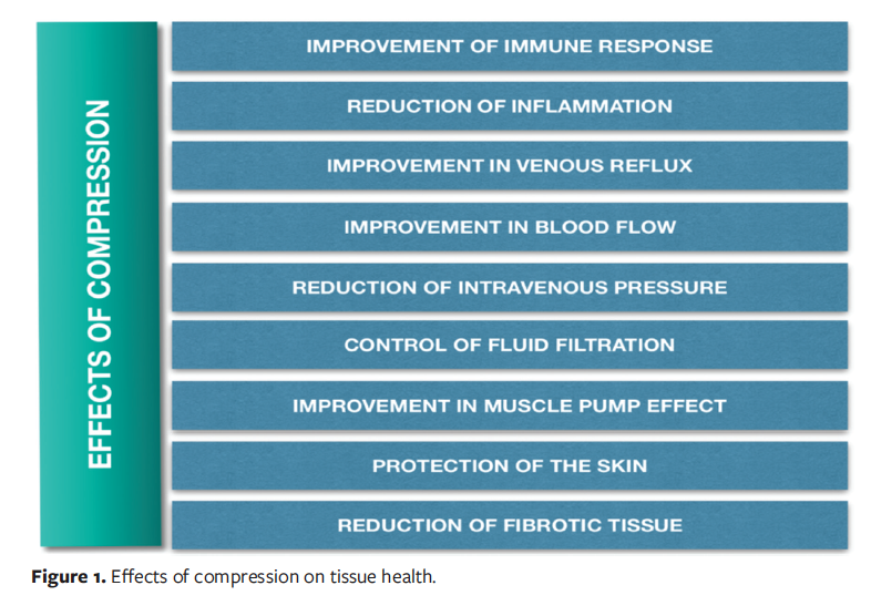
Currently, compression therapy is identified as an essential component of VLU management.13-21 Benefits of compression for tissue health are listed in Figure 1. A multidimensional, interdisciplinary approach to treatment of wounds integrates traditional wound care with lymphedema therapy (LT), specifically, complete decongestive therapy (CDT)—the standard for LT—which confers additional benefits for tissue restoration. However, the utilization of lymphedema services is still suboptimal among health care providers, with patients receiving adequate compression in fewer than 17% of visits,22 indicating a need to rethink current wound healing practices.
Despite the high prevalence of lower extremity lymphedema among community members, with community nurses in 1 study estimating that 43% of their patients had chronic edema of the lower limbs,23 the public recognition and understanding of the negative effect of lower extremity lymphedema on wound healing is nominal. Delayed wound healing in lymphedema involves 2 key mechanisms: pathophysiological and immunological.24
Pathophysiologically, localized edema compresses microvasculature and lymphatic vessels, impairing tissue remodeling.24 The interdependence of the venous and lymphatic systems means that failure of 1 system, such as in CVI, can impair the other, leading to a secondary condition known as phlebolymphedema.25 In CVI, interstitial fluid overload burdens the lymphatic system; when this exceeds its compensatory capacity, lymphatic damage and chronic insufficiency result.25 This can progress to tissue fibrosis, contributing to dystrophic ulcers, infections, and lipodermatosclerosis.25
Immunologically, lymphedema disrupts immune cell differentiation.24 Because the lymphatic system is integral to immunity, patients often have a diminished immune response, thus increasing susceptibility to severe skin infections that impede healing.26 Specifically, suppression of T helper type 1 cells raises infection risk, while excessive T helper type 2–driven responses, such as M2 macrophage polarization, promote fibrosis that disrupts normal wound repair.24
Recognition of compression as an effective agent for chronic wound management has led to the use of certain modalities by vein and wound care specialists (eg, off-the-shelf multilayer elastic compression bandage systems or low-grade compression stockings). However, health practitioners continue to demonstrate a tendency toward treating lymphedema and wounds as separate entities.27 Many physicians have expressed their preference to heal wounds first and then refer patients to lymphedema clinics for management of edema.28 Consequently, delayed referrals for appropriate care may result in worsening of wound condition, leading to infections, pain, copious exudate, foul odor, and other physical and functional limitations. Importantly, LT incorporates manual lymphatic drainage, daily bandaging with a multilayer compression system (short-stretch or inelastic bandages and foam), edema-reducing exercises, vasopneumatic compression, elastic and inelastic compression garments (typically, 30 mm Hg–45 mm Hg), and meticulous skin care.29 The use of an inelastic or short-stretch bandage system with a high static stiffness index promotes high working pressure (eg, during mobility or exercise) and low resting pressure (eg, during sleep or rest).30
Short-stretch bandages are composed of 100% cotton fibers engineered to permit approximately 60% extensibility relative to their resting length.31 When applied as a component of a multilayer compression system, their limited extensibility generates a high working pressure (>60 mm Hg) in response to muscle contraction during ambulation, which enhances the venous and lymphatic pumping mechanism.32 Conversely, during periods of muscular relaxation, the bandages exert low resting pressure, thereby improving comfort, preventing constriction, and avoiding a tourniquet-like effect that could impede physiologic fluid dynamics.32 Moreover, short-stretch compression systems offer a customized approach to lymphedema management, with therapists cutting and fitting the underlying padding and foam to accommodate each patient’s unique anatomical architecture. In contrast, elastic or long-stretch bandages typically used by wound care specialists produce high resting pressure and low working pressure, which significantly decreases the effectiveness of the compression system due to reduced muscle pump effect.30,32 Composed of cotton, polyamide, and polyurethane, long-stretch bandages exhibit an extensibility exceeding 140%, making them appropriate for uncomplicated varicose veins that necessitate medium compression of 30 mm Hg to 40 mm Hg.33,34 However, a recent meta-analysis indicated no statistically significant difference in healing rates between elastic and inelastic compression systems, although only 3 of 12 studies were rated as high quality.35
Knowledge of CDT, combined with a broad range of therapeutic options offered by LT, suggests that referral to skilled lymphedema services will likely achieve higher healing rates than conventional wound care alone. The current study sought out empirical evidence to support the significance of LT as a highly effective ancillary service for management of VLU and to promote evidence-based practice and the development of novel approaches to wound care. The study also aimed to explore the effect of various patient- and treatment-related factors on wound closure, considering the complexity of tissue regenerative processes.
Materials and Methods
This quantitative, cross-sectional, retrospective analytical study with elements of the interrupted time series design examined the dynamics of wound healing and explored potential relationships between various patient- and treatment-related factors and wound healing. After the study proposal was reviewed and approved by the Trinity Health Mid-Atlantic Institutional Review Board (IRB) and Nova Southeastern University IRB, a single investigator (M.S.) traveled to Langhorne, Pennsylvania, and collected a set of pre- and posttreatment observations to assess changes to wound size prior to referral to LT and after the treatment had been initiated. The data were broadly divided into 3 categories: patient-related factors, wound-related factors, and treatment-related factors.
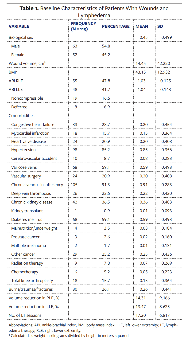
Patient-related data consisted of the patients’ demographic characteristics, including age, body mass index (BMI), ankle-brachial index (ABI), biological sex, and existing comorbidities. For example, cardiovascular comorbidities comprised a history of congestive heart failure, vascular intervention or surgery, hypertension, myocardial infarction, heart valve disease, CVI, cerebrovascular accident, history of deep vein thrombosis, and varicose veins (Table 1). Wound-related data included the wound size and 4 measurements: greatest length, greatest width, greatest depth, and wound volume. Data on Clinical-Etiology-Anatomy-Pathophysiology (CEAP) classification and ambulation index, such as Hauser ambulation index, were unavailable in the retrospective review of electronic medical records (EMRs). Treatment-related data consisted of the percentage of fluid removed from affected limbs during LT and the number of LT sessions patients attended. Some patients received diuretics managed by their nephrology, cardiology, or primary care teams outside of the wound care center (WCC); however, medication use was beyond the scope of the current study.
All wound care providers used a 3-dimensional method of wound size measurement to standardize the assessment of the healing process in relation to all dimensions, including length, width, and depth. A previous study indicated high levels of intrarater and interrater agreement for length and width; however, a low level of reliability was observed for depth, demonstrating an intrarater intraclass correlation coefficient of 0.360 and an interrater intraclass correlation coefficient of 0.649.36 In the current study, wound healing progression was assessed via gross volume healed and percentage volume healed. The wound volume was determined as a dependent variable, and a wound volume of 0 cm3 indicated complete wound closure. The 5-level wound model developed by Smit37 in 2018 was selected as a conceptual model to guide data collection and analysis. This model helps explain how pathologic factors influence non- or slow-healing wound formation and identifies challenges and opportunities in wound treatment.37
Patients
The sample consisted of 115 patients with 178 VLUs treated in a WCC and lymphedema clinic over a period of 3.8 years (June 2016–March 2020). Adults aged 20 years to 99 years with unilateral and bilateral lower extremity ulceration and lymphedema were included in the study. Patients with chronic arterial ulcers, acute cardiovascular conditions, acute respiratory failure, acute infections, and severe kidney failure were excluded from the sample. Additionally, patients with severe arterial insufficiency (ABI ≤ 0.5) and arterial calcification (ABI ≥ 1.4) were excluded, because compression is contraindicated in these cases.38,39 Finally, patients who had not completed a full course of CDT and who had been discharged against medical advice were excluded from the study.
Patients presenting with both lymphatic obstruction and loss of lymphatic channels received a standardized treatment regimen aimed at decongesting the lower extremities, improving skin quality, promoting tissue health, and facilitating wound healing. Patients who do not respond to such treatment should be routinely referred for advanced diagnostic imaging (eg, lymphoscintigraphy or magnetic resonance lymphography) to determine the underlying etiology and guide a targeted treatment plan for the specific lymphatic impairment.
Data collection
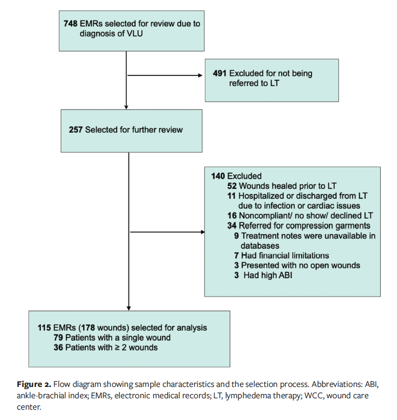
Data collection included EMRs of patients treated in the WCC from June 2016 to March 2020. During this period, 748 patients received treatment for VLUs, and 257 were referred for LT. After examination of 257 EMRs, 142 records were excluded from the analysis, and 115 EMRs of patients with both VLU and lymphedema were selected for further review. The archival deidentified data included patient demographic information, past medical history, progress with treatment, and treatment outcomes. Figure 2 displays the decision flow diagram outlining the sample selection process.
Statistical analysis
To test the hypotheses, the investigators formulated 3 research questions: comparative, correlational, and predictive. The test of normality via the Shapiro–Wilk and Kolmogorov–Smirnov tests, skewness, and kurtosis revealed that normal distribution could not be assumed. In comparative analysis, medians of nonnormally distributed data were compared via the Wilcoxon signed rank test. The associations between patient- and treatment-related factors and wound healing outcomes were estimated using Kendall tau-b correlation coefficients. A 3-step hierarchical regression model was used to assess the unique contribution of various factors in predicting wound healing. Alpha was set at .05 as the level of significance. Data analysis was conducted using SPSS version 28.0 (IBM).
Results
The study included 115 patients with 178 VLUs. Sixty-three patients were male and 52 were female, with the average patient aged 60 years to 69 years (n = 50) and with a mean BMI of 43.1, indicating morbid obesity. Seventy-nine patients (68.7%) had a single wound, and 36 patients (31.3%) had 2 or more wounds. In terms of tissue involvement, 153 wounds were identified as full-thickness wounds (85.9%), 20 wounds were identified as partial-thickness wounds (11.2%), and 5 wounds were unclassifiable (2.8%). The right lower extremity was most affected by the presence of VLU (43.5%), followed by the left lower extremity (34.8%) and bilateral lower extremities (21.7%). The most prevalent comorbid condition was CVI (n = 105), followed by hypertension (n = 98), varicose veins (n = 68), diabetes (n = 68), and chronic kidney disease (n = 42). On average, patients attended 17.2 lymphedema treatment sessions and achieved 14.3% and 13.5% volume reduction in the right and left lower extremities, respectively. Four therapists (including the first author) provided LT to the patients with leg ulcerations and lymphedema. The statistical analysis did not indicate significant therapist effect on the change in wound volume (P = .06). Table 1 provides a summary of health and treatment characteristics of the sample.
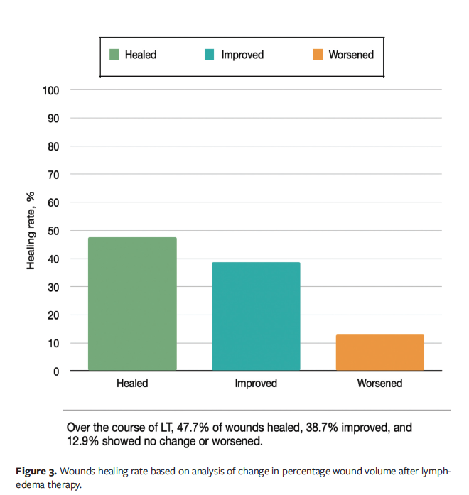
The average (SD) wound volume prior to LT was estimated to be 14.4 (42.22) cm3 (range, 0.009 cm3–379.8 cm3). After LT, the average wound volume decreased to 3.8 (15.07) cm3. Almost half the wounds healed (47.7% [n = 85]), and 38.7% of the wounds (n = 69) improved at the end of the LT course. However, 23 wounds (12.9%) either exhibited no change or worsened, demonstrating an increase in size/volume (Figure 3).
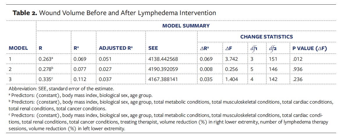
The Wilcoxon signed rank test revealed that wound volume was significantly smaller after LT compared with before LT in the 178 wounds (median = 0.008 and 1.641, respectively; z = -8.976; P < .001), with a large effect size (r = .63). The mean wound volumes before and after lymphedema intervention are displayed in Table 2.
The results of the Kendall tau-b correlation showed no statistically significant association between total percentage change in wound volume and patient age, BMI, biological sex, ABI, comorbidities, and number of LT sessions patients attended. For age, no association with wound size was found (Ʈb = 0.090, P = .114). For BMI, no statistically significant results were indicated (Ʈb = -0.037, P = .114). Likewise, neither biological sex (Ʈb = -0.008, P = .902) nor number of LT sessions (Ʈb = 0.073, P = .161) showed a statistically significant association with wound closure. Comorbidities such as cardiovascular conditions, CVI, and diabetes did not have a statistically significant correlation with total percentage change in wound volume. However, a moderate but statistically significant negative correlation was observed between volume reduction in the right lower extremity and volume reduction in the left lower extremity (Ʈb = -0.577, P < .001). This finding suggests that when the percentage volume of 1 limb increased, the percentage volume of another limb decreased.
Finally, the 3-step hierarchical regression analysis and model summary showed that the first model was statistically significant (F(3, 151) = 3.742, R2 = 0.07; P = .012) (Table 3). BMI emerged as a moderately significant predictor for wound healing (β = .25, t = 2.51; P = .013). In the second model (F(8, 146) = 1.529, R2 = 0.08; P = .152), the addition of comorbidities did not indicate statistical significance in predicting wound healing, although BMI continued to significantly contribute to the prediction (β = .24, t = 2.43; P = .016). The final model (F(12, 142) = 1.498, R2 = 0.11; P = .131), which accounted for treatment-related factors, remained nonsignificant. Again, BMI maintained its moderate predictive value (β = .25, t = 2.50; P = .013). Accordingly, the inclusion of comorbidities and treatment-related factors into the second and third models did not show improvements from the first model (F(5, 146) = 0.256, R2 = 0.08; P = .936, and F(4, 142) = 1.404, R2 = 0.11; P = .236, respectively). Overall, age, BMI, and biological sex predicted approximately 7% of variance in wound healing. After controlling for age, biological sex, and BMI, the second and third models predicted approximately 8% and 11% of variance in wound healing, respectively. However, only BMI significantly predicted wound healing, with greater BMI values being positively associated with higher total percentage change in wound volume. The effect size was small (Cohen f2 = 0.04), indicating that the difference between the model blocks was insignificant.
Analysis of collinearity utilizing various inflation factors revealed no potential issues with predictor variables being highly linearly correlated with each other. No various inflation factor values for predictor variables were greater than 5, and no tolerance values were less than 0.1. Therefore, multicollinearity was not present for this regression model.
Discussion
The current study offers a unique perspective on the dynamics of wound healing through the lens of lymphedema intervention. A preliminary literature review of studies conducted by lymphedema specialists indicated a deficiency in high-quality evidence regarding wound improvement with the addition of LT to the plan of care.40 Unlike studies that have identified advanced age as a significant risk factor for venous ulcer recurrence or delayed healing,41-46 analysis of the data in the current study revealed no correlation between advanced age and slow tissue repair.47,48 Although previous research has suggested that females are at increased risk of wound occurrence45,49 and that wounds are harder to heal in males,41,48 in the current study biological sex showed no significant effect on wound closure. Similarly, despite existing evidence, in the current study no relationship was detected between wound healing and various comorbid conditions, including cardiovascular,41,43,45,50 renal,42,45 metabolic,42,45,51 oncologic,52 and musculoskeletal comorbidities.45 However, a diagnosis of lymphedema was confirmed as a risk factor for delayed healing.12,50 The prevalence of participants with a high BMI (mean of 43.1) aligns with findings in the literature suggesting a relationship exists between obesity and chronic wounds.53 Despite the widely accepted inference regarding the negative effect of a higher BMI on the occurrence of VLU,43-46,49 the effect of both high and low BMI on the speed and quality of wound closure remains unclear and requires further investigation.
In the current study, LT was found to be effective regardless of the wound shape or volume, or the degree of tissue damage, with full-thickness wounds demonstrating high rates of tissue repair after initiation of LT. The assessment of wound dimensions revealed that changes in wound depth were fundamental to the reduction in wound volume, as previously recognized.54 Essentially, only wounds that occurred prior to LT were assessed by the investigator (M.S.). Despite proposed benefits of LT for successful wound closure, several patients developed wounds after the initiation of LT, most likely due to friction from the compression system on often compromised skin (eg, blisters that became wounds) or noncompliance with lymphedema therapists’ recommendations, such as premature removal of compression bandages, resulting in lower extremity volume fluctuation or, vice versa, remaining bandaged when pain or discomfort occurred.
Importantly, most patients receiving treatment in the WCC used some sort of compression modalities, including SpandaGrip (Medi-Tech International Corp), Unna boot, off-the-shelf 3-layer compression bandage systems, or previously issued compression garments. Referral to LT provided patients with the additional benefits of utilizing more advanced compression systems, along with other therapeutic
modalities, which improved the outcomes of wound healing. However, the assessment of wound measurements revealed that wounds tended to worsen after the initiation of LT, most likely due to increase in exudate and skin irritation from short-stretch bandages. The same phenomenon was observed in patients treated with an Unna boot or over-the-counter long-stretch bandages in the WCC when initial application of compression could result in an increase in wound volume.
Although results demonstrated high efficacy of LT for wound healing, the quality of the compression system used and an adequate treatment regimen were essential. In 1 case, a patient’s wound healed in the first week of LT, and 19% volume reduction was achieved in the affected limb. The wound reopened several months after the initial onset, and the patient was referred to the lymphedema clinic for another course of LT. During the second course of LT, the volume of the affected limb was reduced by only 3%, and the wound required 1 month to heal. In another case, a patient was first seen for lymphedema intervention 3 times a week, demonstrating poor treatment outcomes in terms of wound closure. During the second round of LT, the frequency of visits was increased to 5 times a week, and the patient’s wounds healed completely. Although no relationships were found between wound closure, volume reduction in affected limbs, number of LT visits, and treating therapist, the effectiveness and quality of LT services may play an important role in the dynamics and speed of wound healing. For example, patients demonstrated higher healing rates when receiving LT 5 times per week versus only 3 times per week.
A statistically significant negative association between volume reduction in the right lower extremity and volume reduction in the left lower extremity indicated that the percentage volume in 1 limb increased when the percentage volume of another limb decreased. The assessment of data revealed that 15 patients received LT for a single lower extremity versus for bilateral lower extremities. In these cases, only limbs with wounds were treated, while the other limb remained free of compression, although some degree of edema could present in the unaffected limb as well. Thus, therapeutic intervention to a single limb could elicit an asymmetrical decrease in girth of lower extremities, demonstrating greater volume reduction in the limb receiving treatment, whereas the other limb maintained its initial status. Additionally, a positive association between BMI and a total percentage change in wound volume suggested that wounds in patients with higher BMI healed faster compared with patients with lower BMI. However, the complexities of the healing process require a close examination of potential influence of a third variable, such as comorbidities or demographic characteristics, on association between BMI and tissue repair. Further research and a test of interaction effects between variables in the hierarchical model can facilitate understanding of the relationship between higher BMI and wound closure.
Moreover, a solid understanding of venous anatomy is essential in lymphedema management, given the physiological interdependence of the venous and lymphatic systems.25 These systems frequently present with overlapping symptoms and may together give rise to a mixed condition referred to as phlebolymphedema.55 Familiarity with venous pathways enables clinicians to distinguish between venous and lymphatic edema, manage venous hypertension that can overwhelm lymphatic drainage, and apply manual techniques that leverage the anatomical proximity of these vessels.25 CVI, as indicated by a CEAP score of C3 to C6, indicates the presence of a phlebolymphedema component, requiring a treatment approach that addresses both venous and lymphatic dysfunction.56 Additionally, a relationship exists between decreased ambulatory status and wound recurrence56; therefore, evaluation of ambulation index may offer critical insight into comorbidities linked to wound recurrence and delayed healing.
One of the challenges facing modern health care is a shortage of lymphedema specialists, particularly those trained in wound care. It is estimated that only 1 certified lymphedema therapist per 100,000 people practices in the United States.57 Another barrier is related to insurance coverage for LT, with some commercial insurers requiring preauthorization for lymphedema services and allowing only a limited number of visits following an initial evaluation. As the current study showed, patients attended an average of 17.2 sessions during the intensive phase of lymphedema intervention to achieve optimal treatment outcomes. Moreover, the chronic and progressive nature of lymphedema requires patients to adhere to a long-term self-maintenance phase. One approach to long-term management is combining compression garments with daily use of an intermittent pneumatic compression (IPC) device. In addition to its role in lymphedema management, the IPC device has shown efficacy in patients with stasis dermatitis58 and VLU.59 Reported benefits include fewer cellulitis episodes,60 reduced risk of deep vein thrombosis,61 and decreased ulcer surface area.59 However, the IPC device is most effective when used after decongestion of the affected limbs during the intensive phase of treatment in a clinic and in combination with other modalities, including compression bandages, compression garments, and manual lymphatic drainage.
In summary, LT provides a multifaceted, holistic approach to managing lymphedema and VLU, optimizing tissue health while accounting for each patient’s unique clinical situation. Lymphedema therapists conduct comprehensive assessments, evaluating medical history, symptom manifestation, anatomical architecture, functional impairments, psychological status, social support systems, overall well-being, and other relevant factors to develop individualized treatment plans. Use of personalized therapeutic modalities, such as adaptable multilayer compression systems or custom control garments, demonstrates superior efficacy in volume reduction and wound healing compared with standardized, so-called one-size-fits-all alternatives. Although commercially available products (eg, cohesive bandages, over-the-counter compression stockings) are more affordable and widely accessible, their generic design often compromises compression levels, patient comfort, and anatomical fit. Consequently, these off-the-shelf solutions do not achieve the therapeutic outcomes attainable with customized interventions, which are specifically designed to meet the biomechanical and physiological requirements of lymphedema management.
Limitations
The current study has limitations, and they indicate a need for further research. First, the retrospective single-center design with nonprobability sampling limits generalizability and application of the results to large segments of the population. Second, the accuracy of the archival data is dependent upon the quality of documentation entered into the EMR. The investigator (M.S.) conducted a secondary analysis of the preexisting data set and relied on the accuracy of the information recorded by others. Third, the relatively small sample size in the presence of a substantial number of predictor variables (n = 12) may have affected the statistical power and skewed the results. The minimum required subjects per variable in a multivariable regression model is 5; however, 20 subjects per variable would be preferable.62 Due to a potential effect on findings, effort should be made to include a greater number of participants from a diverse range of sources to maximize statistical power. Another approach to managing high dimensionality of data is the use of dimension reduction methods that aim to reduce the number of predictors via data compression while preserving essential characteristics of variables.63 Although socioeconomic status was out of scope for this study due to unavailability in the secondary data set, the literature confirms its significance. Disadvantaged socioeconomic status is associated with poor health status, low health literacy, inadequate health care utilization, reduced quality of life, and risky health behaviors.64 Although the EMR did not include the CEAP classification or specify the etiology of lower extremity edema (eg, postphlebitic syndrome, venous reflux, or obesity-related CVI), incorporating these variables into the analysis could elucidate the effect of venous anatomy on wound healing and tailor lymphedema intervention to the specific needs of different patient groups. Finally, the current study did not assess the quality of lymphedema services or patient compliance. Further research should investigate predictors of treatment outcomes such as therapists’ clinical experience, skill level in applying compression systems and proficiency with various therapeutic modalities, and patient adherence to treatment protocols.
Conclusion
The findings of the current study support the evidence that CDT is highly effective for healing venous ulcers in patients with lymphedema. Thus, all clinicians involved in wound care should consider alternative treatment options, such as LT, when dealing with wounds that exhibit prolonged healing and become chronic, especially in the presence of lower extremity edema. Similarly, all certified lymphedema therapists should gain an understanding of the principles of wound healing and serve patients experiencing VLU with knowledge and confidence. Promising results of the current study can help promote LT as a beneficial and cost-effective treatment for VLUs that supplements and supports a conservative approach to wound care. However, the presence of patients in the sample whose wounds reopened or worsened during LT signifies the need to explore factors influencing occurrence and chronicity of wounds, particularly through the consideration of the patient’s perspective and inclusion of social and psychological variables in the analysis.
Author and Public Information
Authors: Marina V. Shabalow, PhD1; Moya L. Alfonso, PhD2; Dana S. Mills, PhD2; and Nathaniel L. Holzman, MD3
Affiliations: 1Lymphedema Therapy, Physicians Regional Hospital, Naples, Florida, USA; 2Health Science Program, Nova Southeastern University, Fort Lauderdale, Florida, USA; 3Division of Plastic and Reconstructive Surgery, Saint Mary Medical Center, Langhorne, Pennsylvania, USA
Acknowledgments: The authors would like to thank Lauren McCracken, MS, OTR/L, Cinthia Malinowski-Diaz, MPT, Rachael Grover, Jeanette M. Stemen, and Mary Dawson, MS, for their contribution to data collection.
Funding: This study was partially funded through the Patel College of Health Care Sciences Research grant, Nova Southeastern University, Fort Lauderdale, Florida, USA.
Disclosure: The authors disclose no financial or other conflicts of interest.
Ethics Statement: This single-center retrospective study was reviewed and approved by the Trinity Health Mid-Atlantic Institutional Review Board (2023-15) and Nova Southeastern University Institutional Review Board (2023-290).
Correspondence: Marina V. Shabalow, PhD; Physicians Regional-Crossroads, 6003 Pine Ridge Rd, Naples, FL 34119; marina_shabalow@physiciansregional.com
Manuscript Accepted: June 18, 2026
References
1. Alvarez OM, Markowitz L, Parker R, Wendelken ME. Faster healing and a lower rate of recurrence of venous ulcers treated with intermittent pneumatic compression: results of a randomized controlled trial. Eplasty. 2020;20:e6.
2. Terrie YC. Recognizing and treating venous stasis ulcers. US Pharm. 2017;42(2):36-39.
3. Ratliff CR, Yates S, McNichol L, Gray M. Compression for lower extremity venous disease and lymphedema (CLEVDAL): update of the VLU algorithm. J Wound Ostomy Continence Nurs. 2022;49(4):331-346. doi:10.1097/WON.0000000000000889
4. O’Donnell TF Jr, Izhakoff J, Gaebler JA, Niecko T, Iafrati MD. Correlation of disease comorbidity with prescribed treatment among insured U.S. lymphedema patients. J Vasc Surg Venous Lymphat Disord. 2021;9(2):461-470. doi:10.1016/j.jvsv.2020.04.030
5. Moffatt C, Thomas M. Lymphoedema and wounds. In: Price A, Grey JE, Patel GK, Harding KG, eds. ABC of Wound Healing. 2nd ed. John Wiley & Sons Ltd; 2021:64-70.
6. Lymphatic Education & Research Network. Effectiveness Assessment Report 2023-2025. Lymphatic Education & Research Network; 2023. Accessed January 17, 2026. https://lymphaticnetwork.org/images/uploads/Assessment_Report_2023-25.pdf
7. Tzani I, Tsichlaki M, Zerva E, Papathanasiou G, Dimakakos E. Physiotherapeutic rehabilitation of lymphedema: state-of-the-art. Lymphology. 2018;51(1):1-12.
8. Shams P, Malik A, Chhabra L. Heart Failure (Congestive Heart Failure). Updated Feb 26, 2025. In: StatPearls [Internet]. StatPearls Publishing; 2025. Accessed March 15, 2026.
9. Sung C, Wang S, Hsu J, Yu R, Wong AK. Current understanding of pathological mechanisms of lymphedema. Adv Wound Care (New Rochelle). 2022;11(7):361-373. doi:10.1089/wound.2021.0041
10. Todd M, Lay-Flurrie K, Drake J. Managing ulceration and lymphorrhea in chronic oedema. Br J Community Nurs. 2017;22(suppl 5):S34-S41. doi:10.12968/bjcn.2017.22.Sup5.S34
11. Banner L, Cohen A, Patel V, Nikbakht N. A practical approach to the diagnosis of lymphedema: a narrative review. Dermatol Pract Concept. 2023;13(3):e2023132. doi:10.5826/dpc.1303a132
12. Vargo M, Aldrich M, Donahue P, et al. Current diagnostic and quantitative techniques in the field of lymphedema management: a critical review. Med Oncol. 2024;41(10):241. doi:10.1007/s12032-024-02472-9
13. Boersema GC, Smart H, Giaquinto-Cilliers MGC, et al. Management of nonhealable and maintenance wounds: a systematic integrative review and referral pathway. Adv Skin Wound Care. 2021;34(1):11-22. doi:10.1097/01.ASW.0000722740.93179.9f
14. Carr C, Shadwell J, Regan P, Hammett S. An evaluation of short-stretch compression systems for chronic lower-limb leg ulcers. Br J Community Nurs. 2015;Suppl Wound Care:S38-S47. doi:10.12968/bjcn.2015.20.Sup3.S38
15. Cox A, Bousfield C. Velcro compression wraps as an alternative form of compression therapy for venous leg ulcers: a review. Br J Community Nurs. 2021;26(suppl 6):S10-S20. doi:10.12968/bjcn.2021.26.Sup6.S10
16. De Carvalho MR, Peixoto BU, Silveira IA, Oliveria BGR. A meta-analysis to compare four-layer to short-stretch compression bandaging for venous leg ulcer healing. Ostomy Wound Manage. 2018;64(5):30-37.
17. Fulcher E, Gopee N. Effect of different compression bandaging techniques on the healing rate of venous leg ulcers: a literature review. Br J Community Nurs. 2020;25(suppl 6):S20-S26. doi:10.12968/bjcn.2020.25.Sup6.S20
18. Lee M, Wong KW, Chan KK. Retrospective review of the effectiveness of compression therapy in venous leg ulcer healing at a wound care centre in Hong Kong. WCET J. 2019;39(4):24-31.
19. O’Meara S, Cullum N, Nelson EA, Dumville JC. Compression for venous leg ulcers. Cochrane Database Syst Rev. 2012;11(11):CD000265. doi:10.1002/14651858.CD000265.pub3
20. Shi C, Dumville JC, Cullum N, Connaughton E, Norman G. Compression bandages or stockings versus no compression for treating venous leg ulcers. Cochrane Database Syst Rev. 2021;7(7):CD013397. doi:10.1002/14651858.CD013397.pub2
21. Welsh L. What is the existing evidence supporting the efficacy of compression bandage systems containing both elastic and inelastic components (mixed-component systems)? A systematic review. J Clin Nurs. 2017;26(9-10):1189-1203. doi:10.1111/jocn.13611
22. Adequate compression at each visit for patients with venous leg ulcers (VLUs) appropriate to arterial supply. U.S. Wound Registry. Date unknown. Accessed August 30, 2026. https://uswoundregistry.com/wp-content/uploads/2024/01/2024-USWR-32-Adequate-Compression-of-VLUs.pdf
23. Benson M, Gaskin R, Moffatt C, Peach, V, Faull C. P-85: Half the community nurse case load!: estimating the prevalence of lower limb chronic oedema. BMJ Support Palliat Care. 2016;6(suppl 1):A1-A112. doi:10.1136/bmjspcare-2016-001245.108
24. Yoshida S, Koshima I, Hamada Y, et al. Lymphovenous anastomosis aids wound healing in lymphedema: relationship between lymphedema and delayed wound healing from a view of immune mechanisms. Adv Wound Care (New Rochelle). 2019;8(6):263-269. doi:10.1089/wound.2018.0871
25. Lee BB. Revisit to phlebolymphedema as ultimate outcome of dual outflow system failure. J Theor Appl Vasc Res. 2024;9(1):15-23. doi:10.24019/jtavr.189
26. Ter-Ovanesyan I, Tashjian M, Escruceria S, Fernandez R, Estadella B, Mayrovitz HN. An update on the role of lymphatic function in skin inflammatory disorders: a scoping review. Cureus. 2025;17(1):e77981. doi:10.7759/cureus.77981
27. Lymphatics and wounds. Wound Cent. Published September 2, 2021. Accessed August 30, 2026. https://www.jwc-woundcentral.com/content/editorial/lymphatics-and-wounds
28. Dowsett C. Breaking the cycle of hard-to-heal wounds: balancing cost and care. Wounds Int. 2015;6(2):17-21.
29. Wang D, Lyons D, Skoracki R. Lymphedema: conventional to cutting edge treatment. Semin Intervent Radiol. 2020;37(3):295-308. doi:10.1055/s-0040-1713447
30. Principles of compression in venous disease: a practitioner’s guide to treatment and prevention of venous leg ulcers. Wounds Int; 2013. https://woundsinternational.com/best-practice-statements/principles-compression-venous-disease-practitioners-guide-treatment-and-prevention-venous-leg-ulcers/
31. Short stretch compression bandages. Bandages Plus. Accessed March 16, 2026. https://www.bandagesplus.com/bandaging-supplies/bandages/short-stretch-compression-bandages
32. Morales-Labarca MF, Ramirez Castro CN. Benefits of inelastic and short-stretch bandages in leg ulcer: a narrative review. J Wound Manag. 2024;25(1):22-30. doi:10.35279/jowm2024.25.01.06
33. Aboalasaad AR, Kolčavová B, Eldeeb M. Influence of woven bandage composition on its elasticity and durability. J Text Inst. 2022;113(11):2299-2309. doi:10.1080/00405000.2021.1978191
34. Aboalasaad AR, Khan MZ, Sirková BK, et al. Antibacterial easy adjustable woven compression bandage for venous leg ulcers. J Ind Text. 2022;51(1_suppl):931S-953S. doi:10.1177/15280837221095204
35. Patton D, Avsar P, Sayeh A, et al. A meta-review of the impact of compression therapy on venous leg ulcer healing. Int Wound J. 2023;20(2):430-447. doi:10.1111/iwj.13891
36. Anghel EL, Kumar A, Bigham TE, et al. The reliability of novel mobile 3-dimensional wound measurement device. Wounds. 2016;28(11):379-386.
37. Smit HJ. A five-level model for wound analysis and treatment. Wounds UK. 2018;14(4):24-29.
38. Aboyans V, Criqui MH, Abraham P, et al. Measurement and interpretation of the ankle-brachial index: a scientific statement from the American Heart Association. Circulation. 2012;126(24):2890-2909. doi:10.1161/CIR.0b013e318276fbcb
39. Weller CD, Team V, Ivory JD, Crawford K, Gethin G. ABPI reporting and compression recommendations in global clinical practice guidelines on venous leg ulcer management: a scoping review. Int Wound J. 2019;16(2):406-419. doi:10.1111/iwj.13048
40. Borman P, Yaman A, Denizli M, Yüzer A, Terzioğlu F. Complex decongestive therapy in a patient with poliomyelitis, bilateral lymphoedema and a deep wound: a case study. J Wound Care. 2022;31(9):792-798. doi:10.12968/jowc.2022.31.9.792
41. Darwin E, Liu G, Kirsner RS, Lev-Tov H. Examining risk factors and preventive treatments for first venous leg ulceration: a cohort study. J Am Acad Dermatol. 2021;84(1):76-85. doi:10.1016/j.jaad.2019.12.046
42. Marques R, Lopes M, Ramos P, Neves-Amado J, Alves P. Prognostic factors for delayed healing of complex wounds in adults: a scoping review. Int Wound J. 2023;20(7):2869-2886. doi:10.1111/iwj.14128
43. Papanikolaou GE, Gousios G, Cremers NAJ, Peters LJF. Treating infected non-healing venous leg ulcers with medical-grade honey: a prospective case series. Antibiotics (Basel). 2024;13(7):614. doi:10.3390/antibiotics13070614
44. Stanek A, Mosti G, Nematillaevich TS, et al. No more venous ulcers–what more can we do? J Clin Med. 2023;12(19):6153. doi:10.3390/jcm12196153
45. Yohan R. Clinical evaluation of venous leg ulcers, and their underlying pathology in correlation with various complications. Int J Med Sci Curr Res. 2021;4(6):1519-1525.
46. Geisler AN, Taylor N. Venous stasis ulcers: an update on diagnosis and management. Curr Geri Rep. 2020;9:219–228. doi:10.1007/s13670-020-00344-4
47. Aloweni F, Mei CS, Lixuan NL, et al. Healing outcomes and predictors among patients with venous leg ulcers treated with compression therapy. J Wound Care. 2022;31(suppl 3):S39-S50. doi:10.12968/jowc.2022.31.Sup3.S39
48. Parker CN, Johnston S, Bui U, O’Donoghue E, Fletcher B, Finlayson K. Risk factors for delayed healing or non-healing of venous leg ulcers in adults: a systematic review protocol. Wound Pract Res. 2022;30(2):119-122. doi:10.33235/wpr.30.2.119-122
49. Bohn GA. Key concepts in healing venous leg ulcers. Wounds. 2023;35(suppl 5):S1-S6. doi:10.25270/wnds/23022. Accessed August 26, 2026. https://www.researchgate.net/publication/370651562_WOUNDS_R_Key_Concepts_in_Healing_Venous_Leg_Ulcers
50. Pizano A, Bequeaith B, Cifuentes S, et al. Association between cardiac conditions with venous leg ulcers in patients with chronic venous insufficiency. Phlebology. 2023;38(4):281-286. doi:10.1177/02683555231162294
51. Mieczkowski M, Mrozikiewicz-Rakowska B, Kowara M, Kleibert M, Czupryniak L. The problem of wound healing in diabetes–from molecular pathways to the design of an animal model. Int J Mol Sci. 2022;23(14):7930. doi:10.3390/ijms23147930
52. Słonimska P, Sachadyn P, Zieliński J, Skrzypski M, Pikuła M. Chemotherapy-mediated complications of wound healing: an understudied side effect. Adv Wound Care (New Rochelle). 2024;13(4):187-199. doi:10.1089/wound.2023.0097
53. Li J, Jia G, Dong W, et al. Incidence and risk factors of delayed wound healing in patients who underwent unicompartmental knee arthroplasty. Int Wound J. 2023;20(2):508-515. doi:10.1111/iwj.13898
54. Aleksandrowicz H, Placek W, Owczarczyk-Saczonek A. Impact of body mass reduction on the treatment process of chronic venous leg ulcers. Dermatol Rev. 2022;109(1):65-73. doi:10.5114/dr.2022.116735
55. Aranz Medical. The importance of accuracy in wound measurement. Updated June 8, 2021. Accessed September 7, 2024. https://www.aranzmedical.com/news/the-importance-of-accuracy-in-wound-measurement/
56. Barnhart H, Maldonado T, Rockson SG. Various therapies for lymphedema and chronic venous insufficiency, including a multimodal at-home nonpneumatic compression treatment. Adv Skin Wound Care. 2024;37(4):212-215. doi:10.1097/ASW.0000000000000091
57. Ukai K, Yamane H, Ikeoka K, et al. Predictors of wound recurrence after wound healing in patients with chronic limb-threatening ischemia. Eur Heart J. 2025;46(suppl 1):ehaf784.3031. doi:10.1093/eurheartj/ehaf784.3031
58. Finding a lymphedema expert: Lana certified therapist at Allied Services. Allied Services Integrated Health Systems; 2024. Accessed March 18, 2026. https://www.allied-services.org/news/finding-a-lymphedema-expert-lana-certified-thera/
59. Janßen S, Schmölders J, Jansen TM, et al. Intermittent pneumatic impulse compression in the treatment of stasis dermatitis–a monocenter randomized controlled trial. J Clin Med. 2025;14(10):3321. doi:10.3390/jcm14103321
60. Dolibog PT, Dolibog P, Chmielewska D. Analysis of predicted full recovery time for venous leg ulcers treated with intermittent pneumatic compression. Postepy Dermatol Alergol. 2022;39(1):52-58. doi:10.5114/ada.2020.99369
61. Padberg FT Jr, Ucuzian A, Dosluoglu H, Jacobowitz G, O’Donnell TF. Longitudinal assessment of health-related quality of life and clinical outcomes with at home advanced pneumatic compression treatment of lower extremity lymphedema. J Vasc Surg Venous Lymphat Disord. 2024;12(4):101892. doi:10.1016/j.jvsv.2024.101892
62. Dai H, Chai S, Yao Y, et al. Effect of intermittent pneumatic compression on preventing deep vein thrombosis using microfluidic vein chip. Front Bioeng Biotechnol. 2023;11:1281503. doi:10.3389/fbioe.2023.1281503
63. Green SB. How many subjects does it take to do a regression analysis. Multivariate Behav Res. 1991;26(3):499-510. doi:10.1207/s15327906mbr2603_7
64. Nanga S, Bawah AT, Acquaye BA, et al. Review of dimension reduction methods. J Data Anal Inf Process. 2021;9:189-231. doi:10.4236/jdaip.2021.93013
65. Stormacq C, Van den Broucke S, Wosinski J. Does health literacy mediate the relationship between socioeconomic status and health disparities? Integrative review. Health Promot Int. 2019;34(5):e1-e17. doi:10.1093/heapro/day062