Prospective Evaluation of the Clinical Effectiveness of an Innovative Catalytic Wound Care Technology
© 2026 HMP Global. All Rights Reserved.
Any views and opinions expressed are those of the author(s) and/or participants and do not necessarily reflect the views, policy, or position of Wounds or HMP Global, their employees, and affiliates.
Abstract
Background. Chronic wounds present a significant health concern due to their prolonged healing time, risk of severe complications, and the burden they place on patients, health care providers, and the health system. Objective. To prospectively evaluate an advanced wound care treatment that delivers catalytic technology (through polymers engineered to replicate the extracellular matrix) and facilitates copper-dependent cellular signaling pathways involved in wound healing. Methods. This interrupted time series was a multisite, within-patient controlled, clinical evaluation of the catalytic technology involving 25 participants each with at least 1 chronic wound (pressure injury, diabetic foot ulcer, or venous leg ulcer). Participants received standard wound care for 28 days (control phase), followed by 28 days with the catalytic technology replacing the primary dressing (intervention phase). Wound percent area reduction (PAR) was assessed weekly. Results. The average wound age prior to study inclusion was 84 weeks. In the control phase, the average PAR was −1.0%. In the intervention phase, the average PAR was 56.6%, showing that the catalytic technology accelerated wound healing (total overall change, 57.6%; P < .0001; > 95% power). The catalytic technology also demonstrated a safety profile equivalent to that of current state dressings. Conclusion. When used in combination with standard of care practices to promote wound healing, the catalytic technology was shown to be an effective tool in the treatment of chronic wounds compared with other available wound dressings.
Chronic wounds present a major burden at all levels of the health system. For patients, these wounds lead to ongoing pain, decreased quality of life, and increased morbidity and mortality.1–3 For health care providers, treating chronic wounds requires managing multiple competing demands, such as increased workload, time constraints, and limited resources.4 For the health system, these wounds have significant negative consequences on all aspects of the Quadruple Aim, which includes health outcomes, patient experience, provider experience, and cost of care.5 In Canada, an estimated $13 billion is spent annually on managing wounds.6
Characterized by a failure to progress through the normal healing stages, chronic wound development is influenced by a variety of factors, including patient age, mobility, nutrition, and comorbidities (eg, diabetes, obesity, incontinence), as well as clinical setting (eg, hospital, intensive care unit, long-term care).7–9 As part of treatment, wounds may be dressed with a variety of products that are chosen based on the unique needs of each wound. Standard of care for chronic wounds also includes the use of supportive interventions, such as compression, nutrition management, and off-loading. Advanced therapies are also available that are implemented to either augment or replace traditional approaches to wound management.9
Pressure injuries, diabetic foot ulcers, and venous leg ulcers are among the most prevalent types of chronic wounds.10 Notably, stage 3 and 4 pressure injuries acquired after hospital admission were designated as a “never event” by the Canadian Patient Safety Institute in 2015.11 Alberta Health Services (AHS), a Canadian provincial health care delivery system serving more than 5 million people, conducted a provincial point prevalence audit and reported that 1 in 6 acute care patients had a pressure injury, 71% of which were hospital-acquired.12
To address the challenges and shortcomings in the existing solutions and approaches, AHS sought an innovative solution for wound treatment. A market scan identified NanoSALV Catalytic Advanced Wound Care Treatment Matrix (NanoTess Inc; hereafter “CTM technology”), a Class II Health Canada–authorized medical device.13 The product employs a proprietary Catalytic Treatment Matrix (CTM) technology administered to affected tissue through polymers designed to mimic the extracellular matrix. The product is indicated for a broad range of wound types, including chronic wounds, acute wounds, burns, and dermatologic conditions. Composed of copper, titanium dioxide, and silica structures, the CTM technology supports the copper-dependent cellular signaling mechanisms that govern the key stages of wound healing, from the initial phases of hemostasis and inflammation through to proliferation and tissue remodelling.14
The CTM technology’s contributions to healing are wide-ranging. In terms of vascular development, copper helps regulate the expression of vascular endothelial growth factor.14,15 It also plays a structural role in strengthening newly formed tissue, because it acts as a cofactor for lysyl oxidase, the enzyme responsible for cross-linking collagen and elastin. Additionally, copper supports fibroblast function and collagen synthesis by promoting the secretion of types I, II, and V collagen, as well as elastin and fibrillin. Wound closure is further aided by copper’s ability to stimulate keratinocyte migration and proliferation, thus accelerating the reepithelialization process.
Copper also contributes to the inflammatory response by serving as a cofactor for superoxide dismutase, an enzyme that protects against free radicals and oxidative stress, which can otherwise damage tissues.14 It also modulates macrophage activity. From an antimicrobial standpoint, copper offers broad-spectrum protection against a wide range of pathogens, including gram-positive and gram-negative bacteria, fungi, yeast, viruses, resistant strains, and biofilms. This is particularly significant given that infection is often the cause of delayed healing.
Notably, despite the well-documented role of copper in wound care, silver-based products remain more widely used for antimicrobial applications in wound dressings, despite silver’s cytotoxicity.16
To evaluate the clinical effectiveness of the CTM technology, AHS collaborated with a research team at the University of Calgary to generate evidence to inform a procurement decision for the health system.
The clinical evaluation focused on the effect of the CTM technology on wound progression (as measured by percent area reduction [PAR]) compared with current state dressings, and whether there is a difference in the frequency of adverse events or serious adverse events with use of CTM compared with current state dressings.
Methods
Real-World Interrupted Time Series Approach
The present study used a real-world interrupted time series with a within-patient control protocol to demonstrate how the CTM product performs in the routine delivery of care.17 Although randomized controlled trials (RCTs) remain the standard for establishing causality under controlled conditions,18 real-world trials capture the heterogeneous patient populations, comorbidities, and variable care settings that reflect actual clinical practice, thereby enhancing external validity and generalizability of findings.19,20 Real-world evidence is used when there is a need to understand how a product performs in everyday clinical practice, particularly when RCTs may not capture the full picture. In wound healing research specifically, real-world trials can accommodate the complex, multifactorial nature of wound healing in which patient factors such as diabetes, vascular disease, behavior, off-loading compliance, support systems, and nutrition status substantially influence outcomes in ways that may be artificially controlled or excluded in RCTs.21,22
A prospective cohort and, as noted previously, interrupted time series methodology was used for the present study.23 The interrupted time series involved 2 phases, each 28 days long. Phase 1 (control phase) examined wound healing with current standard of care wound care practices (eg, off-loading, nutrition, compression bandages), with participants using wound care dressings as prescribed by their health care provider. In phase 2 (intervention phase), standard of care practices continued to be followed, but with the CTM technology used in place of active wound dressings such as antimicrobial dressings (eg, silver, iodine), collagen dressings, topical antibiotic ointments, and negative pressure wound dressings. This study was conducted in parallel with a human factors evaluation of the CTM technology to evaluate the feasibility of implementation in addition to its effectiveness.24
Outcome Measures
PAR was used as an indicator for wound healing. Per the US Food and Drug Administration (FDA), PAR is accepted as a supportive or surrogate end point, whereas complete wound closure remains the standard primary end point in most regulatory trials.25–27 PAR offers a quantifiable and early indicator of wound healing progression, providing a standardized approach to evaluating treatment efficacy prior to complete closure, particularly in trials in which complete healing may not be achievable within the study timeframe.
Participants
Participants were recruited from 7 sites in Edmonton and Calgary, Alberta, Canada, comprising 4 long-term care facilities, 2 outpatient clinics, and 1 in-patient hospital. This multisite approach ensured that both staff and the settings where the participants were treated were representative of the treatment that patients would typically receive.
Participants provided informed consent prior to enrolling in the study. In addition, inclusion and exclusion criteria were assessed at 3 time points: prior to enrollment, at the beginning of the control phase, and prior to beginning the intervention phase.
Inclusion criteria. Inclusion criteria included age 18 years or older, ability to provide informed consent or have a legal representative consent on their behalf, ability and willingness to adhere to the study requirements, including weekly appointments and required site visits, and ability and willingness to comply with standard of care practices as per the type of wound. Additionally, the suspected chronic wound must been present for at least 14 days prior to recruitment.
The chronic wound had to fall into 1 of the following categories: pressure injury, diabetic foot ulcer, or venous leg ulcer. For pressure injuries, only stage 2, 3, or 4 wounds with post-debridement injury area measuring between 2 cm2 and 50 cm2 were included. For diabetic foot ulcers, patients were required to have a diagnosis of diabetes mellitus type 1 or 2, with an ulcer area post-debridement between 1 cm2 and 25 cm2. Venous leg ulcers were included if the ulcer area post-debridement measured between 2 cm2 and 50 cm2.
Before moving on to the intervention phase, participants had to have a wound PAR of less than 50% at day 28 and had to be abiding by the initial inclusion criteria assessed upon recruitment. This is in line with the FDA recommendation to include additional inclusion criteria that are assessed prior to starting the intervention phase in wound trials in order to minimize variability of aspects of care that affect outcomes when the participant moves to the intervention phase.28
Exclusion criteria. Patients were not recruited to take part in the study if they had a life expectancy of less than 6 months, presence of a life-threatening disease with no current treatment plan, underlying osteomyelitis or sepsis that was not responding to treatment, or complete coverage of necrotic debris and/or gangrene post-debridement; if they were on chemotherapy or prednisone greater than 2.5 mg per day at the time of the study; or if they fell within the contraindications of the intervention product (eg, allergy to any of the ingredients). In line with the FDA guidance, if the patient’s chronic wound was responding to their current wound regimen, defined as achieving greater than 50% wound PAR by day 28, or if the patient was participating in another study that would interfere with the current study or cause overburden to concurrently participate, then the patient was not included in the study.
Discontinuation criteria. At any time during the study, participants were discontinued if they did not follow standard of care medical instructions as specified for the type of wound, did not follow wound dressing change protocols, required other advanced therapy treatments, or voluntarily discontinued.
In addition, participants were discontinued if they missed more than 2 consecutive visits within a 4-day window before or after the scheduled visit, did not complete the day 28 visit within a 4-day window before or after the scheduled visit, had a serious adverse event and received a subsequent principal investigator recommendation for discontinuation, were admitted to a nonparticipating hospital with a length of stay of at least 4 days, or if they were admitted to the intensive care unit at any point during the study.
Data Collection
If a participant had more than 1 wound that met inclusion criteria, then each wound was classified accordingly, and study procedures (ie, processes for data collection and monitoring) for each wound were conducted at subsequent follow-up visits. In the present study, only 1 index wound per participant was included in the analysis specifically to minimize intrapatient correlation and reduce confounding associated with nonindependent wound observations.29,30 Participants with multiple wounds were not analyzed at the wound-cluster level, and no participant contributed more than 1 wound to the primary dataset. This approach was intentionally selected to avoid overweighting outcomes from patients with higher wound burden and to preserve independence of observations within the interrupted time series framework. If a patient had multiple wounds, then data from the most advanced wound as determined by the area of the wound was included in the analysis; however, all wounds were treated according to the intervention protocol.
Wound images were collected weekly using the SilhouetteStar wound imaging camera (Aranz Medical Ltd) throughout the data collection period.31 The company provided training at the beginning of the study to support use of the camera. After wound images were captured, a member of the study team traced each wound within the software to identify the surface area. To address possible variability between team members, a random sampling of wound images from multiple study sites was selected by the study team for review by the study lead (CH).
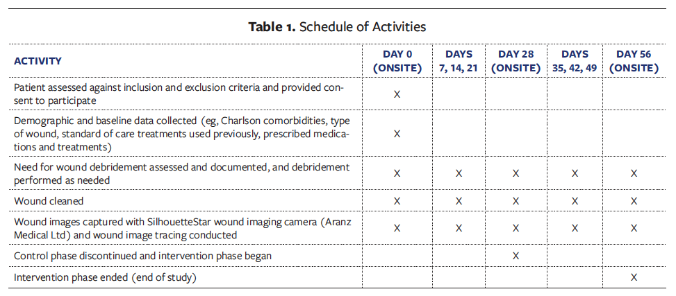
Participation lasted 56 days, as detailed in Table 1. A window of ±4 days was provided for all appointments and/or site visits to accommodate participant schedules. Participants who did not attend within the window for day 56 were followed up for an end of study visit. Whenever possible, day 0, day 28, and day 56 visits were done at the original medical facility from which the participant was recruited. The other visits (ie, days 7, 14, 21, 35, 42, and 49) were done either in the participants’ homes or onsite depending on preexisting appointments the participant had scheduled. For all visits in participants’ homes, the research nurse was present for data collection.
Adverse Events
Starting on day 0 and during each of the weekly follow-up visits, the study team used the Common Terminology Criteria for Adverse Events (version 5.0; US Department of Health and Human Services) to determine if there were any safety concerns, stress from participating in the study, or concerns with the conduct of the study.32 The Canadian Association of Research Ethics Boards’ definitions of adverse events and serious adverse events were used.33
Any adverse events or serious adverse events identified by the study team were reviewed with the study lead and were reported as necessary in compliance with the research institution’s Conjoint Health Research Ethics Board. Part of the review of each adverse event by the study team and study lead (CH) involved determining their relation to the intervention and study procedures, with events classified as definitely related, probably related, possibly related, not related, or unknown.
Statistical Analysis
Descriptive statistical methods were used to summarize the data. These included the number of subjects, mean (standard deviation [SD]) for continuous variable data (median [IQR] for nonnormal distribution of variable data), and frequencies and percentages for categorical variable data. Unless otherwise specified, all statistical testing was 2-sided and performed using an alpha level of .05.
The PAR from baseline was calculated as the percent change in the surface area of the index wound using the following formula: ((A1–A2)/A1) × 100, where A1 is the baseline area (at day 0), and A2 is the area at the specified time point (ie, day 28). The PAR for the intervention phase was calculated using the same formula, but where A1 was the new baseline area (ie, day 28), and A2 was the final time point of the intervention phase (ie, day 56).
All statistical analyses were conducted using SPSS (version 28; IBM) and NCSS 12 (NCSS LLC).
Participant populations for analysis. The population defined for the final analysis was a modified intention-to-treat (ITT) population that excluded participants for whom there were major violations in the control phase or intervention treatment. The ITT population included all subjects who completed the first phase evaluation and received at least 1 treatment with the CTM technology in the intervention phase.
Missing data. Any intermediate data entries (ie, days 7, 14, 21, 35, 42, or 49), excluding final or starting measurements (ie, days 0, 28, 56), that were missed during the study were imputed by the analyst for the ITT population. For PAR, missing data were interpolated or extrapolated using means based on adjacent time points, or last observation carried forward; multiple imputation using maximum likelihood algorithms was also used if data were missing not at random to provide a sensitivity analysis to assess the robustness of outcome data with its assumptions on missing data.34
Results
Demographics
A total of 36 patients were enrolled in the study; however, 11 of these patients were later deemed ineligible due to exclusions such as PAR greater than 50% at 4 weeks (n = 4), hospital admission longer than 4 days prior to the start of the intervention phase (n = 2), infection (eg, cellulitis, osteomyelitis) in the affected limb or in close proximity to the wound (n = 2), not meeting inclusion criteria upon further review (n = 1), and not following the care pathway in either phase (n = 2). As a result of these exclusions, patients from 1 long-term care facility and the inpatient hospital site were not included in the final analysis. Thus, a total of 25 participants from 3 long-term care facilities and 2 outpatient clinics comprised the population for the final analysis. PAR was calculated based on these remaining 25 participants.
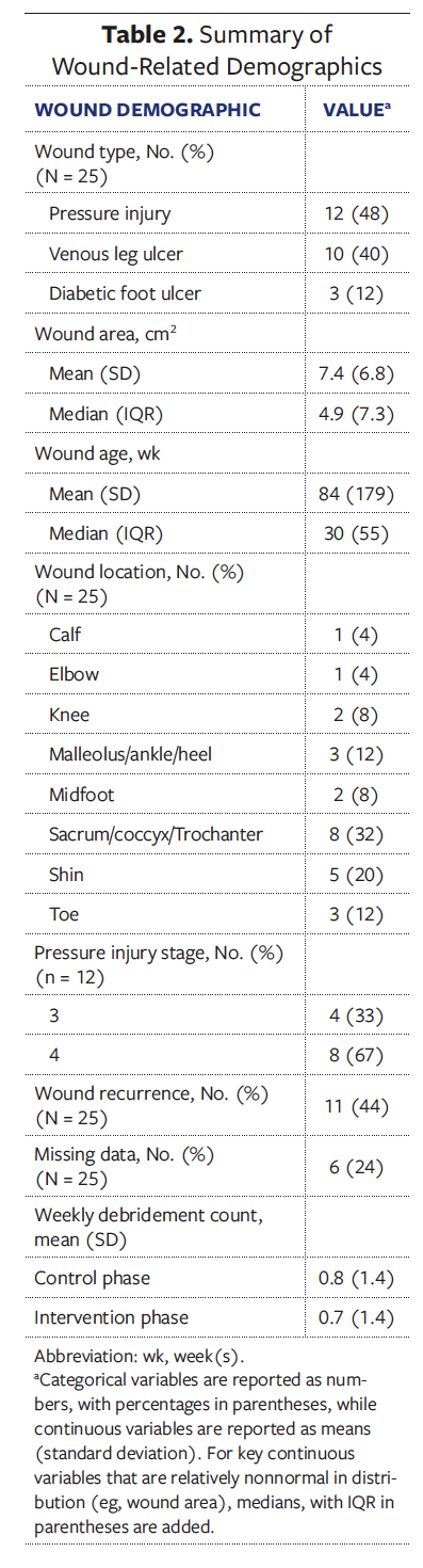
Of the 25 participants, 16 were male (64%) and 9 were female (36%). The most common comorbidities were peripheral vascular disease (64%), hypertension (40%), and diabetes mellitus (44%). Notably, wound duration averaged 84 weeks, or approximately 1.61 years. For a more detailed participant demographic breakdown see Supplementary Table 1. Table 2 provides an overview of the wounds that were included in the full data analysis.
Patient Outcomes
Percent area reduction. All wounds treated with the CTM technology decreased in area. Seven of 25 wounds (28%) had a PAR greater than 80% 4 weeks after intervention. Three wounds (12%) healed to full closure during the 28-day intervention phase.
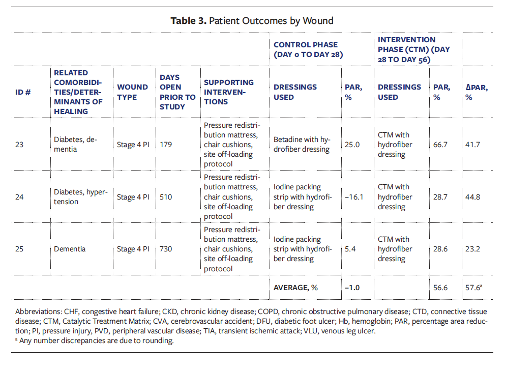
As shown in Figure 1, there was an overall 57.6% PAR with use of the CTM intervention compared with the current state dressing (P < .0001; > 95% power). During the control phase, the average PAR was −1.0%, meaning that on average the wounds increased in size for the 25 included participants. During the intervention phase, the average PAR was 56.6%, meaning that on average the wounds decreased in size.
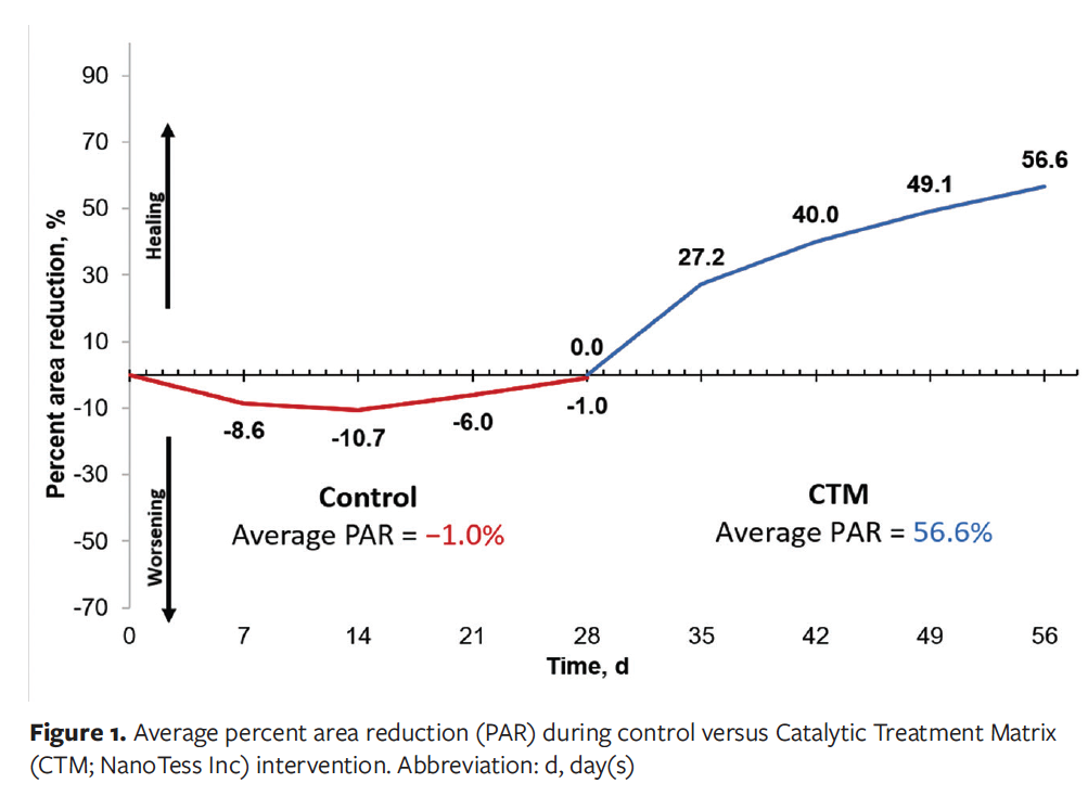
Notably, a large increase in PAR occurred in the first 7 days of implementing the CTM technology, compared with the remainder of the intervention phase. Overall, the average PAR in the first 7 days with the CTM technology was 27.2%. The overall PAR for wounds in the CTM intervention group was 56.6% by day 56; thus, 47% of the total wound closure over the entire intervention phase happened within the first 7 days. There appeared to be a “kick-start” effect when the CTM technology was applied that increased the speed of wound closure.
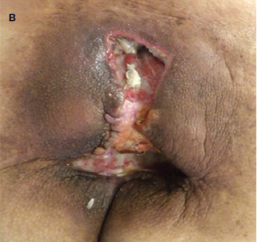
Figures 2 and 3 show examples of wound healing progression of 2 participants over the control and intervention phases.
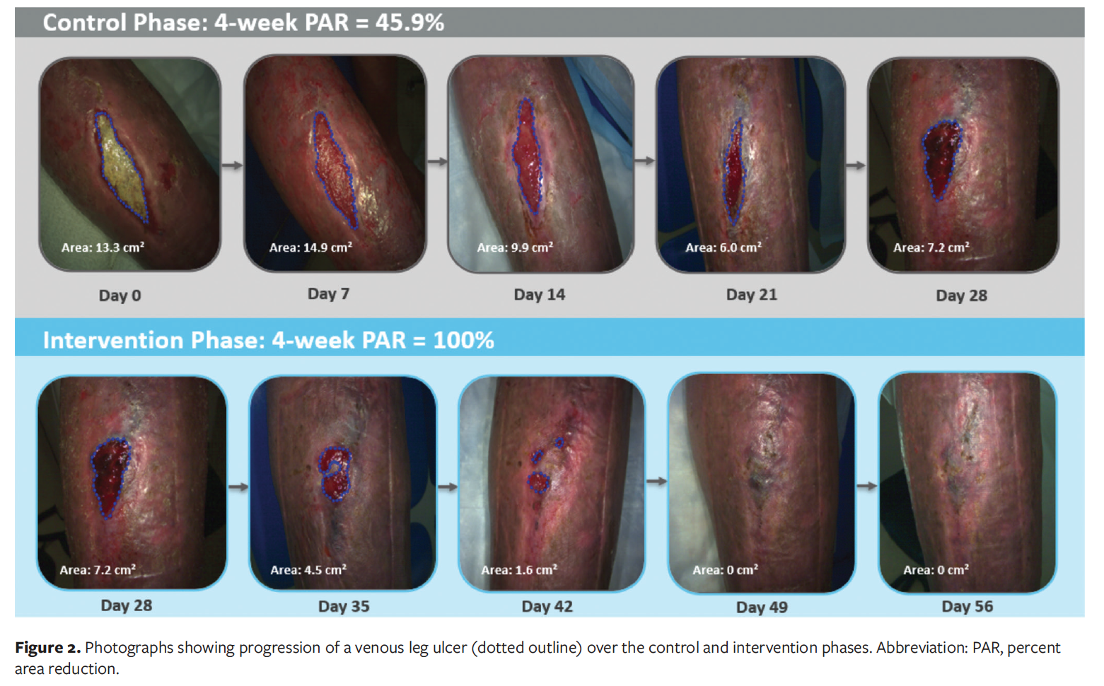
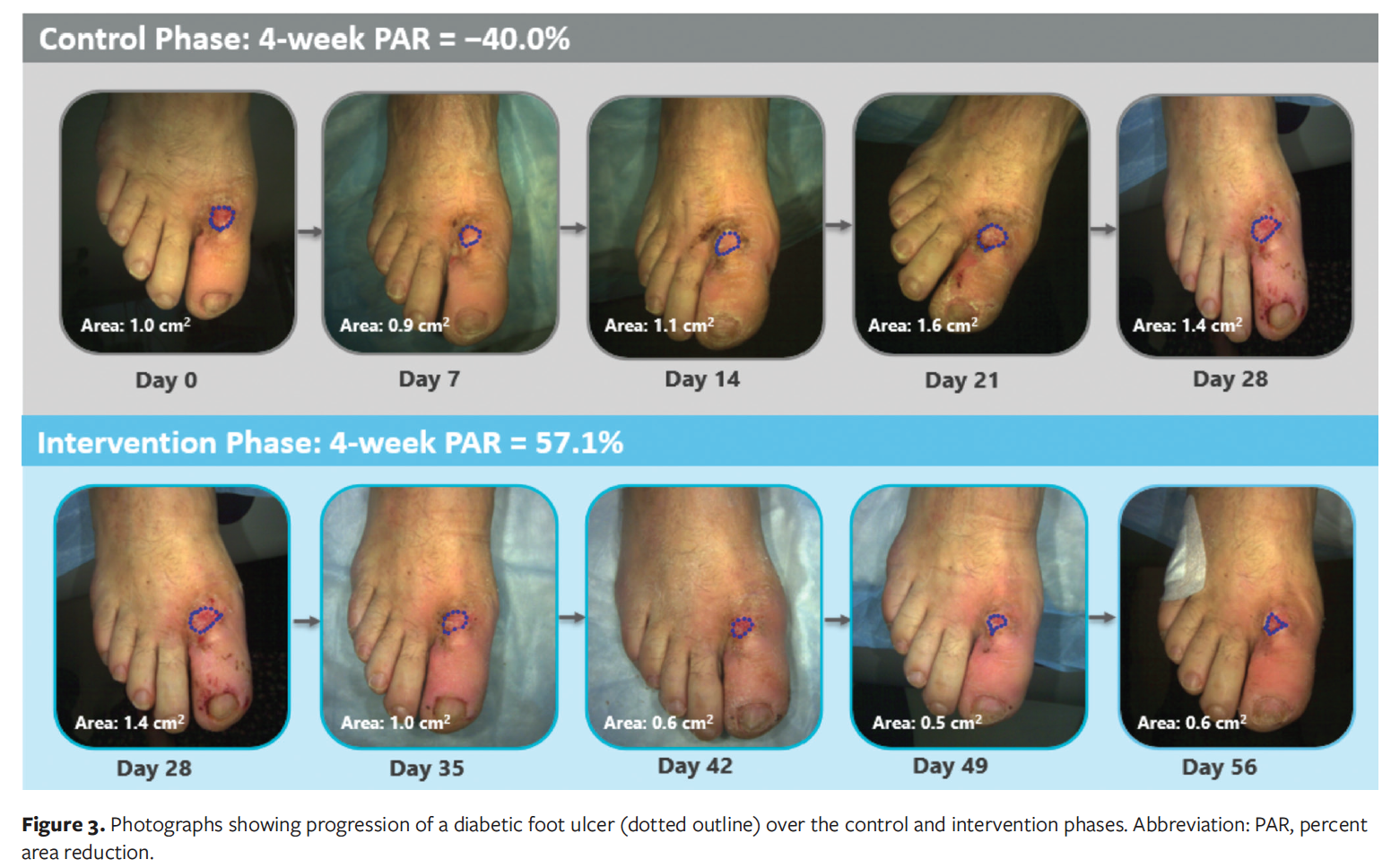
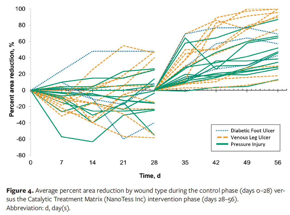
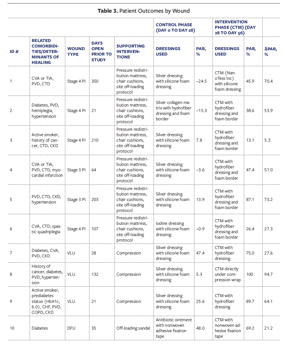
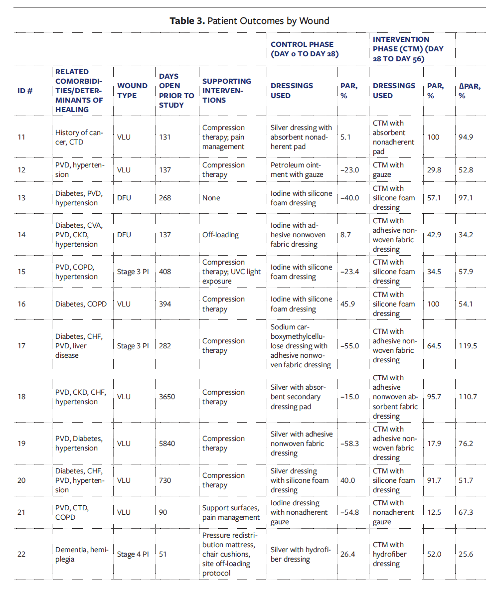
Figure 4 graphically represents PAR for the individual wounds over the 56-day trial phase. After the 28-day control phase, the PAR of individual wounds had an SD of 31.7%, with a minimum PAR of −58.3%, demonstrating an increase in wound size, and a maximum PAR of 48.0%. In contrast, after the 28-day CTM intervention phase, individual wounds had an SD of 29.5%; however, the minimum PAR was 12.5%, demonstrating some degree of healing across all wounds. Most notably, the maximum PAR was 100.0%, demonstrating complete healing.
Figures 5 and 6 show the overall wound healing trajectory for patients whose wounds either decreased in size or increased in size in the first 28 days of the study. Figure 5 graphically shows individual wounds that on average decreased in size in the first 28 days (ie, had a positive PAR value). There was a 34.9% improvement in wound healing with the use of the CTM technology compared with the current state dressings for patients whose wounds had decreased in size in the control phase. Figure 6 graphically shows individual wounds that on average increased in size in the first 28 days (ie, had a negative PAR value). There was a 70.4% improvement in wound healing with use of the CTM technology compared with the current state dressings for patients whose wounds had increased in size in the control phase. While not statistically analyzed by group, there was a positive healing trajectory whether a patient’s wound size increased or decreased in the first 28-day period, and this was even more notable for patients whose wounds had increased in size in those 28 days.
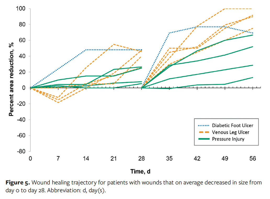
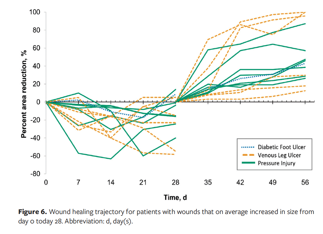
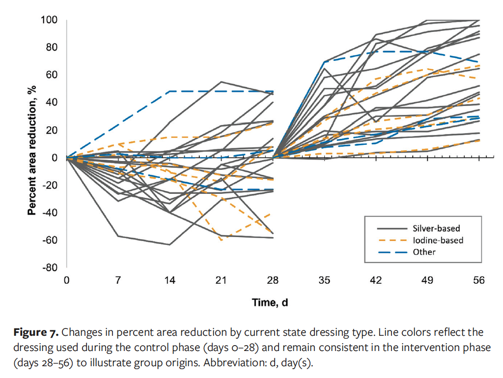
Figure 7 graphically represents an overview of the healing progression by dressing type, specifically silver-based,
iodine-based, and other dressings. Although no statistical analysis was conducted comparing the 2 time periods because of the small sample sizes within the dressing types, the overall improvement seen in the CTM intervention phase appears consistent across the different dressing types replaced.
The design of the trial monitored the established standard of care for each patient within their respective settings during the control phase. This within-
patient design maintained variables inherently constant within each participant. When the intervention was introduced, no variables changed in the care other than the CTM intervention, including compression, off-loading, support services, and nutrition management. A breakdown of patient outcomes by wound is outlined in Table 3.
Safety and adverse events. There were 5 adverse events and 6 serious adverse events reported during the study, 7 during the control phase and 4 during the intervention phase.
No between-group statistical comparisons were made because of the small number of occurrences. In most cases, it was clear that the adverse event was not related at all to the use of the CTM technology. In 2 cases, while unlikely, it was not possible to rule out the CTM as a factor in their occurrence. Both events involved an infection in the wound; however, it is important to note that infections in wounds could also be classified as disease-related events, which are attributable to the disease (ie, the wound) itself. Infections are quite typical within wounds, particularly those that are chronic.
Overall, there does not appear to be a notable difference between adverse events occurring in the control phase compared with the intervention phase. Infections are not uncommon in any wound-related study35; thus, the CTM technology demonstrated a safety profile equivalent to that of the current state dressings.
Discussion
The overall objective of the current study was to assess the clinical effectiveness of the CTM technology using an interrupted time series with a multisite, within-patient control protocol. The CTM demonstrated a noticeable healing effect, achieving an average PAR improvement of 57.6% between the control phase and the intervention phase. For all wounds in the study, wound healing improvement with the CTM technology trended consistently regardless of wound type, dressing type used during the control phase, and whether the wound increased or decreased in size during the control phase.
The chronicity of the wounds makes these results particularly compelling. Notably, most wounds well exceeded the inclusion criteria requirement of being present for at least 14 days. From the recorded date of wound onset up until the date of patient consent to participate in the study, the wounds selected for study inclusion had been present for an average of 84 weeks (1.61 years), and were considered chronic, nonhealing wounds. Moreover, many of the wounds were complex, with 67% of pressure injuries graded as stage 4. On average, these stage 4 wounds had been present for 270 days. This is particularly relevant because stage 4 pressure injury involves full-thickness tissue loss with damage extending into the muscle, bone, and other supporting structures (eg, fascia)36,37; thus, there would be substantial effects on the patient and their quality of life.2,3 These chronic, nonhealing wounds also place a significant burden on caregivers and communities, both emotionally and financially, because active dressings can be expensive and require long-term management.9,38
The current study followed the FDA’s suggestion of excluding patients with wound PAR greater than 50% before starting the intervention phase (ie, day 28) to minimize variability of aspects of care that affect outcomes when the participant moves from the control to the intervention phase.28 Discontinuation of participants whose wounds exhibited PAR greater than 50% by day 28 ensured the inclusion of persistent, chronic wounds that did not demonstrate healing progression with the use of current state dressings or that were potentially self-healing.
All the wounds treated with the CTM technology decreased in surface area, which was not the case for wounds in the control phase. During the intervention phase, most wounds demonstrated greater than 50% PAR, with one-third of wounds exhibiting greater than 80% healing; thus, some wounds may have achieved closure over a longer study period. This possibility was demonstrated in a retrospective evaluation of the clinical effectiveness of the CTM technology.39
As noted previously, the intervention appeared to have a kick-start effect on wounds, with nearly half of total wound healing occurring in the first 7 days of use. Given how quickly the CTM technology appears to begin working, it could be tried before moving on to more costly therapies. Further, health care providers may consider using it earlier in wound treatment to potentially stop wounds from advancing to a chronic state.
The current study demonstrates the value of using an interrupted time series with a within-patient control protocol for evaluating the effectiveness of new wound care technologies. Real-world approaches such as this provide more generalizable evidence than RCTs based on effectiveness within typical patients dealing with complex chronic wounds.21 The diversity and prevalence of comorbidities, wound sizes and types, and age of the participants reflect the complex nature and reality of patients experiencing chronic wounds. Typically, patients with these characteristics would not pass inclusion criteria for RCTs. The real-world findings in the current study demonstrate that the CTM technology has a positive effect on the healing trajectory of complex wounds. Overall, insights gained from these findings can be used to inform health care decision-making, because the results reflect the performance of this novel wound care technology in routine clinical practice.
Limitations
The current study has limitations. As many participants as possible were recruited within the study timeframe; however, many of the sites do not have significant patient turnover (eg, long-term care), and thus, the pool of possible patients to recruit from was limited, resulting in the relatively small sample size of 25 patients. However, the study is still credible because it used a within-patient interrupted time series design, involved long-standing chronic wounds with limited healing during the control period, demonstrated a large and clinically meaningful shift in PAR after CTM integration, and showed quite strong statistical evidence. The total wound burden was not formally analyzed and would be an interesting consideration for future studies.
Awareness of being involved in a study may have influenced participant adherence to wound protocols in both the control phase and the intervention phase, especially because the participants had 8 touchpoints with study team members throughout the study and received ongoing information about their wound progression due to the image capture and measurements done at each session.
Wound tracings using the wound imaging camera software were conducted to measure wound surface area. Although multiple members of the study team performed these tracings throughout the study, all tracings were subsequently reviewed by a single team member to ensure consistency. In cases where wound boundaries were ambiguous, a subset of these tracings was selected for further review in consultation with the study lead. This process helped mitigate interrater variability and improve the reliability of the wound measurement data.
It is important to note that the company that produces the CTM was an active collaborator in the study conceptualization. The company also provided in-service product training at clinical sites, product application training, and guidance on using the image capture camera. This involvement was essential, given the need for proper product education and use. The company had no role in the collection, analysis, or interpretation of the data.
Conclusion
Even with the inclusion of participants with complex comorbidities, the current analysis demonstrated a statistically significant improvement in wound healing with the use of the CTM technology in comparison with standard care. The improvement in wound healing appeared to be consistent across wound type, active dressing type (eg, silver, iodine, topical antibiotic ointments, collagen, and negative pressure dressings), and wound progression prior to the CTM use. In combination with standard of care practices to promote wound healing (eg, nutrition, compression, off-loading), the CTM technology was shown to be an effective tool in the treatment of chronic wounds.
The current study played a key role in informing AHS’s procurement decision by providing evidence-based insights into the product’s effectiveness. As a result, the CTM technology was procured by AHS, and its use has since expanded across geographic regions and practice specialities.
Author and Public Information
Authors: Anna Bradford, MBT1; Jill de Grood, MA, PMP1; Kathryn Ambler, MSc2; Negar Dehghan Noudeha, MBT1; Patty Wickson, eMBA, CHE2; and Chester Ho, MD3,4
Affiliations: 1W21C, O’Brien Institute for Public Health, Cumming School of Medicine, University of Calgary, Calgary, AB, Canada; 2Innovation and Business Intelligence, Alberta Health Services, Calgary, AB, Canada; 3Department of Medicine, Faculty of Medicine and Dentistry, University of Alberta, Edmonton, AB, Canada; 4Glenrose Rehabilitation Hospital, Alberta Health Services, Edmonton, AB, Canada
Acknowledgments: The authors would like to thank Wrechelle Ocampo, MBT, for support with study conceptualization, Mackenzie Murawsky, MScN, for support with data collection, Marissa Carter, PhD, for support with trial design and statistical analysis, and the Innovation and Business Intelligence team members at Alberta Health Services for their support with funding acquisition. A special thanks to all the health care providers who supported the study and patients who took part in the study.
Author Contributions: The final manuscript has been seen and approved by all authors, and all authors accept full responsibility for the design and conduct of the study, had access to the data, and controlled the decision to publish.
Funding: This study was funded by CAN Health West, with in-kind contributions from W21C (study leadership and project management), Alberta Health Services (study leadership and project management), and NanoTess (in-service training for appropriate device use, provision of cameras, and consultation with an independent wound care expert).
Disclosure: The authors disclose no financial or other conflicts of interest.
Ethics Statement: The study was conducted following the research protocol that was reviewed and approved by the Conjoint Health Research Ethics Board on November 13, 2022 (reference number REB22-1394). All procedures were completed in compliance with the ethics approval, and the study followed the ethical standards regarding participant privacy and confidentiality.
Correspondence: Anna Bradford, MBT; W21C GD01, CWPH Building, 3280 Hospital Dr NW, Calgary, AB T2N 4Z6 Canada; anna.bradford1@ucalgary.ca
Manuscript Accepted: June 30, 2026
References
1. Darwin E, Tomic-Canic M. Healing chronic wounds: current challenges and potential solutions. Curr Dermatol Rep. 2018;7(4):302. doi:10.1007/S13671-018-0239-4
2. Roussou E, Fasoi G, Stavropoulou A, et al. Quality of life of patients with pressure ulcers: a systematic review. Med Pharm Rep. 2023;96(2):123. doi:10.15386/MPR-2531
3. Spilsbury K, Nelson A, Cullum N, Iglesias C, Nixon J, Mason S. Pressure ulcers and their treatment and effects on quality of life: hospital inpatient perspectives. J Adv Nurs. 2007;57(5):494-504. doi:10.1111/J.1365-2648.2006.04140.X
4. Barakat-Johnson M, Lai M, Wand T, White K. A qualitative study of the thoughts and experiences of hospital nurses providing pressure injury prevention and management. Collegian. 2019;26(1):95-102. doi:10.1016/J.COLEGN.2018.04.005
5. Waye A, Hughes B, Mrklas K, et al. Innovation pipeline: a framework for value-based decision making. Healthc Manage Forum. 2024;38. doi:10.1177/08404704241271186
6. Douglas Queen, Botros Mariam. True cost of wounds for canadians: annual update (2024). Wound Care Can. 2025;23(2). doi:10.56885/032346qumxzp
7. Falanga V, Isseroff RR, Soulika AM, et al. Chronic wounds. Nat Rev Dis Primers. 2022;8(1):1-21. doi:10.1038/s41572-022-00377-3
8. Orsted HL, Keast DH, Forest-Lalande L, Kuhnke JL, O’Sullivan-Drombolis D, Jin S, et al. Skin: anatomy, physiology and wound healing. In: Kuhnke JL, Burrows CA, Evans RM, Orsted HL, Rosenthal S, eds. Best Practice Recommendations for Skin Health and Wound Management 2025. Wounds Canada; 2025. doi:10.56885/PEWY3827
9. Orsted HL, Keast DH, Forest-Lalande L, et al. Prevention and Management of Wounds: An Overview Chapter 4 Prevention and Management of Wounds: An Overview. In: Kuhnke JL, Burrows CA, Evans RM, Orsted HL, Rosenthal S, eds. Best Practice Recommendations for Skin Health and Wound Management 2025. Wounds Canada; 2025. doi:10.56885/CVEU6924
10. Health Quality Ontario. Community-based care for chronic wound management: an evidence-based analysis. Ont Health Technol Assess Ser. 2009;9(18):1-24. Accessed September 18, 2023. https://www.hqontario.ca/Portals/0/Documents/evidence/reports/rev_smcc_wound_20091019.pdf
11. Canadian Patient Safety Institute. Never Events for Hospital Care in Canada. Published online September 2015. Accessed July 27, 2023. https://www.healthcareexcellence.ca/media/eceoshdc/never-events-for-hospital-care-in-canada.pdf
12. Alberta Health Services. Pressure Injury Prevention in Alberta Hospitals. May 3, 2021. Accessed August 22, 2023. https://www.albertahealthservices.ca/assets/about/scn/ahs-scn-nrv-pressure-injury-prevention-infographic.pdf
13. NanoTess. NanoSALV - Wound Care. Accessed May 25, 2023. https://nanotess.com/?srsltid=AfmBOoqERz77Qq5qPE2A58u-fbFgjc4tOBnhAp9dUZ7HwG2bxACPSKf3
14. Zhang Z, Xue H, Xiong Y, et al. Copper incorporated biomaterial-based technologies for multifunctional wound repair. Theranostics. 2024;14(2):547-570. doi:10.7150/thno.87193
15. Sen CK, Khanna S, Venojarvi M, et al. Copper-induced vascular endothelial growth factor expression and wound healing. Am J Physiol Heart Circ Physiol. 2002;282(5). doi:10.1152/AJPHEART.01015.2001
16. Zhang T, Wang L, Chen Q, Chen C. Cytotoxic potential of silver nanoparticles. Yonsei Med J. 2014;55(2):283. doi:10.3349/YMJ.2014.55.2.283
17. Kim HS, Lee S, Kim JH. Real-world evidence versus randomized controlled trial: clinical research based on electronic medical records. J Korean Med Sci. 2018;33(34). doi:10.3346/JKMS.2018.33.E213
18. Hariton E, Locascio JJ. Randomised controlled trials—the gold standard for effectiveness research. BJOG. 2018;125(13):1716. doi:10.1111/1471-0528.15199
19. Dang A. Real-world evidence: a primer. Pharmaceut Med. 2023;37(1):25-36. doi:10.1007/S40290-022-00456-6/METRICS
20. Kale S, Vatkar A, Gehilot O, Shyam A. Real-world evidence: methodologies for integrating real-world data into clinical research frameworks. J Orthop Case Rep. 2025;15(10):6. doi:10.13107/JOCR.2025.V15.I10.6138
21. Fife C, LeBoutillier B, Taylor C, Marcinek BT. Real-world use and outcomes of hard-to-heal wounds managed with porcine placental extracellular matrix. Cureus. 2024;16(12):e76262. doi:10.7759/CUREUS.76262
22. Stephenson J. Study designs in wound care. WCET Journal. 2022;42(1). doi:10.33235/WCET.42.1.12-15
23. Saldanha IJ, Skelly AC, Vander Ley K, et al. Inclusion of Nonrandomized Studies of Interventions in Systematic Reviews of Intervention Effectiveness: An Update. Agency for Healthcare Research and Quality; 2022. AHRQ publication 22-EHC033. doi:10.23970/AHRQEPCMETHODSGUIDENRSI
24. Bradford A, Arnold K, VanTil K, et al. Human factors evaluation of an innovative wound care technology. Hum Factors Healthc. 2025;8:100108. doi:10.1016/J.HFH.2025.100108
25. Driver VR, Gould LJ, Dotson P, et al. Identification and content validation of wound therapy clinical endpoints relevant to clinical practice and patient values for FDA approval. Part 1. Survey of the wound care community. Wound Repair Regen. 2017;25(3):454-465. doi:10.1111/WRR.12533
26. Driver VR, Gould LJ, Dotson P, Allen LL, Carter MJ, Bolton LL. Evidence supporting wound care end points relevant to clinical practice and patients’ lives. Part 2. Literature survey. Wound Repair Regen. 2019;27(1):80-89. doi:10.1111/WRR.12676
27. Bull RH, Clements D, Collarte AJ, Harding KG. A novel randomized trial protocol for evaluating wound healing interventions. Adv Wound Care (New Rochelle). 2023;12(12):671. doi:10.1089/WOUND.2023.0058
28. U.S. Food & Drug Administration. Chronic Cutaneous Ulcer and Burn Wounds - Developing Products for Treatment. June 2006. Accessed August 26, 2025. https://www.fda.gov/regulatory-information/search-fda-guidance-documents/chronic-cutaneous-ulcer-and-burn-wounds-developing-products-treatment
29. Kim JS. Within-subjects design. In: Salkind NJ, ed. Encyclopedia of Research Design. SAGE Publications Inc; 2010:1639-1644. doi:10.4135/9781412961288.n503
30. Liljequist D, Elfving B, Roaldsen KS. Intraclass correlation – a discussion and demonstration of basic features. PLoS One. 2019;14(7):e0219854. doi:10.1371/JOURNAL.PONE.0219854
31. ARANZ Medical. Silouette. Accessed July 27, 2023. https://www.aranzmedical.com/
32. U.S. Department of Health and Human Services. Common Terminology Criteria for Adverse Events (CTCAE) Version 5.0. Preprint posted online November 27, 2017:1-155. Accessed June 25, 2023. https://ctep.cancer.gov/protocoldevelopment/electronic_applications/docs/ctcae_v5_quick_reference_5x7.pdf
33. Canadian Association of Research Ethics Boards. Guidance on Reporting of Unanticipated Problems Including Adverse Events to Research Ethics Boards in Canada. Published July 2010. Accessed April 22, 2023. https://careb-accer.org/reporting-of-unanticipated-problems-including-adverse-events-to-research-ethics-boards-in-canada/
34. Kang H. The Prevention and Handling of the Missing Data. Korean J Anesthesiol. 2013;64(5):402-406. doi:10.4097/kjae.2013.64.5.402
35. Keitz SA, Young LChronic wound infection. In: Simel DL, Rennie D, eds. The Rational Clinical Examination: Evidence-Based Clinical Diagnosis. McGraw-Hill Education; 2009. Accessed June 15, 2025. https://jamaevidence.mhmedical.com/content.aspx?bookid=845§ionid=61357674
36. Lyder CH, Ayello EA. Pressure ulcers: a patient safety issue. In: Hughes RG, ed. Patient Safety and Quality: An Evidence-Based Handbook for Nurses. Agency for Healthcare Research and Quality; 2008. Chapter 12. Accessed August 19, 2026.
37. Norton L, Parslow N, Ho C, et al. Best practice recommendations for the prevention and management of pressure injuries. In: Kuhnke JL, Burrows CA, Evans RM, Orsted HL, Rosenthal S, eds. Best Practice Recommendations for Skin Health and Wound Management 2025. Wounds Canada; 2025. doi:10.56885/GRYI5585
38. Norton L, Parslow N, Johnston D, et al. Best practice recommendations for the prevention and management of pressure injuries. In: Foundations of Best Practice for Skin and Wound Management. Supplement of Wound Care Canada. Wounds Canada; 2017. Accessed July 30, 2023. https://www.woundscanada.ca/doclink/bpr-prevention-and-management-of-pressure-injuries-2/eyJ0eXAiOiJKV1QiLCJhbGciOiJIUzI1NiJ9.eyJzdWIiOiJicHItcHJldmVudGlvbi1hbmQtbWFuYWdlbWVudC1vZi1wcmVzc3VyZS1pbmp1cmllcy0yIiwiaWF0IjoxNjQyMDk3MTc2LCJleHAiOjE2NDIxODM1NzZ9.uvuc1blTD1CzG5FJ3youuWEeoppQOJGdAxmHt9wPAZg?utm_source=chatgpt.com
39. Bradford A, de Grood J, Ambler K, Dehgan Noudeh N, Wickson P, Ho C. Retrospective clinical effectiveness evaluation of an innovative catalytic wound care technology. Wounds. 2026;38(6):178-183. doi:10.25270/wnds/25123. doi:10.25270/wnds/25123