Use of Negative Pressure Wound Therapy as an Occlusive Dressing on a Skin Graft in a Patient With Hostile Abdomen
© 2026 HMP Global. All Rights Reserved.
Any views and opinions expressed are those of the author(s) and/or participants and do not necessarily reflect the views, policy, or position of Wounds or HMP Global, their employees, and affiliates.
Abstract
Background. In hostile abdomen the abdominal cavity is exposed, with retracted edges compacted into a single block of fibrous tissue that does not allow for adequate dissection or separation. After the inflammation is controlled, definitive closure of the abdomen can be attempted. One closure technique includes the use of skin grafts, which requires an adequate recipient site. Several therapies are used to promote granulation, such as dressings, antiseptic solutions, and negative pressure wound therapy. However, there is limited evidence of the use of these therapies in a hostile abdomen. Case report. A patient with hostile abdomen eventually achieved closure after split-thickness and full-thickness skin grafting in conjunction with use of a lipidocolloidal dressing and negative pressure wound therapy. The findings are reported in line with the Surgical CAse REport criteria. Conclusion. Negative pressure wound therapy may improve the condition of the abdomen for receiving a skin graft. Successful use of a lipidocolloidal dressing as an interface between the graft and negative pressure wound therapy was achieved in this case. Effective and continuous communication among medical staff, the patient, and his relatives contributes to improved outcomes.
In hostile abdomen the abdominal cavity is exposed, with retracted edges compacted into a single block of fibrous tissue that does not allow adequate dissection or separation.1 This condition is secondary to inflammation, sepsis, or chronic radiation, or as a sequela of intestinal leakage, a traumatic injury, or an adhesive process following surgery.2-4
One technique used to address hostile abdomen is open abdomen, which involves intentionally leaving the fascial edges of the abdomen without approximation, thus exposing the abdominal contents.5 After the inflammation is controlled, definitive closure of the abdomen can be attempted with techniques such as primary fascial closure, separation of fascial components, or a controlled ventral hernia (primary skin closure or skin graft).5
A skin graft is a fragment of skin tissue that is separated from its donor site and attached to a recipient site intended for spontaneous revascularization.6 An adequate recipient site without infection is needed for adequate integration of the graft.7 Several therapies are used to promote the granulation process, such as dressings, antiseptic solutions, and negative pressure wound therapy (NPWT).8 However, there is limited evidence on the use of these therapies in a hostile abdomen.5
The present study reports a patient with a hostile abdomen for which a split-thickness skin graft (STSG) and a full-thickness skin graft were used in conjunction with a lipidocolloidal dressing and NPWT for abdominal closure in a public tertiary care hospital. This case is reported in line with the Surgical CAse REport criteria.9
Case Report
A 73-year-old male farmer with a body mass index of 29.1 (weight, 86 kg; height, 1.72 m) with incomplete primary education, and with no comorbidities, allergies, or drug addiction, walked into the emergency department with an intestinal obstruction that had started 48 hours prior.
Clinical findings and diagnostic assessment
Physical examination and laboratory test results led to a diagnosis of acute abdomen, and exploratory laparotomy was performed. The surgeon found a rectosigmoid tumor with peripheral abscesses and performed the Hartmann procedure10 without apparent complications. The histopathology report described severe chronic colitis with ischemic-hemorrhagic infarcts and intestinal perforation. The main challenge to adherence to medical treatment was the patient’s illiteracy, resulting in his complete dependence on his daughter as a caregiver.
On the fourth day, the patient presented fever, tachycardia, and an elevated leukocyte count. Abdominal computed tomography revealed an intra-abdominal abscess in the left colic flexure (Clavien-Dindo grade IIb),11 and laparotomy was performed. After 7 days, the patient was discharged.
Seven months later, surgery for restoration of intestinal transit was performed, with intraoperative findings of multiple loose and firm loop-to-loop and loop-wall adhesions (Mazuji grade 3 and Clavien-Dindo grade III).11 Cold cut adhesiolysis and the Deloyers procedure12 were performed. The patient presented adequate clinical improvement and was discharged 5 days after this surgery on an antibiotic regimen (Ciprofloxacin and Metronidazole).
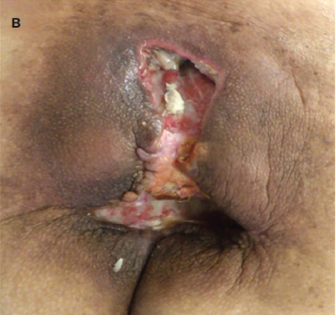
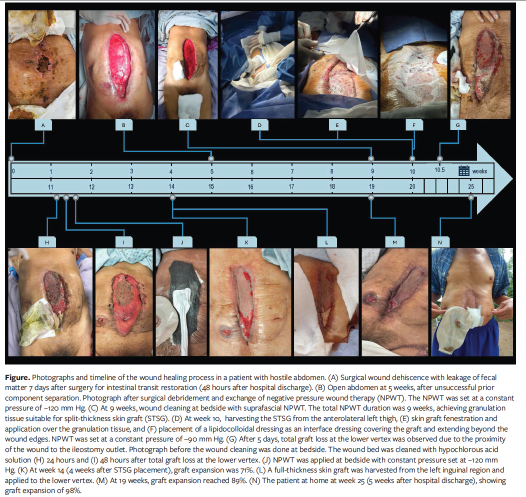
Seven days after surgery (48 hours after discharge), the patient returned to the emergency department with a dehiscent surgical wound (Figure, part A). Urgent exploratory laparotomy was performed, and the anastomosis was found to be dehiscent, with fecal matter in the abdominal cavity (Clavien-Dindo grade III).11 The anastomosis was dismantled, and right hemicolectomy with terminal ileostomy was done; the cavity was cleaned, and the abdominal wall was closed; on the third day the surgical wound was dehiscent, with necrotic edges and with exposure to abdominal contents.
Therapeutic intervention
Surgical cleaning with debridement was scheduled, with the intraoperative discovery of a hostile abdomen (Björck grade 3A and Clavien-Dindo grade III).11 Surgical cleanings were initiated with surgical soap, saline solution, and a hypochlorous acid solution (Vashe; Urgo Medical North America) in conjunction with intra-
abdominal NPWT (AbThera; Solventum) at a constant pressure of −120 mm Hg. Surgical cleanings were scheduled on demand, based on the collection volume of the NPWT device. Seven surgical cleanings with NPWT dressing changes were performed in a 5-week period. Each surgical cleaning lasted approximately 40 minutes.
Component separation was unsuccessfully attempted on 2 occasions (Figure, part B). Thus, it was decided to generate granulation tissue and apply an STSG. After 5 weeks of wound care, the intra-abdominal NPWT was changed to a suprafascial device (Renasys; Smith & Nephew). This change of NPWT device was made for administrative reasons. Wound cleaning was performed weekly for 4 weeks at bedside, using only hypochlorous acid solution. A sterile gauze was used to gently stimulate the granulation tissue, and then the NPWT device was placed. The patient did not experience significant pain, and anesthesia was not required.
The time to achieve adequate granulation tissue with NPWT was 9 weeks (5 weeks intra-abdominal and 4 weeks suprafascial) (Figure, part C). At week 10, the plastic surgeon (ERA) harvested an STSG from the left thigh using an electric dermatome (Padgett Model B; Integra Life Sciences) (Figure, part D). Incisions were made in the graft, and it was sutured with 5-0 nylon. A lipidocolloidal dressing (UrgoTul; Urgo Medical North America) was placed as an interface dressing (Figure, parts E and F). The NPWT was used as an occlusive dressing (over the graft) with a continuous pressure of −90 mm Hg for 5 days. To reduce the risk of contamination of the STSG due to the proximity of the ileostomy outlet, which lay 3 cm from the STSG, a hydrocolloid was used as a barrier method. It was not possible to place the ileostomy at a greater distance. When the site was uncovered 5 days later, total graft loss at the lower vertex due to contamination of the wound was observed, as were areas of hypergranulation and biofilm formation throughout the wound periphery. At the upper vertex, the graft was accepted only in the center and was translucent (Figure, part G). After 24 hours, the graft resembled normal skin (Figure, parts H and I).
To promote granulation tissue suitable for a new skin graft, NPWT was applied at a continuous pressure of −120 mm Hg for 4 weeks, with weekly bedside wound cleanings (Figure, part J). On 2 occasions, the patient reported a slight increase in pain; thus, the pressure was reduced to −90 mm Hg for 1 week. However, the graft became pale and showed no evidence of expansion, which prompted a return to the original pressure of −120 mm Hg with adequate tolerance by the patient.
Wound surface area and skin graft expansions were determined using photo planimetry with the WoundGenius application (imito AG). Four weeks later, the skin graft had expanded 71% (Figure, part K) and was identical to the peripheral skin. At that time, a full-thickness skin graft was harvested (because for administrative reasons an STSG could not be harvested) from the left inguinal region (Figure, part L) and then was applied to the lower vertex and sutured. By the 19th week, 89% epithelialization of the total wound surface was observed (Figure, part M). The remaining defect, which measured approximately 2 cm², was covered with an antimicrobial hydrofiber dressing with silver and EDTA (Aquacel Ag+ Extra; ConvaTec) and plastic film; these were changed every 2 days.
Follow-up and outcomes
The patient and his family were instructed on dressing changes as well as on ileostomy, skin, and wound care, and the patient was subsequently discharged home with follow-up in the outpatient clinic (Figure, part N). The hospital length of stay to close the abdominal breach was 20 weeks.
The patient’s daughter later reported, “Our dad experienced episodes of sadness at various times during his hospitalization, but thanks to the psychological services and the doctors’ care, he was able to overcome these difficulties. Today he is in our house doing most of his daily activities by himself.” In a telephone interview, the patient emotionally commented, “I thank you [the doctors] for taking care of me when I was in the hospital. I am very grateful.” His speech was interrupted by a “lump in his throat” and a cracking voice that conveyed the sincerity of his words.
Discussion
Open abdomen is a strategy to prevent and treat compartment syndrome in patients with abdominal trauma or abdominal wall tissue loss.13 However, it is also associated with early, mid-term, and late complications. Early complications (<1 week) include fluid balance, temperature loss, electrolyte abnormalities, visceral injury, and abdominal compartment syndrome.14 Some mid-term complications (2 weeks-3 weeks) include loss of domain, visceral injury, anastomotic leaks, intra-abdominal abscess, and hospital-acquired infections. Late complications (>3 weeks) include enteroatmospheric fistula, planned ventral hernia, intestinal ileus, and nutritional losses, which may make medical treatment more challenging.14
Although various methods of abdominal coverage have been described, there is scant evidence of methods for abdominal closure.5,15 The patient in the current report required damage control surgery and was subsequently managed as an open abdomen.16 In open abdomen, 2 problems coexist: the wound and the abdominal defect.17 Taking all these factors into account, 2-phase treatment was planned for the patient in the current report, with the first phase consisting of wound management and preparation of the receptor site for STSG, and the second phase focusing on closing the abdominal gap with STSG.
By generating mechanical stress, NPWT increases blood supply, promotes cell reproduction and angiogenesis, and reduces exudate and bacterial colonization.5,18-20 Argenta and Morykwas21 obtained granulation tissue in wounds using continuous pressure of −125 mm Hg and applied a skin graft at a pressure of −50 mm Hg to −75 mm Hg. NPWT is an effective option for temporary closure of the open abdomen, because it adequately drains fluids and improves wound contraction.22,23
The pH changes in the different phases of a wound; when infection is present or debridement is performed, an acidic environment predominates.24 To promote an adequate microenvironment for tissue repair in the patient in the current report, hypochlorous acid solution was used at each wound cleaning. Hypochlorous acid also serves as an antimicrobial adjuvant, aids in odor control, and reduces pain.25,26 For the current case, the solution was used for 60 seconds, which is the average time required to decrease the bacterial load and prevent biofilm formation.27 Dressing changes should occur every 48 hours to 72 hours.21 However, in the current case, due to the decrease in exudate collected in the system, this frequency was reduced in order to minimize trauma and the risk of fistula formation.
Biochemical assessments were completed twice weekly for the first 5 weeks and weekly thereafter. This assessment included complete blood count, blood chemistry, serum electrolytes, lipid profile, liver function tests, procalcitonin, and C-reactive protein. Nutrition plays a key role in recovery in patients with hostile abdomen and chronic wounds.28 For the current case, the Controlling Nutritional status scale29 was used to assess and monitor nutrition status. Initially, the patient was moderately malnourished, and as a result, he was evaluated by a clinical nutritionist who prescribed a high-calorie and high-protein diet supplemented with glutamine, ferrous sulfate, and B complex.28
For the ileostomy assessment and to both avoid electrolytic imbalance and protect the peristomal skin in the current patient, serum electrolyte levels were monitored, as was the daily 24-hour ileostomy output.30 Hemoglobin, procalcitonin, and C-reactive protein levels, as well as the quick sequential (sepsis-related) organ failure assessment scale,31-33 were used as markers of inflammation and sepsis in the current patient.
In the fifth week of treatment for the current patient, the abdominal gap was reduced and the NPWT modality was changed from intra-abdominal to suprafascial.34 This eased the NPWT changes at bedside, which saved time, personnel, and materials costs.35 The patient in the current report required multidisciplinary management involving nursing, general surgery, plastic surgery, nutrition, rehabilitation, and psychology personnel. Constant communication with the patient, his relatives (caregivers), and health care personnel had a positive effect on the patient’s recovery. Applying the Enhanced Recovery After Surgery protocol,5,36 ambulation, basic hygiene activities, oral feeding, and withdrawal of intravenous solutions and medications were achieved in the present patient.
The lipidocolloidal dressing over the STSG serves as an interface between the graft and the NPWT dressing, allowing moisture to pass through, preventing traumatic removal, protecting the graft, and stimulating fibroblast proliferation in vitro and granulation tissue formation.37,38 Chou et al39 evaluated the vacuum-assisted closure system with a silicone-based dressing over the conventional tie-over bolster technique in skin graft fixation and found that the combination of NPWT with silicone dressing in skin grafts was more effective for graft integration compared with a conventional method, using a pressure of −50 mm Hg and removing it on the fourth day. However, that study did not include patients with open abdomen.
Suzuki et al40 used STSG to cover the abdominal contents and NPWT at a continuous pressure of −120 mm Hg as an occlusive dressing. They achieved successful epithelialization in a patient with an enteroatmospheric fistula, but unfortunately, the patient died. In a patient with open abdomen, Kim et al41 achieved abdominal closure using an acellular dermal matrix underneath an STSG placed over the abdominal gap that had been previously prepared with NPWT at a continuous pressure of −120 mm Hg. Cases similar to the one in the present report are scarce (Table).40-42
The Consensus Guidelines on Negative Pressure Wound Therapy for Wound Repair (2025 version) do not include recommendations for NPWT in patients with open abdomen.43 However, despite the presence of open abdomen in both trauma and nontrauma patients, the World Society of Emergency Surgery guidelines state that NPWT can be used in combination with biologic mesh to facilitate granulation and skin closure; a detailed protocol for pressure settings is not described.5 In this challenging clinical scenario, there are currently no comprehensive clinical guidelines regarding the choice of pressure settings in NPWT; thus, the level of suction is based on individual assessment of the wound and patient tolerance.5,43,44
Although there are successful reports using different closure methods, the process for creating an adequate receptor site over the abdominal gap, as well as the use of an intermediate dressing, are not detailed. The present case provides valuable insights into the use of a lipidocolloidal dressing when applying a skin graft in conjunction with NPWT to achieve closure of the abdominal gap in a hostile abdomen.
Limitations
An important limitation of the present case is that a controlled hernia was created, which requires daily use of a girdle, weight control, avoidance of straining, and additional surgical interventions. The main limitation of this report is the lack of generalizability of the methods for achieving epithelialization of incisional wounds in other conditions that generate a hostile abdomen, such as traumatic injuries, abdominal compartment syndrome, and massive intra-abdominal bleeding, which are managed as an open abdomen; these other clinical scenarios should be validated in further studies. Furthermore, the methods described require a multidisciplinary approach, and use of the technique described herein would be difficult at institutions with a limited number of health care providers and limited economic resources. Finally, the method described requires healthy skin to cover the wound with STSG.
Conclusion
A multidisciplinary approach is needed to treat patients with a hostile abdomen. The application of NPWT, combined with a hypochlorous acid solution may improve the condition of the abdomen for receiving a skin graft. A lipidocolloidal dressing can be used as an interface between the graft and NPWT dressing. Effective and continuous communication among medical staff, the patient, and the patient’s family and caregivers contributes to improved outcomes.
Author and Public Information
Authors: Martin Eduardo Morales-Saavedra, MD1; Esperanza Ramírez-Arano, MD2; Juan Carlos Bravo-Soriano, MD2; and Eduardo Alfonso Hernández-Muñoz, MD3
Affiliations: 1General Surgery, Hospital Regional de Alta Especialidad de Veracruz, Veracruz, Mexico; 2Plastic and Reconstructive Surgery, Hospital Regional de Alta Especialidad de Veracruz, Veracruz, Mexico; 3Epidemiology, Unidad de Medicina Familiar No. 181, Instituto Mexicano del Seguro Social, Ixtlahuacán de los Membrillos, Jalisco, Mexico.
Acknowledgment: The authors thank Aidee Alejandra del Toro Hinojosa for her valuable support in reviewing the grammar and syntax of this manuscript.
Author Contributions: Study conceptualization: M.E.M.S. Study design: M.E.M.S., E.R.A., and J.C.B.S. Designing the analytical strategy and interpreting the findings: M.E.M.S. and E.A.H.M. Conducting the literature review and preparing the introduction and methods sections of the text: all authors. Drafting the discussion and final approval of the version to be published: all authors.
Disclosure: The authors disclose no financial or other conflicts of interest.
Ethics Statement: Written informed consent for publication of the case and accompanying photographs was obtained from the patient and 2 family members prior to manuscript submission.
Correspondence: Martin Eduardo Morales-Saavedra, MD; Hospital Regional de Alta Especialidad de Veracruz, Av 20 de Noviembre 1074, Centro, 91700 Veracruz, Veracruz, Mexico; martineduardoms@hotmail.com
Manuscript Accepted: May 11, 2026
References
1. Rivas HAS, Pérez AM, Peña Gio CG, Cervantes AFV, Morales AQV. Hostile abdomen: a review of the current literature. International Journal of Medical Science and Clinical Research Studies. 2023;3(3):337-339. doi:10.47191/ijmscrs/v3-i3-11
2. Lundy M, Ashburn JH. Management of the hostile abdomen. Clin Colon Rectal Surg. 2022;35(3):169-176. doi:10.1055/s-0041-1740043
3. Leppäniemi A. Abdomen abierto en el tratamiento del abdomen catastrófico: evidencia y controversias. Editorial. Cir Esp. 2021;99(8):559-561. doi:10.1016/j.ciresp.2020.06.011
4. Montoya BIM. Surgical management of the hostile abdomen: a comprehensive current review of literature. International Journal of Medical Science and Clinical Research Studies. 2023;3(9):1924-1926. doi:10.47191/ijmscrs/v3-i9-21
5. Coccolini F, Montori G, Ceresoli M, et al. The role of open abdomen in non-trauma patient: WSES Consensus Paper. World J Emerg Surg. 2017;12:39. doi:10.1186/s13017-017-0146-1
6. Depoortère C, Duquennoy-Martinot V. Injertos cutáneos. EMC - Cirugía Plástica Reparadora y Estética. 2020;28(1):1-11. doi:10.1016/S1634-2143(20)43810-5
7. Blatière V. Injertos cutáneos: injertos de piel de grosor variable y total. EMC - Dermatología. 2021;55(1):1-17. doi:10.1016/S1761-2896(21)44711-4
8. Sibbald RG, Elliott JA, Persaud-Jaimangal R, et al. Wound bed preparation 2021. Adv Skin Wound Care. 2021;34(4):183-195. doi:10.1097/01.ASW.0000733724.87630.d6
9. Sohrabi C, Mathew G, Maria N, Kerwan A, Franchi T, Agha RA. The SCARE 2023 guideline: updating consensus Surgical CAse REport (SCARE) guidelines. Int J Surg. 2023;109(5):1136-1140. doi:10.1097/JS9.0000000000000373
10. Barbieux J, Plumereau F, Hamy A. Current indications for the Hartmann procedure. J Visc Surg. 2016;153(1):31-38. doi:10.1016/j.jviscsurg.2016.01.002
11. Golder H, Casanova D, Papalois V. Evaluation of the usefulness of the Clavien-Dindo classification of surgical complications. Cir Esp (Engl Ed). 2023;101(9):637-642. doi:10.1016/j.cireng.2023.02.002
12. Arévalo Guerra OF, Obando Rodallega A. Procedimiento de Deloyers laparoscópico como rescate en anastomosis colorrectal, una opción a la colectomía total. Rev Colomb Gastroenterol. 2023;38(2):117-220. doi:10.22516/25007440.935
13. Kääriäinen M, Kuokkanen H. Primary closure of the abdominal wall after “open abdomen” situation. Scand J Surg. 2013;102(1):20-24. doi:10.1177/145749691310200105
14. Farach S, Ghneim MH, Bruns B, Mowery NT, Diaz JJ. Complications of damage-control abdominal surgery: what you need to know. J Trauma Acute Care Surg. 2025;99(6):819-827. doi:10.1097/TA.0000000000004754
15. Caro A, Olona C, Jiménez A, Vadillo J, Feliu F, Vicente V. Treatment of the open abdomen with topical negative pressure therapy: a retrospective study of 46 cases. Int Wound J. 2011;8(3):274-279. doi:10.1111/j.1742-481X.2011.00782.x
16. Risinger WB, Smith JW. Damage control surgery in emergency general surgery: what you need to know. J Trauma Acute Care Surg. 2023;95(5):770-779. doi:10.1097/TA.0000000000004112
17. Cheesborough JE, Park E, Souza JM, Dumanian GA. Staged management of the open abdomen and enteroatmospheric fistulae using split-thickness skin grafts. Am J Surg. 2014;207(4):504-511. doi:10.1016/j.amjsurg.2013.07.040
18. Turza KC, Campbell CA, Rosenberger LH, et al. Options for closure of the infected abdomen. Surg Infect (Larchmt). 2012;13(6):343-351. doi:10.1089/sur.2012.014
19. Arenas-Márquez H, García-Morales JF, Ruezga KL, et al; Guía de Práctica Clínica: Abdomen Hostil de la Asociación Mexicana de Cirugía General A.C. 2014. Accessed June 17, 2025. https://amcg.org.mx/wp-content/uploads/2023/09/abdomen_hostil.pdf
20. Jones SM, Banwell PE, Shakespeare PG. Advances in wound healing: topical negative pressure therapy. Postgrad Med J. 2005;81(956):353-357. doi:10.1136/pgmj.2004.026351
21. Argenta LC, Morykwas MJ. Vacuum-assisted closure: a new method for wound control and treatment. Clinical experience. Ann Plast Surg. 1997;38(6):563-577. doi:10.1097/00000637-199706000-00002
22. Lindstedt S, Malmsjö M, Hlebowicz J, Ingemansson R. Comparative study of the microvascular blood flow in the intestinal wall, wound contraction and fluid evacuation during negative pressure wound therapy in laparostomy using the V.A.C. abdominal dressing and the ABThera open abdomen negative pressure therapy system. Int Wound J. 2015;12(1):83-88. doi:10.1111/iwj.12056
23. Bruhin A, Ferreira F, Chariker M, Smith J, Runkel N. Systematic review and evidence based recommendations for the use of negative pressure wound therapy in the open abdomen. Int J Surg. 2014;12(10):1105-1114. doi:10.1016/j.ijsu.2014.08.396
24. Schneider LA, Korber A, Grabbe S, Dissemond J. Influence of pH on wound-healing: a new perspective for wound-therapy? Arch Dermatol Res. 2007;298(9):413-420. doi:10.1007/s00403-006-0713-x
25. Mitchell A, Llumigusin D. The assessment and management of hypergranulation. Br J Nurs. 2021;30(5):S6-S10. doi:10.12968/bjon.2021.30.5.S6
26. Niezgoda JA, Sordi PJ, Hermans MHE. Evaluation of Vashe wound therapy in the clinical management of patients with chronic wounds. Adv Skin Wound Care. 2010;23(8):352-357. doi:10.1097/01.ASW.0000383198.35815.a2
27. Harriott MM, Bhindi N, Kassis S, et al. Comparative antimicrobial activity of commercial wound care solutions on bacterial and fungal biofilms. Ann Plast Surg. 2019;83(4):404-410. doi:10.1097/SAP.0000000000001996
28. Shields BE. Diet in wound care: can nutrition impact healing? Cutis. 2021;108(6):325-328. doi:10.12788/cutis.0407
29. Rinninella E, Borriello R, D’Angelo M, et al. COntrolling NUTritional Status (CONUT) as predictive score of hospital length of stay (LOS) and mortality: a prospective cohort study in an internal medicine and gastroenterology unit in Italy. Nutrients. 2023;15(6):1472. doi:10.3390/nu15061472
30. Nightingale JMD. How to manage a high-output stoma. Frontline Gastroenterol. 2021;13(2):140-151. doi:10.1136/flgastro-2018-101108
31. Li S, Renick P, Senkowsky J, Nair A, Tang L. Diagnostics for wound infections. Adv Wound Care (New Rochelle). 2021;10(6):317-327. doi:10.1089/wound.2019.1103
32. Evans L, Rhodes A, Alhazzani W, et al. Surviving Sepsis Campaign: International Guidelines for Management of Sepsis and Septic Shock 2021. Crit Care Med. 2021;49(11):e1063-e1143. doi:10.1097/CCM.0000000000005337
33. Humar R, Schaer DJ, Vallelian F. Erythrophagocytes in hemolytic anemia, wound healing, and cancer. Trends Mol Med. 2022;28(11):906-915. doi:10.1016/j.molmed.2022.08.005
34. Brox-Jiménez A, Díaz-Gómez D, Parra-Membrives P, et al. Sistema de cierre asistido por vacío en heridas complejas. Estudio retrospectivo. Cir Esp. 2010;87(5):312-317. doi:10.1016/j.ciresp.2010.02.001
35. Seternes A, Fasting S, Klepstad P, et al. Bedside dressing changes for open abdomen in the intensive care unit is safe and time and staff efficient. Crit Care. 2016;20(1):164. doi:10.1186/s13054-016-1337-y
36. Ljungqvist O, Scott M, Fearon KC. Enhanced recovery after surgery: a review. JAMA Surg. 2017;152(3):292-298. doi:10.1001/jamasurg.2016.4952
37. Stephen-Haynes J, Callaghan R, Bethell E, Linthwaite A, Bullough L, Armstrong J. Assessing an adherent silicone foam dressing: a clinical evaluation across five NHS trusts. Br J Community Nurs. 2015;20(Suppl 12):S32-S38. doi:10.12968/bjcn.2015.20.Sup12.S32
38. Bernard FX, Barrault C, Juchaux F, Laurensou C, Apert L. Stimulation of the proliferation of human dermal fibroblasts in vitro by a lipidocolloid dressing. J Wound Care. 2005;14(5):215-220. doi:10.12968/jowc.2005.14.5.26775
39. Chou PR, Wu SH, Hsieh MC, Huang SH. Retrospective study on the clinical superiority of the vacuum-assisted closure system with a silicon-based dressing over the conventional tie-over bolster technique in skin graft fixation. Medicina (Kaunas). 2019;55(12):781. doi:10.3390/medicina55120781
40. Suzuki S, Yamamoto R, Hori S, Kitago M, Kitagawa Y, Sasaki J. Incisional wound closure by sequential partial split-thickness skin grafting following failure of primary abdominal fascia closure after open abdomen management: a case report. Int J Surg Case Rep. 2022;101:107802. doi:10.1016/j.ijscr.2022.107802
41. Kim Y, Kim TA, Hahn HM, Kang BH. Delayed open abdomen closure using a combination of acellular dermal matrix and skin graft in Korea: a case report. J Trauma Inj. 2023;36(2):152-156. doi:10.20408/jti.2022.0024
42. Imaoka K, Yano T, Choda Y, et al. Successful use of negative pressure wound therapy for abdominal wall necrosis caused by a perforated ascending colon using the ABThera System. Case Rep Surg. 2020;2020:8833566. doi:10.1155/2020/8833566
43. Greater Bay Area Chronic Wound Care Standardization Alliance, Chinese Journal of Science and Technology-Editorial Board of Dermatologic Surgery, Guangtao H, Yating W. Consensus guidelines on negative pressure wound therapy for wound repair (2025 version). Regenesis Repair Rehabilitation. 2026;2(1):73-79. doi:10.1016/j.rerere.2026.01.002
44. Vowden P. Ten top tips... using negative pressure wound therapy effectively. Wounds International. 2014;5(2):13-15.