Performance of a Novel Negative Pressure Wound Therapy Filler in Wound Contraction
© 2026 HMP Global. All Rights Reserved.
Any views and opinions expressed are those of the author(s) and/or participants and do not necessarily reflect the views, policy, or position of Wounds or HMP Global, their employees, and affiliates.
Abstract
Background. Negative pressure wound therapy promotes wound healing through fluid removal, edema reduction, and mechanical contraction. A novel thermoplastic elastomer (TPE) dressing has been developed to improve exudate evacuation and reduce tissue ingrowth, but its effect on wound contraction compared with reticulated open cell foam (ROCF) remains unclear. Objective. To compare the degree of wound contraction with the TPE dressing, ROCF, or no filler at different wound depths and negative pressure settings in a porcine explant model. Materials and Methods. The effect of the materials or lack thereof on wound contraction was investigated at 2 wound depths (shallow, deep) and 3 negative pressure settings (−50 mm Hg, −80 mm Hg, and −125 mm Hg). Wound width was measured at 3 standardized locations before and during therapy. Univariable and multivariable linear regression analyses were performed to identify factors associated with contraction. Results. A total of 810 measurements were obtained. Both the TPE dressing and ROCF demonstrated statistically significantly greater contraction than no filler. In deep wounds, greater contraction was noted with the TPE dressing compared with ROCF. Higher negative pressure and increased wound depth were independently associated with greater contraction. Absolute differences in contraction between dressings were small (<3 mm across all conditions). Conclusion. The TPE dressing provides equivalent or greater wound contraction compared with ROCF, particularly in deep wounds, without impairing macrodeformation. Given the small magnitude of differences, the clinical relevance of this finding likely depends on additional factors, which should be evaluated in future in vivo studies.
Negative pressure wound therapy (NPWT) has become a standard of care for many wound types and has been increasingly utilized over the past 3 decades.1 The technique involves the application of controlled negative pressure (NP) to a sealed wound in order to accelerate wound healing.2 Studies suggest that NPWT promotes the drainage of wound fluid and reduction of edema, while simultaneously stimulating cellular proliferation and wound contraction.3-6
Most NPWT systems use reticulated open cell foam (ROCF) dressings that are believed to stimulate both wound micro- and macrodeformation.7,8 In microdeformation, microscopic interactions between the dressing and the wound bed affect cellular activity, local perfusion, and edema control. In contrast, macrodeformation, or wound contraction, consists of the gross contraction of the wound cavity when NP is applied. Macrodeformation has been shown to trigger cytoskeletal reorganization at the wound margins and stimulate granulation tissue formation.9 This mechanical contraction additionally facilitates the gradual approximation of wound edges in large soft tissue defects, thus reducing the area that needs to heal by traditional secondary intention.10
Wound fillers are used to evenly distribute NP across the wound bed. Some data suggest that ROCF yields increased macrodeformation compared with alternatives (eg, gauze), but other data have shown that both gauze and foam provide similar degrees of wound contraction.7,8 Although the porous structure of ROCF enables drainage of exudate, it also allows tissue ingrowth, which can make dressing changes painful and malodorous (as foam becomes saturated with bacteria-laden exudate) and may result in trauma to newly formed granulation tissue.11,12
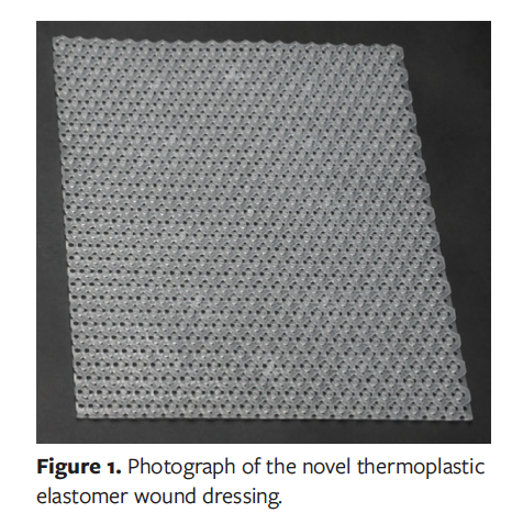
A new wound contact layer made from thermoplastic elastomer (TPE), Prevent (Clear Choice Therapeutics, Inc; hereafter “TPE dressing” or “novel TPE dressing”), has been cleared by the US Food and Drug Administration.13 It was developed to specifically address and overcome the aforementioned limitations of other types of wound contact layers.13 The TPE dressing has multiple potential clinical benefits. Its wound contact layer is transparent, nonabsorptive, pliable, and nonporous, thus preventing tissue ingrowth, reducing dressing pain and/or odor related to removal at dressing changes, and enabling extended therapy intervals between dressing changes. The TPE design is noncompressible and has markedly improved flow characteristics, which enable its clinical use with lower NP settings (−50 mm Hg) while still effectively removing exudate. However, the novel TPE dressing’s ability to facilitate macrodeformation comparable to that of traditional ROCF dressings under NP has not been tested. Given the generally accepted belief that macrodeformation is a key mechanism of action in NPWT, it is important to evaluate the macrodeformation capability of any new NPWT wound filler.
The purpose of the present study was to compare the degree of wound contraction achieved using the novel TPE dressing with that of a traditional ROCF dressing and a negative control with no wound filler during NPWT in a porcine explant model across different wound depths and several NP conditions.
Materials and Methods
Specimen preparation and wound creation
Five full-thickness dorsal porchetta cuts were harvested from mature standard land swine (approximately 100 kg body weight). Specimens were kept refrigerated (not frozen) until the time of testing, then brought to room temperature. Each porcine quarter measured approximately 12 to 18 in cranial to caudal, 18 in dorsal to volar, and 3 in deep to include underlying musculature.
The TPE wound dressings used in the present study were prepared in standardized sizes corresponding to wound dimensions (Figure 1).
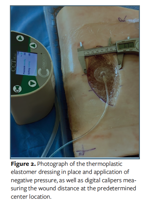
A 15-cm × 7.5-cm elliptical paper template was used to standardize wound creation and dressing dimensions. Using this template, an elliptical wound was excised in the center of each specimen using a scalpel and pickups. The initial wound depth was approximately 0.5 cm to 1 cm (shallow, subcutaneous, or full dermal). After completion of shallow wound testing, the wound was deepened to 3 cm to 4 cm (deep, intramuscular). Three measuring points were marked across each wound: one central, one 3.75 cm cranial, and one 3.75 cm caudal. The width of each wound was recorded at all 3 locations before dressing placement to establish baseline wound dimensions (Figure 2).
Dressing application
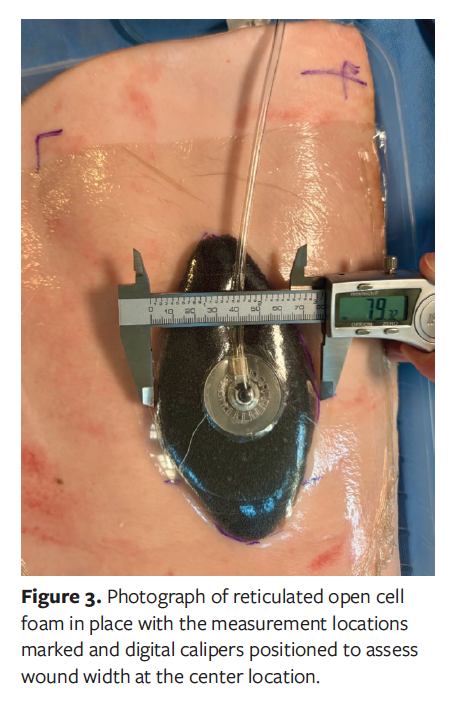
Three types of NPWT dressings were tested: ROCF, the novel TPE dressing, and a no-filler control (drape and dome only). Each dressing was cut to the same 15-cm × 7.5-cm elliptical dimensions as the wound. Dressings were applied sequentially to each wound. After placement, an adhesive film was applied to create an airtight seal. For the no-filler control, no material was inserted into the wound prior to sealing. A 1-cm × 1-cm hole was then cut over the wound center, and suction tubing was connected to a pump to apply NP.
NP experimental design
NP was applied at 3 clinically relevant settings: −50 mm Hg, −80 mm Hg, and −125 mm Hg. For each pressure and dressing condition, 3 consecutive trials were performed. In each trial, NP was applied for approximately 2 minutes until a steady state was reached, followed by release of pressure and disconnection of the tubing. At each pressure setting, wound width was measured at the central, cranial, and caudal positions using digital calipers accurate to 0.1 mm.
Wound contraction and interaction with the wound filler were analyzed at 4 time points: (1) prior to dressing placement and prior to NP, representing the original wound width, (2) after dressing placement but prior to NP, (3) during active NP, and (4) after NP was discontinued while the dressing remained in place. Because dressing placement can widen the wound cavity, the measurement obtained after dressing placement but before NP does not represent the true baseline wound width for assessing wound contraction produced by the NPWT dressing. Therefore, wound contraction was defined as the difference between the original wound width at time point 1 and the wound width during active NP at time point 3, in order to capture true macrodeformation independent of dressing-induced widening. Alpha level less than .05 was considered statistically significant.
This sequence was repeated for all 3 dressing types at both wound depths. After completion of shallow wound testing, wounds were deepened to 3 cm to 4 cm, and the full protocol was repeated. In total, 810 measurements were collected, calculated as follows: 5 specimens × 2 wound depths × 3 dressing types × 3 pressure settings × 3 measurement positions × 3 repeated trials = 810 measurements.
Results
Overall, the TPE dressing resulted in similar wound macrodeformation in shallow wounds and greater macrodeformation in deep wounds compared with traditional ROCF. Other variables significantly associated with increased wound contraction included higher levels of NP, measurement position within the wound (ie, central measurements were larger than cranial/caudal measurements), and the initial wound width prior to NP application.
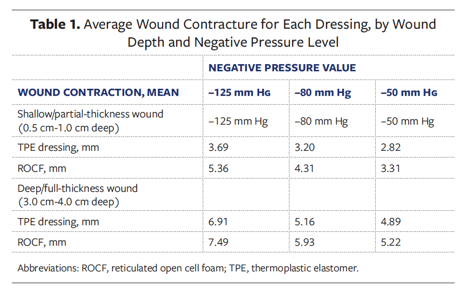
This explant study generated a total of 810 discrete wound measurements, obtained across 5 porcine specimens at varying wound depths, NP settings, and dressing conditions. Measurements were performed at 3 wound positions (center, cranial/right, and caudal/left) at 3 NP levels (−50 mm Hg, −80 mm Hg, and −125 mm Hg) for each filler type (TPE, ROCF, or no filler). The mean contraction values for each dressing at each NP level are presented in Table 1.
The experimental setup, including the wound template, measurement points, and NPWT dressing application, is shown in Figure 2 and Figure 3.
Shallow wounds
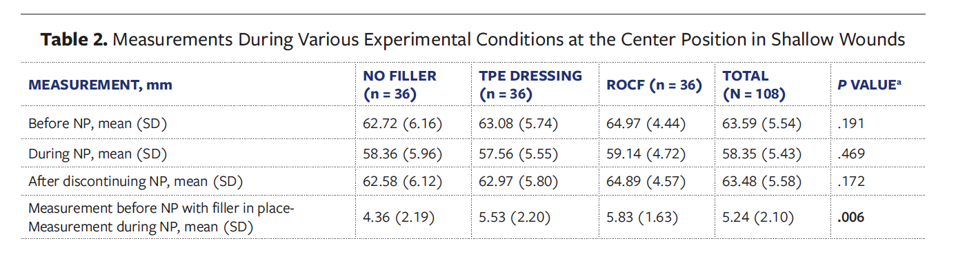
Measurements at the center position of shallow wounds were collected and aggregated for various wound filler and NP conditions and are presented in Table 2. This was also done for the cephalad and caudal measurements. There was a significant difference between filler conditions in wound width before and after NP applications across all 3 positions (cephalad, caudal, center) on univariable analysis (mean, SD measurements for no filler, TPE, and ROCFs at center position 4.36, 2.19, and 5.53, 2.20, and 5.83, 1.63 mm, P < .05).
On multivariate analysis, in shallow wounds only, both the TPE dressing and ROCF demonstrated a statistically significant increase in wound contraction (negative estimate [standard error], −1.58 [0.38] and −2.98 [0.38], respectively; P < .05) compared with no filler when controlling for measurement position, degree of NP, and original wound width. Measurement position, higher NP, and smaller original wound width were each independently associated with greater wound contraction.
Deep wounds
Measurements at the center position of deep wounds were collected and aggregated for various wound filler and NP conditions and are presented in Table 3. There was a significant difference between filler conditions with regard to wound width before NP application with filler in place compared with after NP application in the center position (at the center position, mean [SD] measurements 4.50 [1.91] mm for no filler, 5.83 [1.58] mm for TPE, and 5.53 [1.92] mm for ROCFs [P < .05]). At higher NP, the TPE dressing trends toward greater contraction compared with either ROCF or no wound filler.
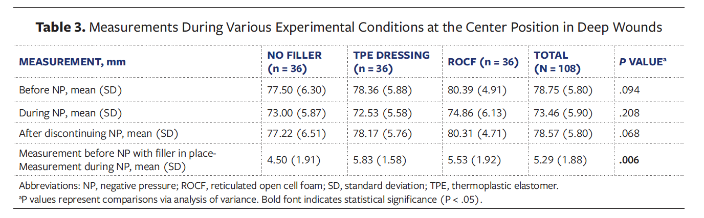
On multivariable analysis, in deep wounds only, the TPE dressing demonstrated a statistically significant increase in wound contraction (greater negative estimate [standard error], −0.72 [0.35]; P = .038) compared with no filler when controlling for measurement position, degree of NP, and original wound width. Greater applied NP and smaller initial wound width were also independently associated with increased wound contraction when controlling for measurement position.
Multivariable analysis
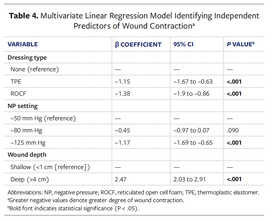
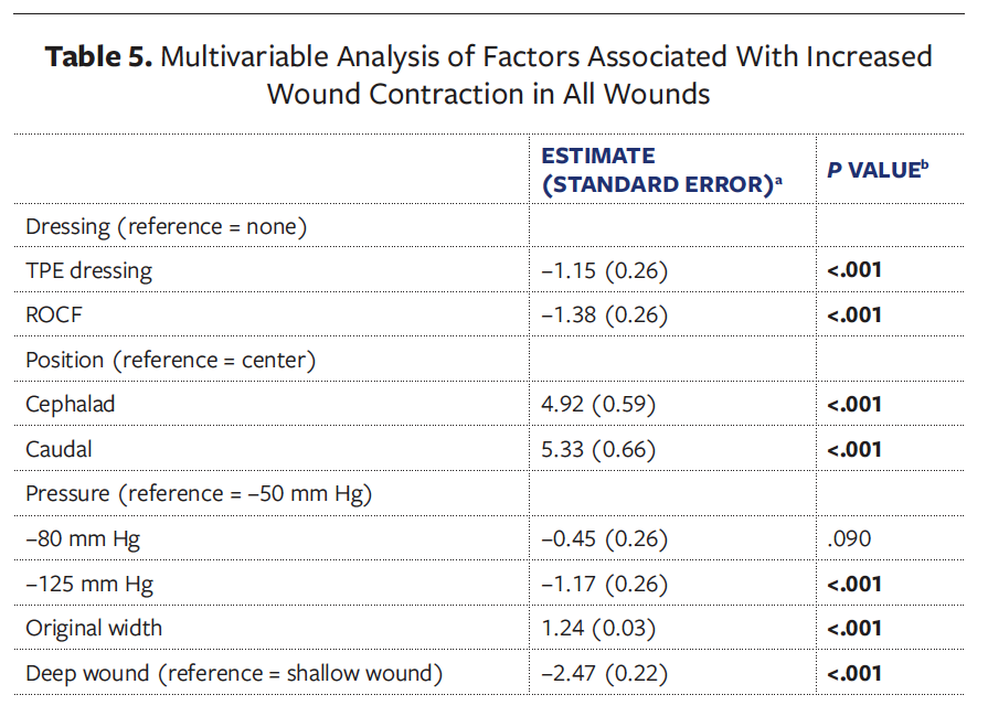
A final multivariable analysis combining shallow and deep wounds showed that both the TPE dressing and ROCF produced statistically significant increases in wound contraction (estimates of −1.15 and −1.38, respectively; P < .05) vs no filler when controlling for measurement position, NP level, and original wound width and depth. Measurements taken at the center of the wound, higher NP, and deeper wounds were each independently associated with greater contraction. The full model is presented in Table 4 and Table 5.
Discussion
NPWT systems have emerged as a mainstay of therapy for a variety of wounds, including large defects that cannot be managed with primary closure, as well as chronic or nonhealing wounds.14,15 The goal of the current study was to compare macrodeformation, or wound contraction, using a novel TPE dressing, a traditional ROCF, or no wound filler in a porcine model at several wound and NP conditions. To adequately assess the performance and versatility of all 3 dressings, varying wound depths and varying levels of NP were used. This diverse set of experimental conditions facilitated assessment of uniquely optimal conditions for both ROCF and the novel TPE dressing performance.
Overall, the findings indicate that in deep wounds, the TPE dressing is associated with a statistically significant increase in wound contraction compared with no filler. Because no established minimal clinically important difference exists for wound contraction, the clinical relevance of these small differences is uncertain. Multivariable analysis additionally demonstrated that deep wounds were associated with an overall greater degree of wound contraction compared with shallow wounds. Contracture was significantly greater at NP of −125 mm Hg than at lower NP of −50 mm Hg. Both of these findings are consistent with previous studies on wound contracture.2,5-8,16,17
From a clinical relevance perspective, the present study demonstrates that while there are statistically significant differences in the magnitude of wound contraction for different fillers under different conditions (which often favored the TPE dressing), the absolute (average) magnitude of this difference is very low (<3 mm for any condition [Table 1], which represents <4% of the original width of the wound). Although a minimal clinically important difference has not been previously established, it would seem that this low a difference in contraction does not rise to the level of being clinically impactful. Therefore, the best application of the results of the present study on clinical practice is that there is no clinically relevant difference in the degree of macrodeformation generated by the TPE or ROCF dressings and that the TPE dressing is noninferior to ROCF dressings with regard to this specific metric related to wound healing.3,18 Additionally, the contracture seen with the TPE dressing at low NP (−50 mm Hg and −80 mm Hg) are not statistically or clinically different from ROCF at traditional NP (−125 mm Hg).
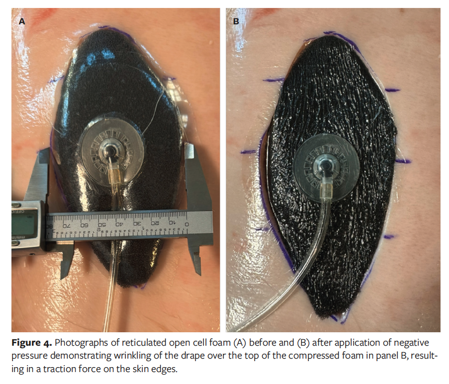
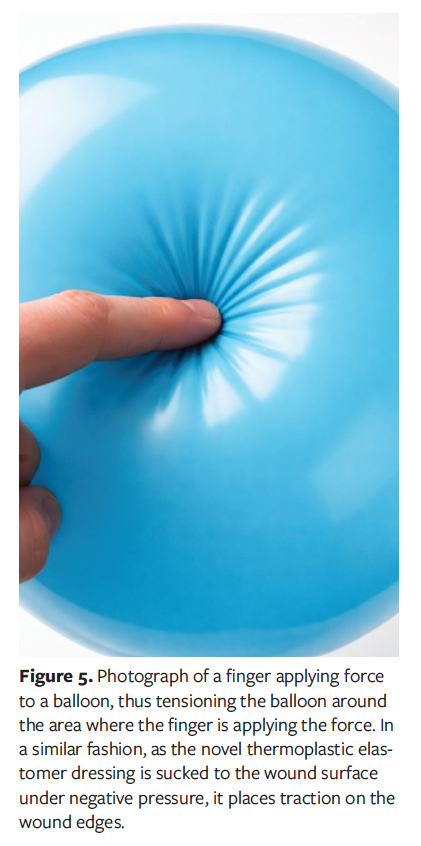
The mechanism of action for the 2 dressings is likely different based on the physical characteristics of the material from which the dressings are made. ROCF allows for up to 80% compression when placed under NP of −125 mm Hg.5 As the foam compresses, the drape that is attached to the foam is pulled tight, resulting in a wrinkling of the drape over the top of the foam (Figure 4). By contrast, the TPE dressing does not compress when NP is applied. However, the TPE dressing measures only 6 mm in depth, whereas typical ROCF can be up to 3 cm in height. The low durometer TPE dressing conforms to the wound surface and is pushed into the wound cavity by the downward force of the NP. As the dressing is sucked into the wound, the drape is pulled into the mouth of the wound, applying traction to the wound edges similar to the effect of pushing a finger into a balloon (Figure 5). The deeper the wound, the more traction is placed on the edges.
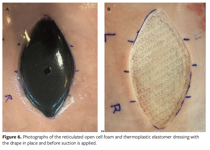
Lastly, a previously unreported occurrence was noted with placement of the ROCF dressing. There was a small but reproducible expansion of the wound from initial measurements before the dressing was placed and after the ROCF was placed, but before NP was applied.The average wound expansion across all wounds was 1.77 mm for ROCF vs 0.39 mm for the TPE dressing. The wound expansion is likely associated with ROCF recoil as the drape is placed. The drape places some small level of compression of the ROCF, causing an outward or expansive force on the wound edges. Because the TPE dressing does not compress and is only 6 mm in height, this phenomenon was not observed with the TPE (Figure 6).
Limitations
The current study has limitations that are important to consider. First, although a porcine model is a reasonable surrogate and has been previously described in the literature, there may be differences in the quality of human and porcine cadaveric skin and soft tissue that may result in these data lacking external validity in human models.7,16 Future research should seek to better describe the performance of the TPE dressing vs conventional wound fillers in in vivo models. Nonetheless, these data provide an important initial exploration of this topic. Second, the experiments were performed on a limited number of specimens (n = 5); however, despite this sample size limitation, significant differences between the TPE dressing and traditional wound filler conditions with regard to wound contraction were detected. Therefore, overall, these data provide important information regarding various options for wound filler type, as well as a foundation for future studies examining the relative performance of these various wound filler types.
Conclusion
The current explant study provides important insight into the performance of a novel TPE dressing in facilitating wound contraction and macrodeformation during NPWT. Wound contraction was influenced by wound depth and the amount of NP applied. Even at low NP (−50 mm Hg), the TPE dressing achieved wound contraction equivalent to or greater than ROCF, especially in deep wounds, confirming that the nonporous and noncollapsible structure of the TPE dressing does not impede macrodeformation. Macrodeformation in each dressing likely occurs through different mechanisms of action. These findings indicate the need for additional studies.
Author and Public Information
Authors: Colton C. Mowers, MD1; Joe H. Hilsman, BS2; Meera M. Dhodapkar, MD, MHS3; Michael S. Shuler, Jr4; James Coleman Fleming2; Mohamed Sarraj, MD5; Brett A. Freedman, MD3; and Michael S. Shuler, Sr, MD4
Affiliations: 1Rush University Medical Center, Chicago, IL, USA; 2University of Georgia, Athens, GA, USA; 3Department of Orthopedic Surgery, Mayo Clinic, Rochester, MN, USA; 4Athens Orthopedic Clinic, Athens, GA, USA; 5Department of Neurologic Surgery, Mayo Clinic, Rochester, MN, USA
Disclosure: Drs Michael S. Shuler, Sr and Brett A. Freedman are cofounders of Clear Choice Therapeutics, the company that developed the novel TPE dressing evaluated in this study. Their spouses own shares in the company
Ethics Statement: The porcine tissue used in the study was obtained via commercial sources. No ethics approval required.
Correspondence: Michael S. Shuler, MD; Athens Orthopedic Clinic, 1765 Old West Broad St, Athens, GA 30606; Mshuler@ccthera.com
Manuscript Accepted: June 8, 2026.
References
1. Argenta LC, Morykwas MJ, Marks MW, DeFranzo AJ, Molnar JA, David LR: Vacuum-assisted closure: state of clinic art. Plast Reconstr Surg. 2006;117(7 Suppl):127S-142S. doi:10.1097/01.prs.0000222551.10793.51
2. Borgquist O, Ingemansson R, Malmsjö M: Wound edge microvascular blood flow during negative-pressure wound therapy: examining the effects of pressures from −10 to −175 mmHg. Plast Reconstr Surg. 2010;125(2):502-509. doi:10.1097/PRS.0b013e3181c82e1f
3. Shuler MS, Mowers CC, Dhodapkar MM, Greenberg M, Ngendahimana D, Freedman BA: Contact pressure under traditional foam and novel thermoplastic elastomer negative pressure wound therapy interfaces. Wound Repair Regen. 2026;34(2):e70157. doi:10.1111/wrr.70157
4. Kairinos N, Voogd AM, Botha PH, et al: Negative-pressure wound therapy II: negative-pressure wound therapy and increased perfusion. Just an illusion? Plast Reconstr Surg. 2009;123(2):601-612. doi:10.1097/PRS.0b013e318196b97b
5. Borgquist O, Ingemansson R, Malmsjö M: The influence of low and high pressure levels during negative-pressure wound therapy on wound contraction and fluid evacuation. Plast Reconstr Surg. 2011;127(2):551-559. doi:10.1097/PRS.0b013e3181fed52a
6. Torbrand C, Anesäter E, Borgquist O, Malmsjö M: Mechanical effects of negative pressure wound therapy on abdominal wounds – effects of different pressures and wound fillers. Int Wound J. 2017;15(1):24-28. doi:10.1111/iwj.12810
7. Anesäter E, Borgquist O, Hedström E, Waga J, Ingemansson R, Malmsjö M: The influence of different sizes and types of wound fillers on wound contraction and tissue pressure during negative pressure wound therapy. Int Wound J. 2011;8(4):336-342. doi:10.1111/j.1742-481X.2011.00790.x
8. Borgquist O, Gustafsson L, Ingemansson R, Malmsjö M: Micro- and macromechanical effects on the wound bed of negative pressure wound therapy using gauze and foam. Ann Plast Surg. 2010;64(6):789-793. doi:10.1097/SAP.0b013e3181ba578a
9. Glass GE, Murphy GF, Esmaeili A, Lai LM, Nanchahal J. Systematic review of molecular mechanism of action of negative-pressure wound therapy. Br J Surg. 2014 Dec;101(13):1627-36. doi: 10.1002/bjs.9636. Epub 2014 Oct 8. PMID: 25294112.
10. Putnis S, Khan WS, Wong JM-L: Negative pressure wound therapy - a review of its uses in orthopaedic trauma. Open Orthop J. 2014;8:142-147. doi:10.2174/1874325001408010142
11. Borgquist O, Gustafson L, Ingemansson R, Malmsjo M: Tissue ingrowth into foam but not into gauze during negative pressure wound therapy. Wounds. 2009;21(11):302-309.
12. Upton D, Andrews A: Pain and trauma in negative pressure wound therapy: a review. Int Wound J. 2015;12(1):100-105. doi:10.1111/iwj.12059
13. Clear Choice Theraputics. Accessed July 27, 2026. https://clearchoicetherapeutics.com
14. Webster J, Liu Z, Norman G, et al: Negative pressure wound therapy for surgical wounds healing by primary closure. Cochrane Database Syst Rev. 2019;3(3):CD009261. doi:10.1002/14651858.CD009261.pub4
15. Ludolph I, Fried FW, Kneppe K, Arkudas A, Schmitz M, Horch RE: Negative pressure wound treatment with computer-controlled irrigation/instillation decreases bacterial load in contaminated wounds and facilitates wound closure. Int Wound J. 2018;15(6):978-984. doi:10.1111/iwj.12958
16. Torbrand C, Ugander M, Engblom H, Arheden H, Ingemansson R, Malmsjö M: Wound contraction and macro-deformation during negative pressure therapy of sternotomy wounds. J Cardiothorac Surg. 2010;5:75. doi:10.1186/1749-8090-5-75
17. Orgill DP, Manders EK, Sumpio BE, et al: The mechanisms of action of vacuum assisted closure: more to learn. Surg. 2009;146(1):40-51. doi:10.1016/j.surg.2009.02.002
18. Mowers CC, Shuler MS, Freedman BA. Negative pressure wound therapy and the perfusion paradox: reconciling clinical success with foundational principles of tissue healing. Int J Orthop Trauma. 2026;2(1):1-2.