Precision PCI in the Contemporary Era: NIRS-IVUS Optimized PCI
© 2026 HMP Global. All Rights Reserved.
Any views and opinions expressed are those of the author(s) and/or participants and do not necessarily reflect the views, policy, or position of Cath Lab Digest or HMP Global, their employees, and affiliates.
Rushi Parikh, MD, FACC, FSCAI
Associate Clinical Professor of Medicine/Cardiology
Director, Interventional Cardiology Research
Clinical Director, UCLA Biodesign
David Geffen School of Medicine at UCLA,
Los Angeles, California
This case is supported by Nipro Medical.
Case Presentation
A 68-year-old man with hypertension and hyperlipidemia (untreated) presented with crescendo angina over the past week and resting chest pressure radiating to the jaw and left arm. Physical exam and electrocardiogram were unremarkable.
Pertinent labs included an LDL of 139 mg/dL, lipoprotein(a) of 10 mg/dL, hemoglobin A1C of 5.8%, and a troponin I peak of 1.48 ng/mL (normal <0.04 ng/mL). An echocardiogram showed normal left ventricular ejection fraction with no regional wall motion abnormalities. The patient was initiated on aspirin, ticagrelor, atorvastatin, and an intravenous heparin infusion, and urgent cardiac catheterization in the setting of non-ST elevation myocardial infarction (NSTEMI) was performed.
Diagnostic Evaluation
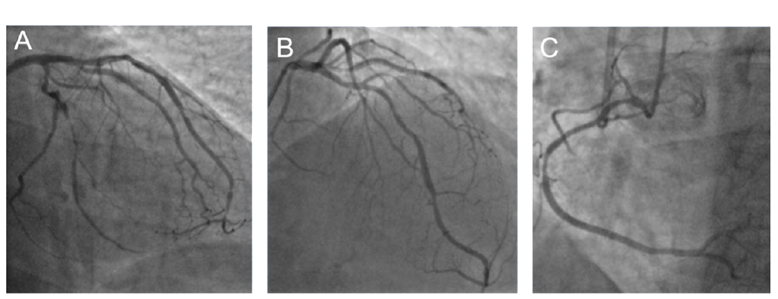
Coronary angiography showed 90% proximal stenosis of the obtuse marginal 2 (OM2) consistent with acute plaque rupture and 70-80% severe diffuse mid-OM2 disease distal to the culprit lesion. The proximal-mid left circumflex (LCx) had moderate 50-60% disease, while OM1 was a moderate-caliber vessel without significant disease (Figure 1A). The left anterior descending (LAD) (Figure 1B) and right coronary artery (RCA) (Figure 1C) had moderate disease.
Percutaneous Coronary Intervention (PCI)
Access: 6 French right radial artery
Wire: Sion Blue (Asahi Intecc)
Pre-dilation: 2.5 x 20 mm semi-compliant balloon
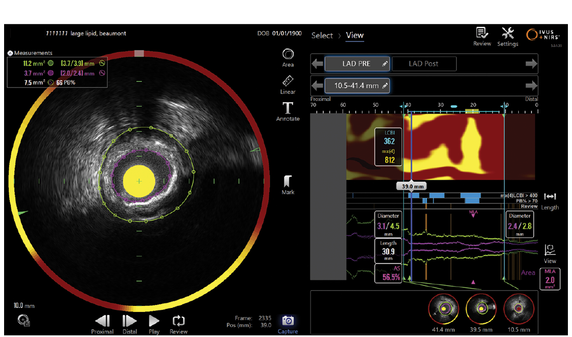
Near infrared spectroscopy-intravascular ultrasound (NIRS-IVUS) pre-stent: Proximal and distal reference vessel diameter (media-media) were 4.0 mm and 2.7 mm, respectively. No significant calcium was observed. The culprit lesion had a plaque burden of 78% and minimum lumen area (MLA) of 1.6 mm² (Figure 2, left-hand panel); the lipid core burden index (LCBI 4-mm segment) was 892, and there was additional lipid-rich plaque in the proximal LCx (Figure 2, right-hand panel).
Stents: Overlapping 3.0 x 28 mm and 2.5 x 33 mm drug-eluting stents (proximal to distal) were placed from the ostial LCx to the mid OM2, post-dilated from 2.5 mm distally gradually to 3.75 mm proximally.
Post-PCI Data
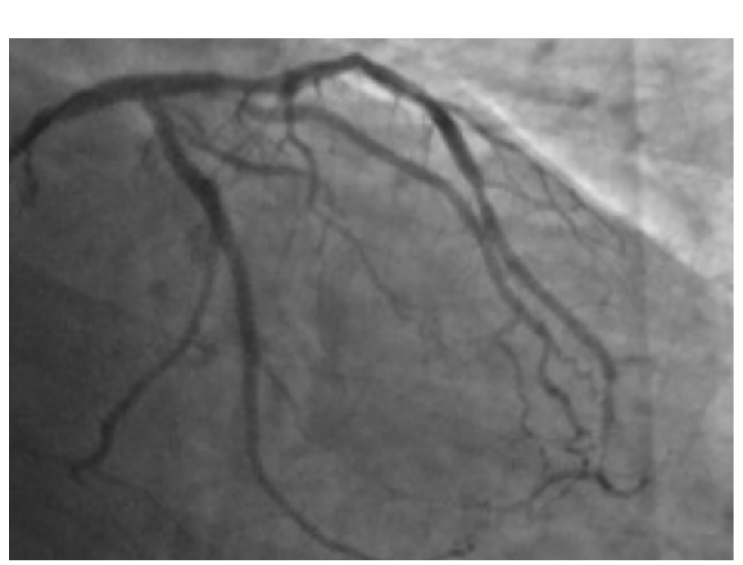
Post-PCI IVUS showed stents were well-expanded and well-apposed. Final angiography demonstrated no residual stenosis in the LCx-OM2 system (Figure 3). Physiologic assessment of the non-culprit vessels showed a fractional flow reserve (FFR) of 0.89 in the LAD and 0.91 in the RCA.
The Impact of NIRS-IVUS on Clinical Decision-Making
The Makoto HD NIRS-IVUS (Nipro Medical) supported a more precise and optimized PCI strategy in several ways:
1. Its exceptional resolution and visualization provided enhanced operator confidence in balloon and stent sizing.
2. Optimized stent landing zones (i.e., covering lipid-rich plaque, if feasible): Lipid-rich plaque in the stent margins is associated with a higher risk of in-stent restenosis. Thus, the overlapping proximal stent was adequately sized to ensure coverage of the lipid-rich plaque in the proximal LCx.
3. Selective escalation of anti-lipid therapy: Given the patient’s very high LCBI and evidence of moderate plaque throughout the entire coronary tree, aggressive lipid-lowering therapy with a PCSK9 inhibitor in addition to a statin was initiated.
Find More
Beyond Angiography: The Power of IVUS + NIRS in PCI
CLD talks with Dhananjay Chatterjee, MD
UCLA Health, Los Angeles, California