From Patient Selection to 3D ICE: Best Practices From a High-Volume LAAO Program
© 2026 HMP Global. All Rights Reserved.
Any views and opinions expressed are those of the author(s) and/or participants and do not necessarily reflect the views, policy, or position of EP Lab Digest or HMP Global, their employees, and affiliates.
EP LAB DIGEST. 2026;26(11).
Interview by Jodie Elrod
In this interview, Alok Gambhir, MD, discusses the structured approach that helped Northside Hospital Heart Institute’s high-volume left atrial appendage occlusion (LAAO) program surpass 2000 implants. He also shares how standardized workflows and 3D intracardiac echocardiography (ICE) have expanded access and improved efficiency, as well as how new devices and imaging technologies may advance LAAO care.
Can you start with a brief introduction about yourself?
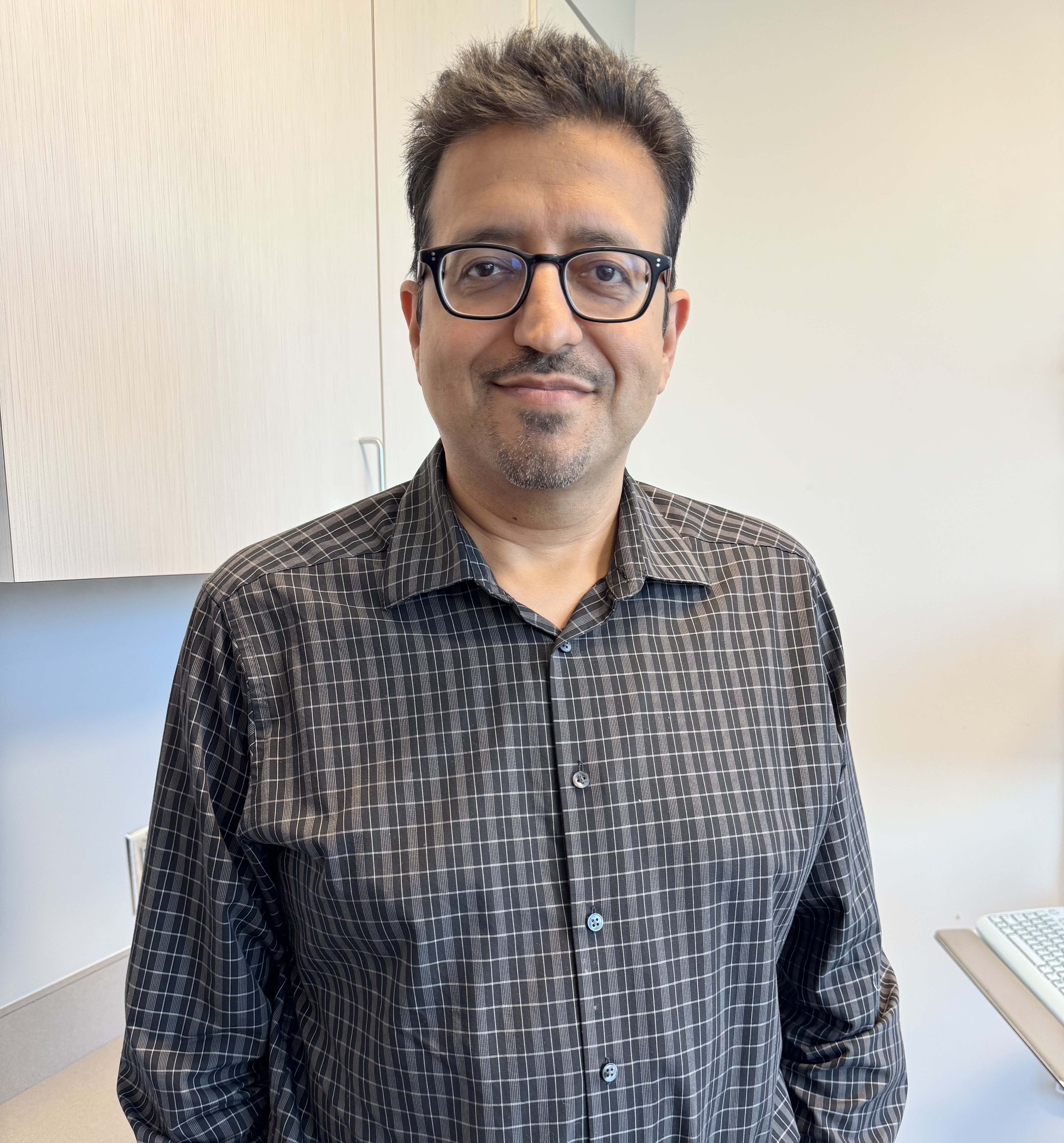 My name is Alok Gambhir. I am a cardiac electrophysiologist, the section chief of cardiac electrophysiology (EP) and executive medical director of EP at Northside Hospital Heart Institute.
What do you think have been the biggest drivers of your program’s sustained growth and consistently strong outcomes?
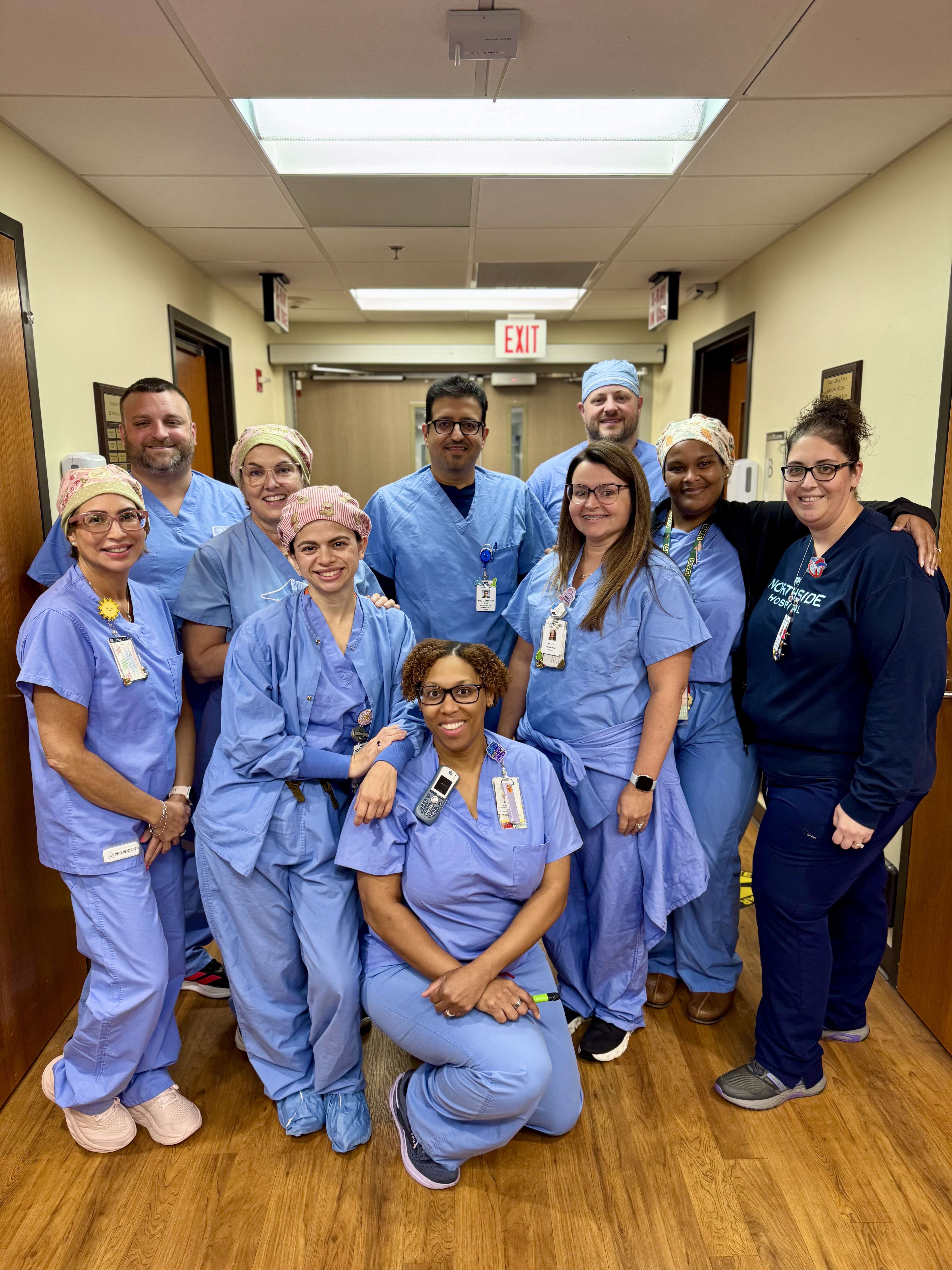
Reaching this 2000-procedure milestone has been very important for us. I would say our biggest driver of growth has really been democratizing the procedure while maintaining safety and strong outcomes. Rather than concentrating LAAO procedures in the hands of 2 to 4 operators, we have developed a program in which 10 physicians can perform the procedure. As a result, we perform 50 to 60 of these procedures per month, and that volume continues to grow—not because of any individual operator, but because of the program as a whole.
The key to this democratization is having a very structured framework. We standardized our approach to patient selection, preprocedural evaluation, imaging, the implant itself, and postprocedural management. We have a clear framework outlining what is expected of everyone.
Once we established those expectations, we began evaluating everything based on quality and efficiency. That has allowed us to expand access because we are not limited to one geographic location. We have more than 100 cardiologists across multiple locations, which expands our reach and awareness without sacrificing outcomes.
When you think about democratizing the procedure among multiple physicians, the most important thing is having a highly experienced operator base who can take on the more difficult cases. For example, someone might not be able to perform a procedure effectively with 3D ICE, but ICE is more appropriate for a patient. There may be a failed implant due to challenging anatomy. These cases could be referred to more experienced operators. We learn from each other, and the more experienced operators take on or assist with the complex cases. Eventually, when you perform this many cases, there are going to be some peri-device leaks. You must have a subset of physicians who are very experienced at managing these leaks. For those difficult cases, we have narrowed it down to certain operators.
We set up the program to position every physician for success, but sometimes a physician is unable to build their practice within that framework. If that happens, we can pivot to some of our younger proceduralists, give them an opportunity to grow their practice, and help them understand our expectations so they can develop within the program. I think that approach really helps. As long as you have a framework centered on quality and efficiency, you can build a successful program with multiple operators.
As director of a high-volume LAAO program, what best practices or workflow changes have had the greatest impact on patient selection, procedural efficiency, and team success?
We have streamlined and standardized the entire patient journey, not just the procedure itself. That journey begins with patient selection and the shared decision-making process. We use the American College of Cardiology’s CardioSmart shared decision-making tool, a validated booklet provided to each patient. The tool also includes an online video that patients can watch with their family members. This gives patients and their families the opportunity to understand stroke risk, anticoagulation, and LAAO within a broader framework, while allowing us to provide a consistent message during shared decision-making. We do not simply use a document that we check off once shared decision-making is complete, but use this one standardized tool for all of our patients.
We also have some standardization around preprocedural imaging. Some patients come from far away, and while we prefer every patient to undergo preprocedural imaging, that is not always feasible. We take the broader perspective that we do not want to require imaging for every patient, but we recognize that some patients may have had their LAA clipped or oversewn during an open-heart procedure. We have found sometimes there is no note in a surgical operative report of oversewing the LAA, but it was actually performed. Therefore, we require preprocedural imaging for any patient who has undergone an open-heart procedure, even if they are coming from far away. Overall, we try to standardize preprocedural imaging for as many patients as possible.
After that, it is about standardizing the workflow at the time of the procedure, including whether procedures are performed with TEE or 3D ICE imaging and which patients are appropriate for each approach. Some physicians are more comfortable performing procedures using only TEE. We determine the imaging need for the upcoming week for all our procedures: TEE or 3D ICE based on patient- and operator-specific needs. When we do perform TEE, we have a select group of noninvasive imaging physicians and a select group of cardiac anesthesiologist imagers.
How has 3D ICE changed your approach to imaging and navigation during LAAO compared with traditional TEE guidance?
I would say 3D ICE has significantly changed how we think about intraprocedural imaging for our LAAO procedures. We mainly use the NUVISION ultrasound catheter with the Vivid system (GE HealthCare) and sometimes use Philips ICE imaging.
Traditional TEE is an excellent modality that provides great information, and as an operator, your hands are free to focus on the procedure with another physician in the room. However, coordinating and reserving time with another physician can be challenging. We primarily work with cardiac anesthesiologists, who are excellent imagers. We also have a select group of structural imaging physicians who do a great job with more complex structural cases and assist with peri-device leaks and similar issues.
So, 3D ICE has significantly improved efficiency for us because we are not relying on another operator to be there. That is even more important for our rapidly growing number of concomitant procedures. You cannot have an imager there the entire time while you perform the transseptal puncture and the atrial fibrillation portion of the procedure and then move on to the LAA. The timing is also variable because you do not know when the ablation component will be finished. Having the ability to use a 3D ICE catheter has therefore been a game changer for us. Almost all of our concomitant procedures within our system are performed with 3D ICE. It provides an efficient workflow and allows us to visualize the LAA well and assess for any leaks.
Compared with TEE, 3D ICE is also more comfortable for the patient because there is no need to place a tube down the esophagus, reducing the risk of complications. Certain patients are also better suited for ICE, including those at high risk of bleeding or with esophageal varices, in whom placing a TEE probe could potentially cause bleeding. For those patients, it is safer to perform the procedure with 3D ICE.
Looking ahead, where do you see LAAO evolving over the next 5 years, and what opportunities are you most excited about for improving stroke prevention and patient care?
With the CHAMPION trial results, there are mixed feelings in the EP community regarding the utility of the results. Where I am excited about further growth in LAAO is with concomitant procedures. The OPTION trial certainly suggests that we can perform these procedures safely and effectively, and we are seeing that in real-world settings as well. As the devices become better, and with the big push to perform more procedures in ambulatory surgical centers (ASCs), I think we are going to see a subset of LAA closure procedures expand into the ambulatory surgical environment or even into nonsurgical hospitals—those without open-heart surgery programs. That could expand care for many patients, who would not have to travel as far to undergo their procedures. Based on what we have with current technology, I think we can safely treat a substantial subset of patients at noncardiac surgical hospitals and ASCs. I think these procedures have become much safer than ever before, and there will be an evolution in that direction.
Another consideration, beyond safety, is the regulatory aspect. The national coverage decision still calls for these procedures to be performed in hospitals with open-heart surgical backup. I think there will be some regulatory changes as the technology continues to improve.
The other major area I see is improvement in devices. Device-related thrombus is something I believe we are now very adept at managing. With the postprocedural protocol of using half-dose direct oral anticoagulation and improvement in implant technique, this has become an extremely rare event in our experience. However, we all still deal with peri-device leaks. We have learned that even small peri-device leaks may not be completely benign. We used to think the cutoff was perhaps 5 millimeters; now, we are saying it should be 3 millimeters. But I think most of us believe that what we really want is a complete seal since many of the leaks are more slit-like leaks. We have not yet achieved that consistently with our devices.
So, I’m really excited about newer technologies, including the WATCHMAN Omni (Boston Scientific) device and the Amulet 360 Left Atrial Appendage Occluder (Abbott), which will most likely receive FDA approval very soon. Having these options, which could provide a better seal and help us achieve zero leaks, is where I think we want to be with these devices. Once we can safely reach that point with zero leaks, this procedure will be used even more often in the right patients.
As we continue to also improve 3D ICE and preprocedural imaging, that will help guide us in determining which device is right for each patient so we can achieve a full seal. n
Disclosures: Dr Weiner has completed and returned the ICMJE Form for Disclosure of Potential Conflicts of Interest. He reports consulting fees from Boston Scientific, Abbott, and Zywie; payment or honoraria for lectures, presentations, speakers’ bureaus, manuscript writing, or educational events from Boston Scientific; and participation on a Data Safety Monitoring Board or Advisory Board for Boston Scientific and Abbott.
Related Content