Baptist Health Lexington
© 2026 HMP Global. All Rights Reserved.
Any views and opinions expressed are those of the author(s) and/or participants and do not necessarily reflect the views, policy, or position of EP Lab Digest or HMP Global, their employees, and affiliates.
EP LAB DIGEST. 2026;26(8):14-22.
Gery Tomassoni, MD, and Rachelle Whitaker, RT(R)(CT)
Lexington, Kentucky
When was the cardiac electrophysiology (EP) program started at your institution, and by whom?
Michael Rukavina, MD, performed the first catheter ablation at our institution in 1993. Subsequently, the EP program including dedicated EP lab personnel was established in 1999 under the leadership of Gery Tomassoni, MD. Since its inception, the program has expanded significantly and has grown into one of the busiest EP labs in Kentucky. It continues on a strong trajectory of growth to meet the needs of an ever-increasing population of patients requiring advanced arrhythmia management.
What drove the need to implement an EP program?
The decision to implement a cardiac EP program was driven by the growing recognition that our patient population needed more advanced, specialized care for complex arrhythmias. Prior to the program’s establishment, patients often had limited local access to arrhythmia evaluation and treatment, resulting in delays in care or the need to travel significant distances for specialized procedures. Developing an EP program allowed the institution to expand its cardiovascular services, improve timely access to advanced therapies such as ablation and device implantation, and meet the increasing demand for arrhythmia management within the region.
What is the size of your EP lab facility?
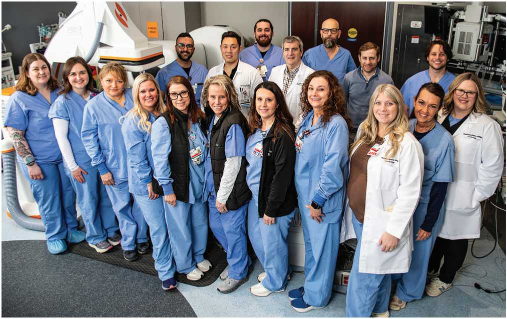
Our EP facility currently includes 3 dedicated EP labs and 1 swing lab shared with the cardiac catheterization lab. This configuration provides both dedicated EP capacity and flexible procedural space to support our high volume program. In addition to the current footprint, the institution has active plans to add a fourth dedicated EP lab.
Does your institution offer EP-related procedures in an ambulatory surgical center (ASC)?
Our institution presently performs all EP procedures within the hospital setting, which utilizes an outpatient ambulatory center for appropriate same-day patient care and recovery. We are currently evaluating the need and location for an ASC for EP.
Who manages your EP laboratory, and what is the mix of credentials and experience?
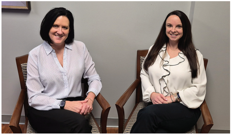
The EP laboratory is managed by Rachelle Whitaker, RT(R)(CT), who oversees daily operations, staffing, workflow coordination, and quality initiatives. With 25 years of EP experience, Rachelle has been instrumental in guiding the program’s growth and development since its early stages. She is supported by Ruth Johnson, BSN, RN, the Invasive Cardiology Director, who brings 20 years of experience in this specialty area and provides strategic leadership and program oversight.
The clinical team is further strengthened by Brandy Reed, BSN, RN, CV BC, the charge nurse, who has 12 years of EP experience. Brandy manages day-to-day clinical workflow, patient care activities, and procedural readiness, ensuring smooth and efficient operations.
What is the number of staff members?
The EP lab is staffed by a multidisciplinary team of 10 registered nurses (RNs) and 8 radiologic technologists (RTs). Many team members bring extensive experience in EP, cardiac catheterization, and advanced cardiovascular procedures.
What types of procedures are performed at your facility?
Our EP program provides a comprehensive range of advanced arrhythmia procedures, including:
- Catheter ablations for atrial and ventricular arrhythmias, utilizing radiofrequency (RF), cryoablation, and pulsed field ablation (PFA) technology
- Stereotaxis-guided supraventricular tachycardia (SVT) and ventricular tachycardia (VT) ablations
- Advanced mapping technologies, including Vektor Medical and Volta Medical
- Device implantation, including the transvenous implantable cardioverter-defibrillator (ICD) and subcutaneous ICD (S-ICD), transvenous and leadless pacemakers, and implantable loop recorders (ILRs)
- Cardiac device extractions
- Both stand-alone and concomitant left atrial appendage occlusion (LAAO)/ablation procedures
- Cardiac resynchronization therapy (CRT) implantation and management
- Phrenic nerve stimulation for central sleep apnea
- Cardiac contractility modulation (CCM) therapy
- Internal cardioversion for rhythm restoration in select arrhythmia patients
Approximately how many ablations (for all arrhythmias), device implants, lead extractions, and LAAOs are performed each week?
Our EP program maintains a high procedural volume each week. On average, the team performs:
- 25-30 catheter ablations for both atrial and ventricular arrhythmias
- 20-25 device implants, including ICDs, pacemakers (transvenous and leadless), CRT devices, and ILRs
- LAAO procedures performed during dedicated LAAO days, held 6 to 8 times per month, with 4 to 6 procedures per session
- 2 blocked days each month for device/lead extractions
What types of EP equipment are commonly used in the lab?
Our EP laboratory utilizes a wide range of advanced equipment, catheters, devices, and mapping technologies from leading industry manufacturers. Commonly used systems and products include:
- Carto 3 (Johnson & Johnson MedTech) 3D electroanatomic mapping system
- EnSite X EP System with EnSite Omnipolar Technology (OT) (Abbott)
- RHYTHMIA HDx high density mapping system (Boston Scientific)
- Genesis robotic magnetic navigation (RMN) system (Stereotaxis)
- AI-enhanced mapping technology for arrhythmia localization (Vektor Medical)
- AI-driven mapping and signal interpretation platform (Volta Medical)
- Affera™ Mapping and Ablation System with the Sphere-9 catheter (Medtronic)
- ARTIS Q.zen fluoroscopy (Siemens), with Prucka with CardioLab recording system (GE HealthCare) and Bloom 2 stimulator (Fischer Medical)
Ablation and diagnostic catheters:
- ThermoCool SmartTouch (Johnson & Johnson MedTech) ablation catheters and PFA-compatible catheters
- TactiCath Contact Force Ablation Catheter, Sensor Enabled, and FlexAbility Ablation Catheter (Abbott)
- IntellaNav and IntellaMap Orion catheters, plus FARAPULSE™ PFA system (Boston Scientific)
- Arctic Front cryoballoon catheters, Affera Mapping and Ablation System, and Sphere-9 catheter
Device manufacturers (implants and extractions):
- ICDs, CRT devices, and pacemakers from Medtronic, including the Micra leadless pacemaker
- ICDs, CRT devices, the Confirm Rx insertable cardiac monitor (ICM), and pacemakers from Abbott, including Aveir single- and dual-chamber leadless pacemakers
- Pacemakers, ICDs, S-ICDs, CRT devices, and ICMs from Boston Scientific, including the LUX-Dx ICM
- Pacemakers, ICDs, CRT devices, and implantable monitoring systems from Biotronik
- Lead extraction tools from Philips, including the GlideLight and TightRail
- Evolution system (Cook Medical)
Specialty therapies:
- remedē System (ZOLL Medical Corporation) – phrenic nerve stimulation for central sleep apnea
- Optimizer Smart (Impulse Dynamics) device that delivers CCM therapy
What new technologies or techniques have been introduced in your lab, and how have they changed procedures?
Our EP laboratory has incorporated several new technologies and techniques that have significantly enhanced procedural efficiency, precision, and patient throughput.
One of the most impactful advancements has been the adoption of PFA for AF cases. Presently, we utilize the FARAPULSE, Afferra, VARIPULSE™ (Johnson & Johnson MedTech), and Volt™ (Abbott) PFA systems. These technologies have shortened procedural times and enabled us to increase the number of patients scheduled each day without compromising safety or quality.
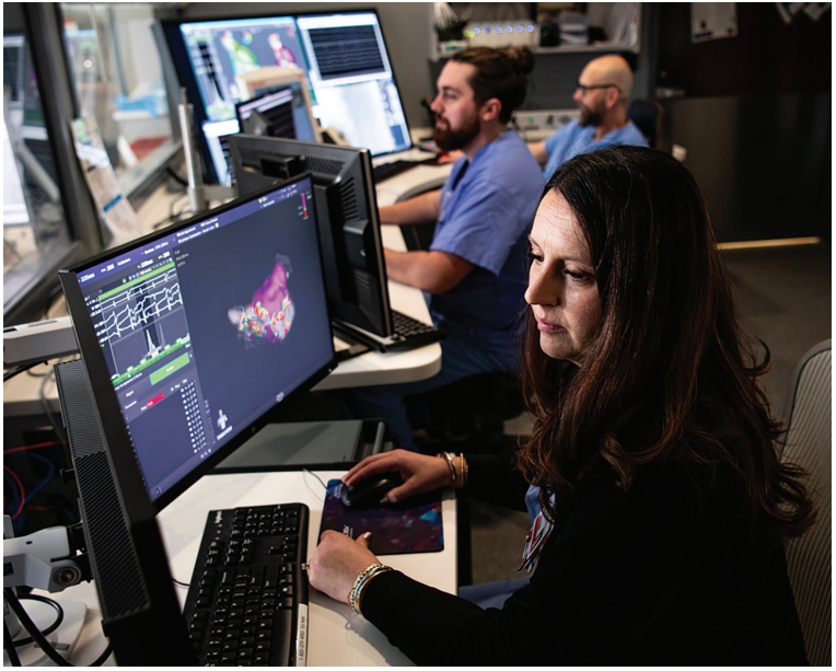
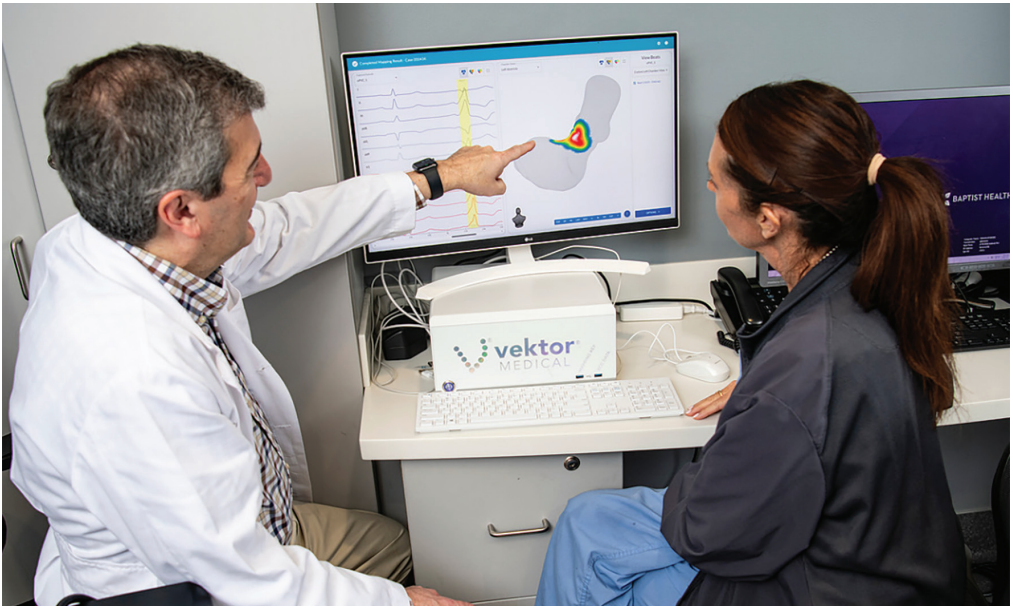
We have also expanded our use of Vektor Medical mapping from premature ventricular contractions (PVCs)/VT ablations to atrial arrhythmia cases as well. Vektor’s rapid signal analysis and mapping capabilities have improved arrhythmia localization and streamlined case workflows.
Discuss your technique for preventing esophageal injury during AF ablation.
We employ multiple strategies to minimize the risk of esophageal injury during AF ablation, tailored to the energy source being used.
For RF ablation, we utilize either the ensoETM (Haemonetics) system to provide proactive cooling or the esolution device (S4 Medical) to mechanically deflect the esophagus away from the posterior LA. In cases where the cooling catheter is not used, continuous esophageal temperature monitoring is used to ensure early detection of temperature rise and guide real-time adjustments in power delivery, catheter position, or lesion duration.
With the transition to PFA, our approach has evolved. Because PFA is associated with less risk of esophageal injury, our focus has shifted away from the use of esophageal catheters to close monitoring of the number and distribution of applications.
Tell us about your use of RMN.
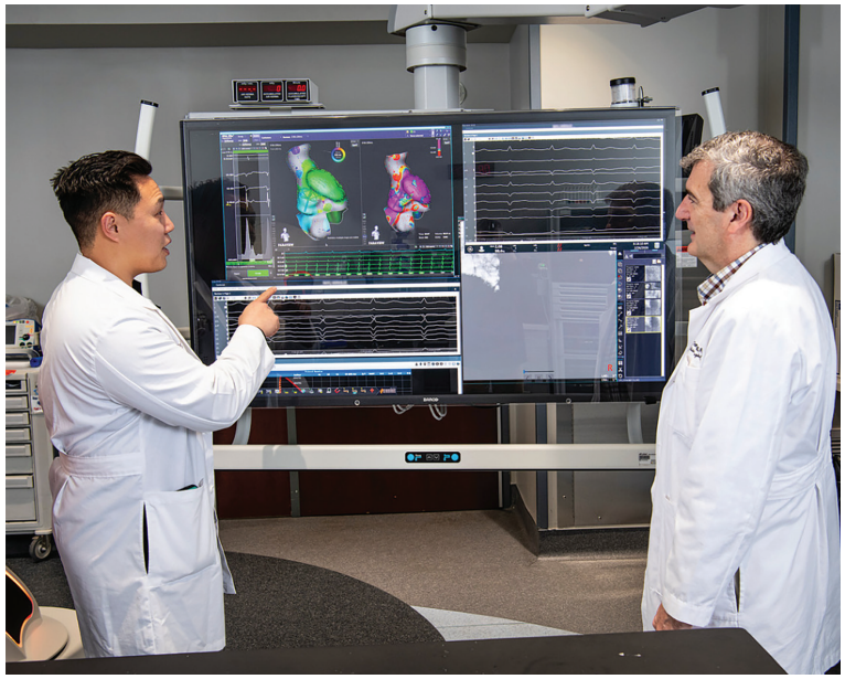
We performed our first RMN catheter ablation in 2004. Presently, the Genesis RMN system (Stereotaxis) is utilized in one of our dedicated EP labs. The system is especially helpful in PVC/complex VT ablations, where the enhanced catheter stability and precise navigation provide significant advantages.
How is inventory managed in your laboratory?
Inventory is managed through a dedicated, structured process. We have 2 full-time inventory management specialists who support both the EP and cath labs. They oversee ordering, stocking, tracking, and rotation of all EP supplies, including catheters, devices, mapping system disposables, and specialty equipment. This shared model allows for efficient coordination across both service lines while supporting the high procedural volume of our EP program.
Tell us about your device clinic.
Our device clinic operates with a structured, team-based model designed to ensure timely follow-up, accurate device monitoring, and comprehensive patient support. The clinic is staffed by a combination of 2 specially-trained device nurses, 1 device technician, 1 administrative assistant, and EP physicians/advanced practice clinicians who work together to manage both in-person and remote device evaluations. We also utilize a hybrid home/clinic work structure that assists in decreasing interruptions, increases productivity, and improves job satisfaction.
Day-to-day operations include routine device interrogations, remote monitoring review, troubleshooting alerts, and coordinating follow-up for patients with pacemakers, ICDs, CRT devices, or ILRs. Staff members manage incoming transmissions, triage actionable findings, and communicate directly with patients and providers to ensure continuity of care.
Our clinic uses Murj, an industry standard device management platform and remote monitoring software, to track transmissions, document findings, and maintain accurate records integrated with Epic.
Discuss your approach to remote monitoring of arrhythmias.
Our device clinic team reviews alerts daily and prioritizes actionable findings with the assistance of our remote monitoring platform, Murj. This platform helps organize high-priority transmissions related to arrhythmias or device function, allowing both staff and physicians to triage them promptly and ensure timely follow-up. Murj also supports the growing volume of data from cardiovascular implantable electronic devices by providing tools for triage, communication, advisories and recalls, patient compliance, heart failure (HF) monitoring, in-office interrogation, physician compliance, and a wide range of analytics—enabling a comprehensive remote monitoring program.
Tell us what a typical day might be like in your EP lab.
A typical day is fast paced, highly coordinated, and consistently busy. Our team includes 4 EP physicians, with 2 to 3 physicians working in the lab on most days. When 2 physicians are scheduled, we aim to staff 3 active EP labs, allowing us to run parallel cases and maximize efficiency.
The office team frequently schedules device implants between ablation cases, and we often flip physicians between 2 labs to maintain momentum and minimize downtime. This approach allows us to keep cases moving smoothly while ensuring that each lab is utilized to its fullest capacity.
Add on procedures are accommodated daily, integrated into the schedule as clinical needs arise.
Can you describe the extent and use of vascular closure devices? Tell us about your approach for same-day discharge (SDD).
The EP physicians use a variety of vascular closure devices to support safe and efficient postprocedural recovery. Commonly used closure methods include Perclose (Abbott), Mynx Control (Cordis), and Vascade (Haemonetics) vascular closure devices, along with figure-of-8 sutures when preferred by the operator.
These closure strategies play an important role in maintaining a SDD rate of ~95%. By using reliable closure devices and standardized postprocedural monitoring, we are able to mobilize patients sooner, reduce recovery area time, and safely discharge them home the same day.
Has your lab recently undergone a national accrediting inspection?
Our program is accredited by the American College of Cardiology in both EP and AF, including a recently completed second round of accreditation. We also have a dedicated accreditation coordinator who oversees compliance, data reporting, and ongoing quality initiatives to ensure we consistently meet and exceed national standards.
How do you ensure timely case starts and patient turnover?
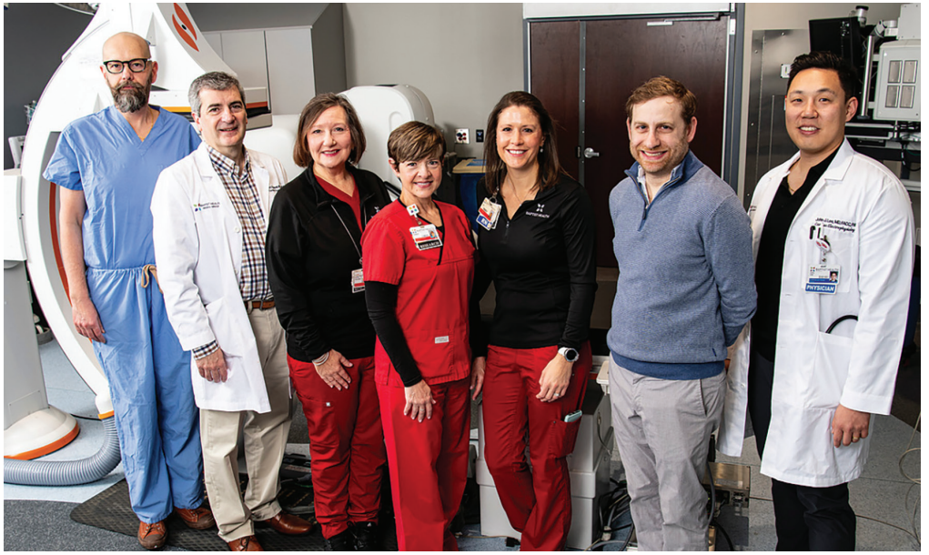
We closely track case start times and routinely review opportunities for improvement to keep the schedule running smoothly. Our charge nurse conducts a daily coordination call with the office scheduling team to ensure that the next day’s cases are organized in a way that maximizes efficiency for physicians, staff, and patients.
We are often fortunate to have a floating staff member available to support room turnover, provide break and lunch coverage, and assist wherever needed. This additional flexibility helps maintain momentum throughout the day and contributes to consistently efficient transitions between cases.
How does your laboratory schedule team members for call?
Our EP laboratory rarely requires call coverage, but when call is needed, we begin by requesting volunteers, allowing team members to sign up based on their availability and preferences. Call responsibilities typically amount to only a few days per year and are coordinated around holidays and the physicians’ personal schedules to ensure fairness and adequate coverage.
Do you have flexible or multiple shifts? How do you handle slow periods?
Our EP laboratory offers flexible staffing with multiple shift options to support workflow demands while promoting staff work–life balance. Team members work a combination of 10-hour and 12-hour shifts, while the charge nurse works an 8-hour schedule. We also use self scheduling, which allows staff to build schedules that align with both personal needs and operational coverage requirements.
During slower periods, we maintain productivity and staff engagement through several strategies. Many team members are cross-trained to the cath lab and frequently pick up shifts there when EP volume is lighter. Others use this time to complete required education, competencies, and professional development activities, ensuring ongoing readiness and continued skill advancement.
How are vendor visits managed?
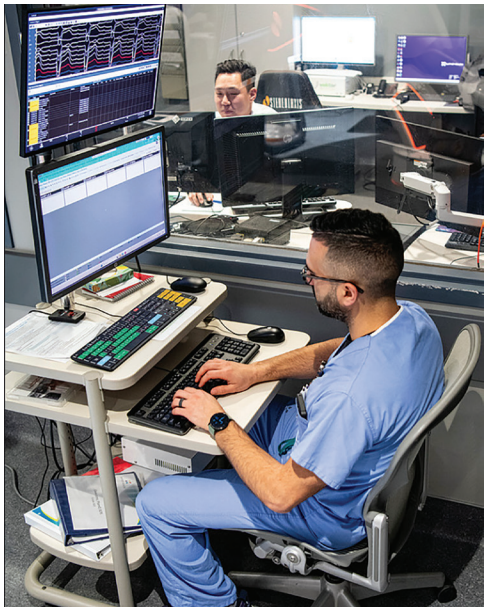
Vendor access to our EP laboratory is tightly controlled to ensure safety, compliance, and minimal disruption to clinical operations. All vendor activity is managed through Green Security, which requires representatives to request appointments in advance. These requests must be approved by management before a visit is permitted.
Once on-site, vendors are required to check in to ensure proper credentialing, documentation, and tracking.
What are the best features of your EP laboratory’s layout or design?
One of the best features of our EP laboratory is its central core layout, which is positioned close to both our preprocedural and postprocedural areas. This design allows for smooth patient flow, efficient communication, and quick transitions between phases of care. Our supply areas are conveniently located within this core, making it easy for staff to access equipment and disposables without disrupting case progress.
We also have a designated charge desk within the central core that supports real-time coordination, structured team assignments, and rapid response to changing needs throughout the day. Additionally, our parking garage is located directly parallel to the lab, providing easy access for staff and physicians and helping streamline arrival and departure during busy shifts.
What measures has your laboratory implemented to cut or contain costs?
We routinely send eligible catheters for reprocessing, which significantly reduces disposable supply expenses. We also leverage bulk purchasing to secure more favorable pricing on high-use items.
Each morning, we review our first case on-start time (FCOST) metrics, which are tracked for efficiency and used to identify opportunities to improve workflow, reduce delays, and optimize resource utilization. By focusing on both supply management and operational efficiency, we support responsible cost stewardship and long-term financial sustainability for the program.
What quality control measures are practiced in your laboratory?
Our EP laboratory utilizes a prominently displayed huddle board that is updated daily, weekly, and monthly to keep the entire team informed and aligned with current performance metrics and improvement goals.
We actively track a wide range of quality indicators, including hand hygiene compliance, time-out adherence, universal protocol compliance, fire risk assessments, medication handoff accuracy, documentation compliance of American Society of Anesthesiology physical status and Mallampati (airway) scores, preprocedural and postprocedural pain assessments, staff turnover metrics, missed break tracking, and productivity index.
By reviewing these metrics regularly and engaging the team in discussions around performance, we ensure that our laboratory consistently meets high standards for patient safety, workflow efficiency, and overall quality of care.
What works well in your laboratory for onboarding new team members?
New RN team members begin with a 12-week sedation orientation, during which they work as part of a 4-person team to learn workflow, safety protocols, and procedural expectations. After completing this phase, they transition to a 3-person team structure for an additional 6 weeks, always paired with another experienced nurse. This extended period provides added support and ensures safety as they continue developing their skills before moving on to monitor training.
New RT team members typically complete a 10- to 12-week scrub orientation, beginning as part of a 3-person team to learn sterile technique, equipment handling, and procedural flow. Once they have established a strong foundation in the scrub role, they also progress to monitor training.
What continuing education (CE) opportunities are provided for staff members? How do staff typically maintain and renew credentials?
Our EP laboratory supports ongoing professional development by providing multiple CE opportunities throughout the year. Many of our industry partners and vendors offer on-site education sessions, allowing staff to stay current with new technologies, best practices, and evolving procedural techniques.
While it is ultimately each staff member’s individual responsibility to obtain and maintain their required credentials, we make a strong effort to ensure that ample CE opportunities are available. This includes vendor-led education, in-house training sessions, and access to hospital-supported learning resources.
Discuss the role of mid-level practitioners in your laboratory.
Mid-level practitioners play an important supportive role within our EP program. They assist physicians by seeing patients in preop, evaluating consults throughout the hospital, and seeing patients in the office as part of ongoing outpatient care. While they contribute significantly to patient assessment and continuity of care, they do not hold procedural privileges within the EP laboratory.
This structure allows mid-level providers to enhance workflow efficiency and patient access while ensuring that all invasive procedures are performed by credentialed EP physicians.
Share a memorable case from your EP laboratory and how it was addressed.
One memorable and impactful case in our EP lab involved a patient who experienced an unexpected poor outcome following a device lead extraction. This event highlighted opportunities to strengthen our approach to patient safety during complex extraction procedures.
In response, our EP physicians, management team, administration, staff, and cardiothoracic (CT) surgeons came together to collaboratively redesign our extraction protocol. This multidisciplinary effort resulted in a more structured, safety-focused process that continues to be followed today. The updated protocol has led to significantly improved patient outcomes, enhanced team readiness, and a more coordinated approach to managing high-risk cases.
Does your lab use a third party for reprocessing or catheter recycling? How has it impacted your lab?
Yes, our EP laboratory utilizes third-party reprocessing services through Stryker Sustainability Solutions and Sterilmed. Partnering with these vendors has had a positive impact on our lab by reducing supply costs, decreasing waste, and supporting more sustainable resource utilization. Reprocessed devices that meet safety and quality standards allow us to maintain high-quality patient care while managing expenses responsibly.
Discuss your program’s approach to conduction system pacing (CSP).
Our program recognizes the growing evidence supporting CSP—particularly left bundle branch area pacing—as strategies that preserve physiologic activation and may reduce pacing-induced cardiomyopathy in appropriate patients.
Our physicians evaluate each case individually to determine whether CSP is the most beneficial option based on patient anatomy, underlying conduction disease, and long-term pacing needs. When CSP is selected, the team follows a structured workflow that emphasizes careful mapping, precise lead placement, and thorough intraprocedural testing to ensure stable capture and optimal thresholds.
Tell us about your primary approach for LAAO.
Our program’s approach to LAAO is supported by a structured workflow and a strong emphasis on patient coordination. A key component of this process is our designated LAAO nurse navigator, who plays a central role in guiding patients through evaluation, preprocedural imaging, scheduling, education, and follow-up. This dedicated position ensures continuity, enhances communication among the care team, and helps patients move smoothly through each step of the LAAO pathway.
The nurse navigator works closely with physicians, imaging teams, and procedural staff to maintain consistency in patient selection, preparation, and postprocedural monitoring.
Does your program have a dedicated AF clinic and is it considered an AF Center of Excellence?
Yes, our EP program has a dedicated AF clinic and is recognized as an AF Center of Excellence.
Our AF clinic has significantly improved patient access by offering rapid appointments and timely evaluations, which has helped reduce unnecessary emergency department (ED) visits. Patients benefit from quick symptom assessment, medication adjustments, and expedited follow-up after procedures such as ablation or cardioversion.
A key strength of our program is the involvement of a designated AF nurse navigator. This role supports patient education, scheduling, communication with providers, and adherence to evidence-based treatment pathways.
We have also developed clear goals for our AF program, including:
- Improving symptom control and quality of life
- Reducing AF-related hospitalizations and ED visits
- Ensuring timely access to specialized care
- Standardizing treatment pathways to promote consistency and safety
- Enhancing long-term rhythm management and stroke prevention strategies
Our comprehensive care team includes EP physicians, nurse practitioners, the AF nurse navigator, RNs, pharmacists, imaging specialists, and outpatient cardiology partners. This multidisciplinary structure ensures that each patient receives individualized, coordinated care.
Discuss your approach to lead extraction and management.
We utilize a risk-based grading system to determine the appropriate setting and level of support required for each case. High-risk extractions are performed in our hybrid operating room with a CT surgeon immediately available for backup, ensuring rapid intervention capability if complications arise. Low-risk extractions are performed in our EP laboratory, where staff are trained and equipped to manage routine extraction procedures safely and efficiently.
Our physicians bring a range of perspectives to lead management, with some taking a more proactive or aggressive approach to extraction and others favoring a more conservative strategy. This diversity of clinical judgment allows for individualized decision-making tailored to each patient’s history, device needs, and long-term risk profile. Collaborative discussion among the EP team ensures that each case is evaluated thoroughly and managed according to best practices and patient-specific considerations.
Discuss your program’s approach to lifestyle risk factor modification (RFM) for reduction of AF. What impact has this had on patient outcomes, and what best practices can you offer to other programs?
Our EP program incorporates lifestyle RFM as a central component of AF management. We use structured care pathways that address obesity, weight loss, physical activity, smoking cessation, alcohol moderation, hypertension, sleep apnea, and other comorbidities known to influence AF burden. Each patient receives clear education on these factors, supported by our AF nurse navigator, who reinforces goals, coordinates referrals, and helps patients stay engaged in their care plan.
We partner closely with primary care, nutrition services, sleep medicine, and cardiology to ensure that RFM is consistent and comprehensive. Patients are encouraged to achieve weight loss targets, safely increase physical activity, monitor blood pressure at home, and reduce alcohol and tobacco use. These expectations are introduced early—often at the time of diagnosis—and revisited throughout follow-up.
Discuss your approach to treatment of AF in patients with HF.
Generally, our EP physicians are very aggressive in achieving and maintaining sinus rhythm in patients with HF, especially those with heart failure with reduced ejection fraction. Based on data from previous ablation trials, our approach is to pursue AF ablation early (within 1 year of diagnosis) in this patient population. In our experience, successful AF ablation can result in improved functional status, increased left ventricular ejection fraction, and better cardiovascular outcomes.
How does your EP laboratory handle radiation protection for physicians and staff?
Radiation protection for physicians and staff is a top priority in our EP laboratory. All team members are provided with properly fitted lead, and staff may request protective eyewear and radiation-shielding scrub caps for additional safety. We conduct biannual lead inspections and require all staff to wear dosimeters, which are monitored monthly to track individual exposure.
We also have a radiation physicist who plays an important role in our safety program by monitoring exposure trends, providing education to new staff, and reinforcing best practices for minimizing radiation risk. Our fluoroscopy units are regularly maintained and kept up to date, with settings optimized to remain as low as reasonably achievable while still supporting procedural needs.
What approaches has your lab taken to reduce fluoroscopy time? What percentage of cases are done without fluoroscopy? How do you record fluoroscopy times/dosages?
Our EP laboratory has implemented several strategies to reduce fluoroscopy exposure for both patients and staff. The use of advanced 3D mapping systems has significantly decreased our reliance on fluoroscopy for ablations, and our fluoroscopy units are consistently operated at low-dose settings to minimize radiation whenever imaging is required.
Also, cases are performed with zero fluoroscopy, though this varies by physician based on training, comfort level, and case complexity. Regardless of technique, each patient’s fluoroscopy time and radiation dose are documented individually in the procedural log, allowing for accurate tracking and ongoing quality monitoring.
What are some of the dominant trends you see emerging in the practice of EP?
One of the dominant trends we are seeing in the practice of EP is the increasing influence of lifestyle-related risk factors on arrhythmia development. Conditions such as obesity, sleep apnea, alcohol use, and drug use are becoming more prevalent and are contributing to higher arrhythmia burdens across our patient population.
We are also seeing a noticeable rise in younger patients presenting with AF and other rhythm disorders. Many of these individuals have modifiable risk factors that are driving earlier onset of disease, underscoring the importance of early intervention and comprehensive RFM.
In addition, our program has observed an increase in ventricular arrhythmias, often linked to underlying structural heart disease, cardiomyopathy, or lifestyle-related contributors. This trend has reinforced the need for timely evaluation, advanced imaging, and coordinated care pathways to ensure early detection and appropriate management.
Is your EP lab involved in clinical research?
Our EP program is highly engaged in clinical research since the inception of our Clinical Research Center in 1999. We are presently enrolling patients in AF ablation trials (IMPRoVED-AF, COHERENT-AF, OPTIMIZE), device trials (SYNCHRONICITY, AIM HIGHer, BACKBEAT), and LAAO trials (SIMPLAAFY, LAAOS-4).
We are fortunate to have a dedicated research team exclusively focused on EP, allowing us to support complex protocols, maintain high-quality data collection, and ensure patients have access to innovative therapies and technologies. This commitment to research strengthens our program, enhances patient care, and keeps us at the forefront of advancements in EP.
What is considered historic about your EP program or hospital? Has your program or hospital recently experienced any “firsts”?
Our EP program has achieved many “firsts” in our history. Central Baptist Hospital was the first to enroll a patient in the LAAO PREVAIL trial in United States in 2010. Our EP physicians also performed the first RMN catheter ablation of SVT using the Niobe magnetic system (Stereotaxis). In addition, our program received the first AF accreditation in the United States. Finally, the world’s first Nanostim leadless pacemaker (Abbott) in the LEADLESS II IDE trial was implanted at Central Baptist Hospital.
Describe your city or general regional area. How is it unique?
Lexington, Kentucky, is unique because it blends Southern tradition with modern growth. Widely known as the “Horse Capital of the World,” the city is surrounded by scenic bluegrass farms and a strong equine culture. It is also part of Kentucky’s bourbon heritage, with distilleries and tasting experiences woven into the area’s identity. The University of Kentucky adds energy and pride—especially during basketball season—while local restaurants, festivals, and historic neighborhoods give the city a friendly, community-focused feel. Lexington offers a balance of rural beauty, cultural richness, and urban convenience, making it a distinctive place to live.
Please tell our readers what you consider special about your EP laboratory and staff.
Our EP laboratory is special because of the unique blend of personalities and talents that come together to form a truly cohesive team. The staff work exceptionally well together, creating an environment that feels more like family than a workplace. We function much like a well-coordinated pit crew—efficient, focused, and always aligned around one priority: patient safety.
What sets our team apart is not only how smoothly we operate during procedures, but also how much we genuinely enjoy one another’s company. That camaraderie extends beyond the lab, where we regularly celebrate each other’s milestones, joys, and life events. This strong sense of connection and mutual support creates a positive culture that enhances teamwork, communication, and the overall care we provide to our patients.