





Dermoscopy With UV Imaging May Help Differentiate Mycosis Fungoides, Psoriasis, and Atopic Dermatitis
Dermoscopy combined with ultraviolet (UV) imaging may help distinguish early-stage mycosis fungoides (MF), psoriasis, and atopic dermatitis (AD) by identifying disease-specific vascular and scaling patterns, according to findings from a prospective cohort study.
Investigators evaluated 159 patients, including 55 with MF, 53 with psoriasis, and 51 with AD. Participants underwent conventional dermoscopy and UV imaging to assess vascular patterns, scaling characteristics, background color, structureless areas, and vessel density per square millimeter.
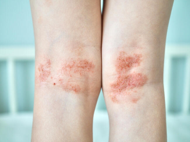
The study identified distinct dermoscopic features across the 3 conditions, which can present with overlapping clinical manifestations and contribute to diagnostic delays.
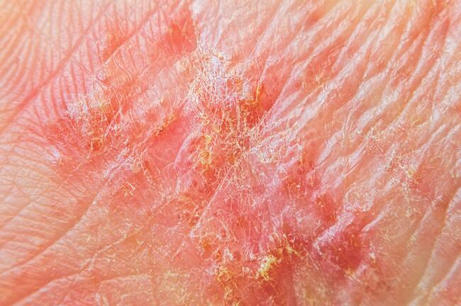
MF was characterized by linear and spermatozoa-like vessels, scaling along skin furrows, focal structureless areas, and the lowest vessel density among the 3 conditions.
Psoriasis demonstrated uniformly distributed dotted vessels against a bright-red background, accompanied by diffuse scaling and the highest vessel density.
In contrast, AD primarily exhibited dotted vessels with additional linear vessels, yellow-orange scaling, ulceration, dystrophic hairs, and intermediate vessel density.
UV imaging provided additional diagnostic information by improving visualization of structureless areas and scaling edges, potentially helping clinicians recognize features that distinguish these inflammatory and neoplastic conditions.
The findings suggest that combining conventional dermoscopy with UV imaging may provide a useful noninvasive approach when evaluating patients with clinically similar skin lesions.
However, the investigators acknowledged several limitations, including the single-center study design, relatively small sample size, and restriction to early-stage disease. These factors may limit the generalizability of the findings to broader patient populations and more advanced presentations.
“This approach may support early, noninvasive differentiation,” the authors wrote.
Reference
Balan K, Lallas A, Akdogan N. Patterns and pitfalls: dermoscopic differentiation of early-stage mycosis fungoides, atopic dermatitis, and psoriasis. J Am Acad Dermatol. 2026;94(4):1193-1198. doi:10.1016/j.jaad.2025.12.049