





Embolization of Hypervascular Tumors Using the Embrace® Hydrogel Embolic System
Introduction
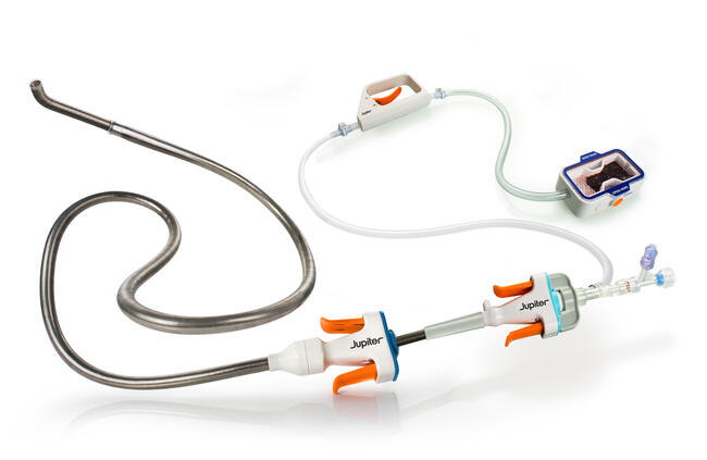
Embrace® Hydrogel Embolic System (HES, Instylla) consists of 2 low-viscosity liquid precursors that crosslink intravascularly when simultaneously injected through a coaxial microcatheter system, forming a soft, water-based polyethylene glycol hydrogel. Embrace HES is intended to embolize hypervascular tumors in peripheral arteries ≤ 5 mm. The hydrogel is nonadhesive, eliminating the possibility of catheter entrapment, contains no dimethyl sulfoxide, and does not require sizing to the vessel diameter.
Case 1: Enlarging Liver Neuroendocrine Tumor
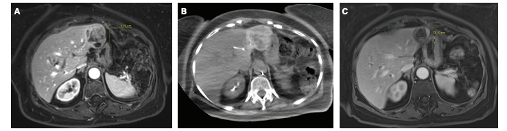
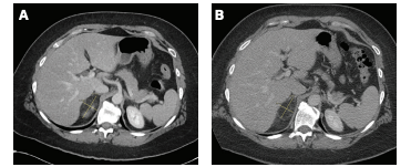
A 78-year-old female with a history of adenocarcinoma was initially diagnosed in 2013. She had a complete response to treatment; however, in 2023 she developed new liver lesions on surveillance imaging. The largest tumor was biopsied, which showed a well-differentiated neuroendocrine tumor. The lesions remained stable over subsequent exams until the middle of 2025, when the segment III lesion started to grow up to a size of 5.25 cm over subsequent exams (see Figure 1A and 1B). The decision was made at Tumor Board to perform liver-directed therapy to the progressive enlarging lesion.
In 2026, the patient underwent transarterial embolization (TAE) with Embrace HES under total intravenous anesthesia. Access was gained via a left distal radial approach. A 2.8F 150 cm Progreat® microcatheter (Terumo) was placed in a distal segment 3 branch of the left hepatic artery. The Embrace HES was prepared using Omnipaque™ 350 contrast (GE Healthcare), and the coaxial catheter system was assembled by placing the 1.7F 162 cm Instylla microcatheter through the Progreat microcatheter. The embolic was delivered to vessel stasis. The coaxial system was retracted and additional embolic was delivered until all vessels supplying the tumor were occluded. A total of 6.2 mL of hydrogel was delivered to achieve complete vessel stasis.
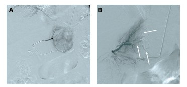
Postembolization angiography (Figure 2) demonstrated:
- Complete occlusion of the neuroendocrine tumor vascular bed
- Absence of residual tumoral enhancement
- Preservation of uninvolved hepatic perfusion
- No evidence of nontarget embolization
Follow-up magnetic resonance imaging was performed at 90 days post procedure (Figure 1C).
Imaging demonstrated:
- Reduction in the treated lesion from 5.25 cm to 3.10 cm
- No evidence of revascularization
- No local tumor progression
- No evidence of treatment-related complications
Case 2: Renal Angiomyolipoma Treated With Transarterial Embolization Using the Embrace Hydrogel Embolic System
A 44-year-old female was referred to Interventional Radiology following the incidental discovery of a right renal mass on cross-sectional imaging. Follow-up computed tomography renal mass protocol performed in 2025 demonstrated imaging characteristics consistent with a right renal angiomyolipoma measuring approximately 5.4 x 3.3 cm.
Review of prior imaging demonstrated interval growth from 3.7 cm in 2019 to 5.4 cm in 2025. Given the lesion size of > 4 cm and documented interval enlargement, the patient was considered at increased risk for spontaneous hemorrhage and was referred for prophylactic TAE.
The patient underwent selective right renal angiography and embolization of the upper pole renal angiomyolipoma in 2026 utilizing the Embrace HES.
The procedure was performed under moderate sedation. Selective angiography demonstrated a hypervascular upper pole right renal angiomyolipoma supplied by dedicated tumor-feeding arterial branches arising from the right renal artery. Superselective catheterization of the tumor-feeding vessels was achieved utilizing a 2.8F 130 cm Progreat microcatheter. The coaxial microcatheter system was assembled using the 1.7F 142 cm Instylla microcatheter.
Embrace HES was delivered into the target arterial supply until complete angiographic stasis was achieved, requiring 2.5 mL of Embrace HES.
Post-embolization angiography (Figure 3B) demonstrated:
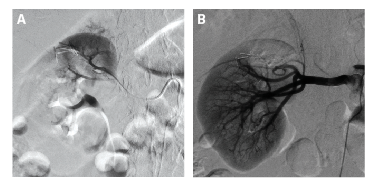
- Complete exclusion of the angiomyolipoma vascular bed
- Absence of residual tumoral enhancement
- Preservation of uninvolved renal parenchymal perfusion
- No evidence of nontarget embolization
The procedure was completed without immediate complication.
Post-treatment computed tomography renal mass protocol (Figure 4) was obtained approximately 5 weeks following embolization. Imaging demonstrated:
- Interval reduction in size of the angiomyolipoma from 5.4 x 3.3 cm pre-treatment to 4.5 x 2.9 cm
- Complete loss of internal enhancement
- Persistent devascularization of the upper pole lesion
- No evidence of residual tumoral perfusion
- No evidence of treatment-related complications
Overall, imaging findings were consistent with a successful therapeutic response characterized by both substantial volumetric reduction and complete angiographic devascularization. n
Affiliations and Disclosures
Edgar St. Amour, MD, is from the Central Arkansas Radiation Therapy Institute, Little Rock, Arkansas; Joseph S. Bittle, MD, is from Prisma Health Greenville Memorial Hospital, Greenville, South Carolina.
Dr St. Amour is a consultant for Instylla and has a small equity interest in the form of stock options with the company. Dr Bittle is a consultant for Instylla.