







Preoperative Embolization of Cerebellar Hemangioblastomas Using N-Butyl Cyanoacrylate: Techniques, Efficacy, and Safety Profile
Key Summary
- A retrospective case series at a tertiary center in South India included 9 patients with cerebellar hemangioblastomas who underwent preoperative embolization and surgery between 2001 and 2024. N-butyl cyanoacrylate was used in 8 cases.
- Angiography showed a mean estimated 70% reduction in tumor vascularity. All 9 tumors were completely resected; mean operative blood loss was 450 mL. One patient had a cerebellar artery infarct without significant neurologic deficit, and one had an access-site complication.
- The authors consider preoperative embolization a potentially useful surgical adjunct, but the small sample and lack of a surgery-only control group prevent conclusions about its benefit.
© 2026 HMP Global. All Rights Reserved.
Any views and opinions expressed are those of the author(s) and/or participants and do not necessarily reflect the views, policy, or position of Vascular Disease Management or HMP Global, their employees, and affiliates.
VASCULAR DISEASE MANAGEMENT. 2026;23(11):E167-E177
Abstract
Purpose: To evaluate the safety, efficacy, and surgical impact of preoperative embolization using N-butyl cyanoacrylate (NBCA) in patients with cerebellar hemangioblastomas (CHBs). Method and Materials: This retrospective study reviewed records of patients with CHBs who underwent preoperative embolization followed by surgery between January 2001 and April 2024. Of 12 patients, 9 with complete records were included. Data analyzed included demographics, angiographic features, embolized arteries, embolic agents, complications, timing of surgery, operative time, blood loss, extent of resection, and status at discharge. Results: Nine patients (8 men, 1 woman) were included; 1 had von Hippel-Lindau disease. Five tumors were previously operated elsewhere. Angiography showed a mean vascularity reduction of 70% (range, 50%-95%) post embolization. NBCA (10%-25%) was used in 8 cases for pial supply. Embolization was uneventful in 8 patients; 1 developed a right superior cerebellar artery infarct without significant clinical deficit. Surgery followed within 24 hours of embolization in 8 patients. Mean blood loss was 450 mL (range, 50-1200 mL), and mean operative time was 4.8 hours (range, 2.5-6 hours). Neurological status (Modified Rankin Scale score) at discharge was stable or improved in 8 of 9 cases. Conclusion: Preoperative embolization for CHBs using NBCA appears to be a safe and useful adjunct to surgical management. NBCA is a feasible and economical embolic agent when used by experienced operators. In this case series, embolization was associated with reduced intraoperative blood loss and facilitated more controlled tumor resection, particularly in recurrent or residual lesions.
Introduction
Cerebellar hemangioblastomas (CHBs) are highly vascular posterior fossa tumors that present significant surgical challenges, largely due to the risk of substantial intraoperative hemorrhage. CHBs are histologically benign lesions that account for 1% to 2% of primary central nervous system tumors and 8% to 12% of posterior fossa lesions in adults, occurring either sporadically or as part of von Hippel-Lindau (VHL) disease.1,2 Their rich vascularity and location within tight neuroanatomical compartments make surgical resection technically challenging.
Preoperative embolization has been proposed as an adjunct to reduce tumor vascularity and potentially facilitate safer, more controlled tumor excision. However, its role remains contentious. While several studies report decreased intraoperative blood loss and improved surgical handling following embolization, particularly with liquid agents such as N-butyl cyanoacrylate (NBCA),3-5 a major systematic review and other publications argue that the evidence is inconsistent and embolization may introduce additional procedural risk without demonstrating clear surgical advantage.6 Thus, the literature is distinctly divided on the utility of preoperative embolization in CHB management.
In the background of this ongoing controversy, we report our institutional experience with NBCA-based embolization for CHBs, representing, to the best of our knowledge, the largest case series from India on this topic.
Materials and Methods
This retrospective study was conducted at the authors’ institution. Electronic medical records of all patients who underwent preoperative embolization for CHBs between January 2001 and April 2024 were reviewed. Approval was obtained from the Institutional Review Board (IRB Min. No. 2409172). This retrospective study was conducted in accordance with the Declaration of Helsinki and its later amendments. Patient consent for participation and publication was not required as no identifiable data or images are included.
Patient selection criteria
Patients were considered for preoperative embolization based on predefined radiologic and clinical factors. Embolization was recommended in the following situations:
- Large tumors (≥3 cm) on magnetic resonance imaging (MRI) where significant intraoperative bleeding was anticipated.
- Tumors located deep within the posterior fossa or in regions where early vascular control during surgery was difficult.
- Previously operated cases where altered anatomy increased vascular risk.
A comparative control group of patients with CHBs treated with surgery alone was not available, and this represents a methodological limitation of the study.
Study population
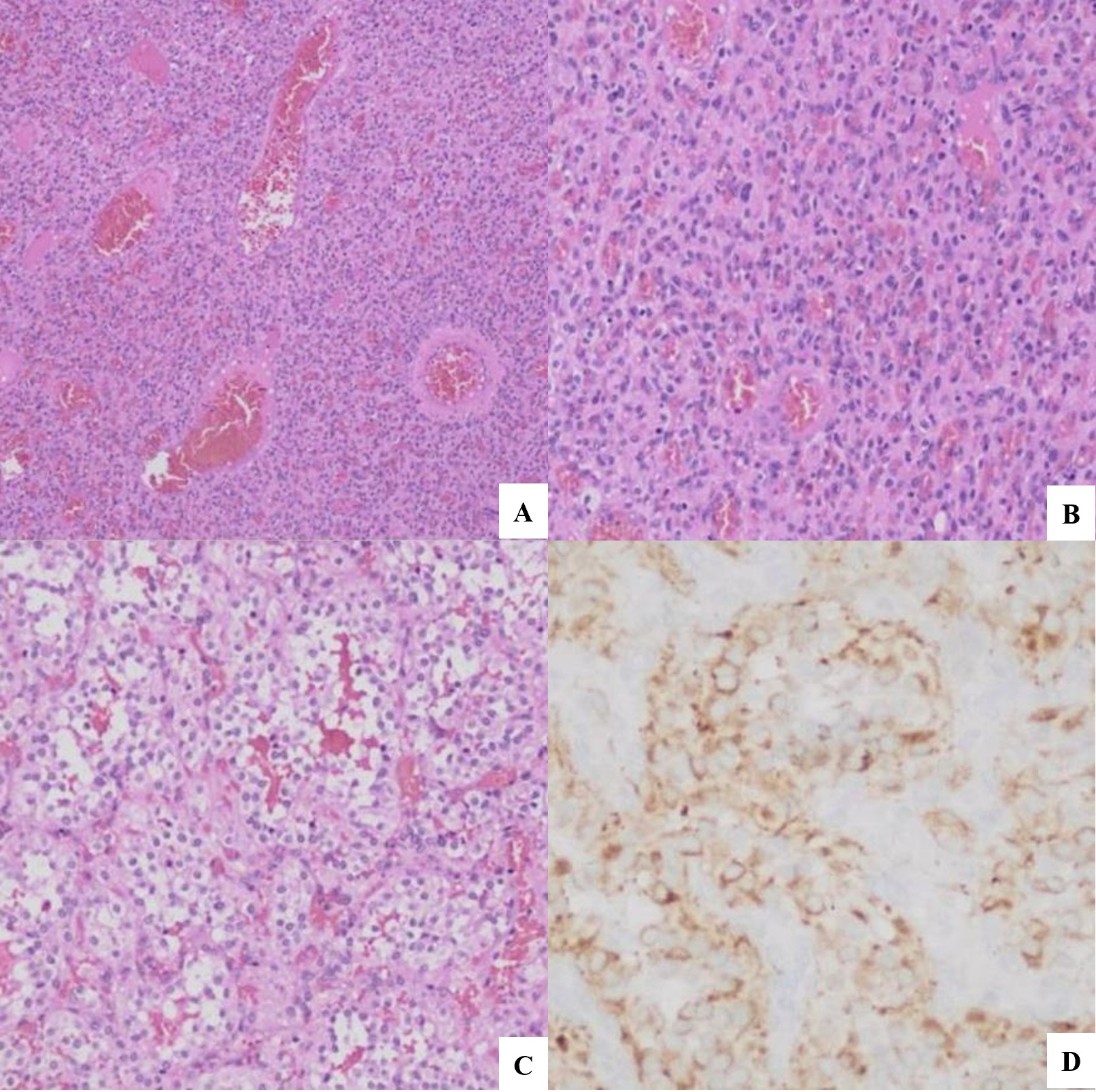
A total of 12 patients were identified who had undergone preoperative embolization followed by surgical resection and had histopathological confirmation of CHB (Figure 1). Electronic data were incomplete for 3 patients; therefore, 9 patients were included in the final analysis.
Embolization technique
Femoral arterial access was utilized in all patients because of its lower radiation exposure compared to radial access and its superior catheter maneuverability for intracranial navigation. A diagnostic cerebral angiogram was performed 2 to 3 days prior to embolization to evaluate tumor vascularity, identify feeding arteries, and assess the presence of any internal carotid artery-external carotid artery (ECA) anastomoses.
All embolization procedures were performed under general anesthesia. The dominant vertebral artery was used to access the posterior circulation; in cases with codominant vertebral arteries, the artery providing preferential filling of the tumor feeders was selected. The procedural setup typically included an 8F short sheath, a long sheath (eg, Neuron MAX 088 [Penumbra]), an intermediate catheter (eg, Fubuki 043 guiding catheter [Asahi Intecc]), and a microcatheter system (straight or angled, depending on vascular anatomy). Liquid embolic agents (NBCA, 10%-25% concentration) were used as the primary material. For feeders arising from the ECA territory, polyvinyl alcohol (PVA) particles (300-500 μm) and Gelfoam (Pfizer) were used selectively.
N-butyl cyanoacrylate embolization technique
The microcatheter was advanced selectively into the tumor-feeding artery. Before embolization, a super-selective angiogram was performed to confirm exclusive opacification of tumor blush without any normal parenchymal supply. NBCA (10%-25%) was prepared according to tumor blood flow characteristics and injected after flushing the microcatheter with 5% dextrose. Glue injections were performed under continuous fluoroscopic roadmap guidance (free-flow injection).7 The injection time was typically less than 1 minute.
Polyvinyl alcohol particle and Gelfoam embolization technique
Contour PVA embolization particles (Boston Scientific) sized 350 to 500 μm were mixed with contrast medium prior to injection. PVA or Gelfoam was used only for feeders arising from the ECA branches and not for vertebrobasilar system feeders due to the risk of inadvertent non-target embolization.
Endpoint of embolization
In case of multiple arterial feeders to the tumor, it is advisable to embolize the tumor as extensively as possible but at the same time to not increase the risk of complications. Patients were given steroids post embolization until surgery to decrease the edema associated with the tumor.
Timing of preoperative embolization
It is advisable to do the embolization within 24 hours of operation as delayed surgery may increase the risk of tumoral edema, which may lead to urgent decompressive craniotomy. In our institution, the patient is shifted to the intensive care unit post embolization until surgery is scheduled for high-intensive monitoring.
Assessment of tumor vascularity reduction
Tumor vascularity reduction was assessed qualitatively by comparing pre- and post-embolization digital subtraction angiography. Two independent neurointerventional radiologists categorized the reduction in tumor blush as mild (0%-30%), moderate (30%-70%), and substantial (>70%) reduction on an eyeballing basis. Disagreements were resolved by consensus.
Surgical technique
All patients underwent surgery under general anesthesia in prone position within 24 hours after completion of embolization. A midline suboccipital craniectomy or craniotomy was performed with excision of the posterior arch of C1 (in 3 of 9 cases). The dura was opened widely to expose the lesion. The site where the tumor reached the pial surface was identified, and dissection started superficially, disconnecting the pial feeders one by one. Dissection was continued circumferentially by separating the tumor from the adjacent cerebellum or brain stem surface, dividing the pial vessels, and entering it from the dorsal and lateral surfaces circumferentially, taking care not to coagulate the draining vein. Once the dorsal and lateral aspects of the tumor were devascularized and dissected, the ventral surface was dissected off the cerebellum by mobilizing the tumor from the pole distant from the draining vein. As the last step, the draining vein was coagulated and divided to excise the tumor in toto.
Estimation of intraoperative blood loss
Intraoperative blood loss was estimated using standard surgical methods, including suction canister volume (after subtracting the volume of fluid used for irrigation from the total volume collected) and visual estimation of field loss, with a consensus achieved between the surgical and anesthesia teams.
Data collection
The following data were collected: patient demographics, tumor characteristics, feeding arteries identified on angiography, arteries embolized, embolic agents used, embolization-related complications, time interval between embolization and surgery, operative duration, estimated intraoperative blood loss, extent of tumor resection, postoperative complications, and clinical follow-up outcomes.
Results
Patient characteristics
A total of 9 patients (8 men, 1 woman) were included in the study. Ages of the patients were 16 to 68 years (mean, 46 years). Predominant presenting clinical symptoms were headache (n = 5), ataxia (n = 8), visual disturbances (n = 4), and decreased hearing (n = 1). One patient was found to have CHB as a part of VHL disease. Five the 9 patients were previously operated cases. Eight patients had a single lesion, and 1 patient had 2 lesions, of which the larger lesion was embolized and operated (Table 1).
Table 1. Clinical and radiological characteristics
| Patient number | Age/sex | Previously operated/ fresh case | Clinical details | VHL disease | No. of lesions | Tumor location | MRI dimensions (cm) | Consistency (MRI) |
| 1 | 40/M | Fresh | Headache, ataxia, visual disturbance × 2 years | No | Single | Fourth ventricle | 5.2 × 3.5 × 3.4 | Solid-cystic |
| 2 | 30/M | Previously operated | Headache, vomiting, ataxia × 4 months | No | Single | Right cerebellar hemisphere | 6.8 × 6.4 × 4.9 | Solid |
| 3 | 53/M | Fresh | Headache, visual obscuration, imbalance × 2 months | No | Single | Left cerebellar hemisphere | 4.2 × 3.2 × 3.8 | Solid-cystic |
| 4 | 41/M | Previously operated | Hearing loss, visual loss, ataxia × 7 months | No | Two | Right CPA | 4.3 × 5.5 ×3.6 | Solid-cystic |
| 5 | 68/M | Previously operated | Right-sided ataxia × 1 year | No | Single | Right cerebellar hemisphere | 4.0 × 3.1 × 3.2 | Solid-cystic |
| 6 | 67/M | Fresh | Ataxia, dysarthria × 5 months | No | Single | Right cerebellar hemisphere | 4.0 × 3.1 × 3.2 | Solid-cystic |
| 7 | 16/M | Fresh | Headache, diplopia, imbalance × 3 months | No | Two | Fourth ventricle | 5.1 × 4.2 × 4.0 | Solid |
| 8 | 48/F | Previously operated | Giddiness, imbalance × 2 months | Yes | Single | Vermis | 3.4 × 2.5 × 2.6 | Solid |
| 9 | 42/M | Previously operated | Headache, giddiness × 6 months | No | Single | Right CPA | 4.2 × 2.6 × 2.9 | Solid |
Abbreviations: VHL, von Hippel-Lindau; MRI, magnetic resonance imaging; CPA, cerebellopontine angle.
Tumor characteristics
The mean largest diameter of tumor on MRI was 4.5 cm (range, 3.4-6.8 cm). The tumors were along the tentorial surface of the cerebellum (n = 1), suboccipital surface (n = 2), and petrosal surface (n = 1). Five of 9 patients were abutting the fourth ventricular floor.
Preoperative angiogram showed tumor vascularity from the posterior inferior cerebellar artery (PICA) (n = 4), anterior inferior cerebellar artery (AICA) (n = 2), and superior cerebellar artery (SCA) (n = 4). More than 1 artery supplied the tumor in 8 of 9 cases.
Results of embolization
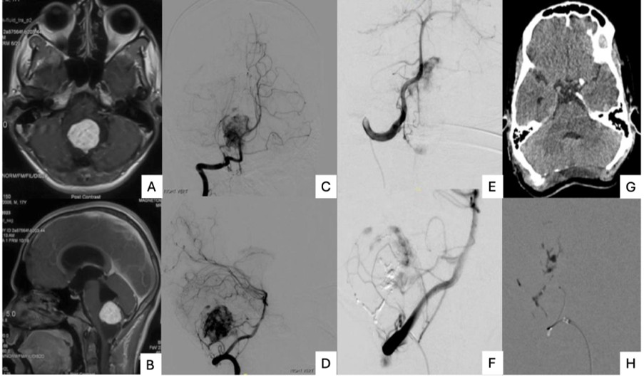
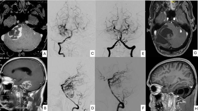
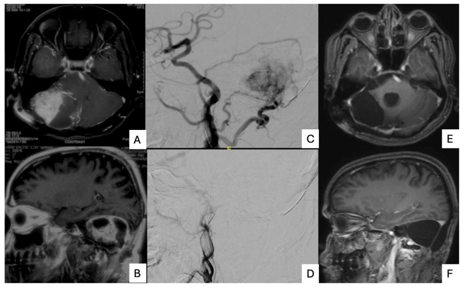
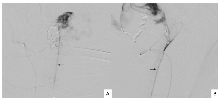
Mean reduction in vascularity of 70% was achieved on post-embolization angiograms (range, 50%-95%). A total of 14 arteries were embolized. The most embolized arteries were the PICA and SCA (n = 4 each), followed by the AICA and occipital artery (n = 2 each) and the posterior auricular artery (n = 1). The main embolizing agent used was glue in 8 cases (10%-25% concentration) (Figures 2 and 3). Other embolizing agents used were Gelfoam (1 case) and PVA (1 case). Gelfoam and PVA were used to embolize branches from the ECA only (dural supply) (Figure 4).
Complications of embolization
Eight of 9 patients with preoperative embolization underwent the surgery without any complications. One patient developed a right SCA territory infarct detected on follow-up MRI. However, no significant neurologic deficits were seen. One patient had an access site complication. The foot plate of the Angio-Seal occlusion device (Terumo) dislodged, which was removed surgically (Table 2). In our case series, all 9 patients post embolization developed no clinically significant neurological deficits. In Patient 9, tortuous vascular anatomy led to difficulty in selectively cannulating the right SCA. During attempted catheterization, thrombus formation occurred in the right SCA and extended into the distal basilar artery. Mechanical thrombectomy was performed, successfully restoring basilar artery flow; however, the right SCA remained occluded. Despite this, a significant reduction in tumor vascularity was achieved due to iatrogenic clot. The remaining arterial feeders were small and therefore not embolized. Post-procedure MRI demonstrated a right SCA territory infarct without any resulting clinical neurological deficits.
Table 2. Angiographic, embolization, and surgical details
| Patient number | Arterial supply (angiography) | Arteries embolized | Reduction in tumor vascularity (%) | Embolic agent | Post-embolization complication | Interval to surgery (hours) | Surgery time (hours) | Blood loss (mL) | Extent of resection | Status at discharge | Preop mRS score | Postop mRS score |
| 1 | Left AICA, right PICA | Left AICA | Moderate (60) | Glue | None | <24 | 4 | 400 | Total excision | Doing well | 3 | 3 |
| 2 | Multiple posterior circulation arteries | Right PICA, occipital | Moderate (60) | PVA, glue | CFA occlusion | 48 | 6 | 500 | Total excision | Doing well | 2 | 2 |
| 3 | Left SCA | Left SCA | Moderate (60) | Glue | None | <24 | 6 | 350 | Total excision | Doing well | 2 | 2 |
| 4 | Right PCA, SCA, AICA | Right SCA, AICA | Moderate (50-60) | Glue | None | <24 | 6 | 200 | Total excision | Doing well | 3 | 2 |
| 5 | Occipital, PA, MMA, SCA, PICA | Occipital, PA, MMA | Substantial (75-80) | Gelfoam | None | <24 | 6 | 350 | Total excision | Doing well | 2 | 2 |
| 6 | ICA, PCA, SCA | Left SCA | Substantial (65-70) | Glue | None | <24 | 5 | 300 | Total excision | Doing well | 3 | 2 |
| 7 | Bilateral PICA | Right PICA | Moderate (50-60) | Glue | None | <24 | 4 | 1200 | Total excision | Doing well | 3 | 2 |
| 8 | Bilateral PICA | Bilateral PICA | Substantial (90) | Glue | None | <25 | 2.5 | 50 | Total excision | Doing well | 2 | 2 |
| 9 | SCA, AICA, occipital | Right SCA | Substantial (70) | Glue | SCA infarct | <24 | 4 | 700 | Total excision | Doing well | 1 | 2 |
Abbreviations: mRS, Modified Rankin Scale; AICA, anterior inferior cerebellar artery; PICA, posterior inferior cerebellar artery; PVA, polyvinyl alcohol; CFA, common femoral artery; SCA, superior cerebellar artery; PCA, posterior cerebral artery; PA, pulmonary artery; MMA, middle meningeal artery; ICA, internal carotid artery.
Results of surgery
Duration between preoperative embolization and surgery was less than 24 hours in 8 of 9 patients. One patient was operated 24 to 48 hours later because of access site complications. The mean operative time was 4.8 hours (range, 2.5-6 hours). The mean blood loss was 450 mL, and the surgeon’s estimate of the extent of embolization achieved to facilitate surgery was satisfactory in all patients. One patient (Patient 7), in whom only a partial embolization of 50% to 60% was done in view of a spinal artery arising from the feeder vessel, had blood loss of 1200 mL intraoperatively (Figure 5).
All 9 patients underwent total excision of the tumor without any residue. Gross total resection was achieved based on intraoperative inspection and confirmed by postoperative neuroimaging. In all patients except one, neurological status was better or similar during the discharge (Modified Rankin Scale scores are included in Table 2). Patient 9 had mild worsening of cerebellar symptoms at discharge.
Discussion
HGBs are rare central nervous system tumors, accounting for 1% to 2% of intracranial neoplasms and 8% to 12% of posterior fossa tumors, with a predilection for the cerebellar hemispheres.1,2,8 Sporadic lesions typically demonstrate male predominance, whereas VHL disease-associated tumors occur earlier, are more often multifocal, and show female predominance.8 Our cohort mirrors this distribution, consisting predominantly of sporadic, male-predominant lesions with a single VHL disease case.
Vascular anatomy and angiographic patterns
CHBs derive blood supply from pial branches of the vertebrobasilar circulation, including the posterior cerebral artery, SCA, AICA, and PICA. Previously operated cases or those with dural involvement may recruit additional feeders from the external carotid system or meningeal branches of the vertebral artery.9 Consistent with these patterns, 3 previously operated patients in our series demonstrated significant dural feeders, while the remainder showed classical vertebrobasilar supply. These findings highlight the essential role of comprehensive pre-embolization vascular mapping.
Effectiveness of preoperative embolization
In our cohort, preoperative embolization achieved a mean devascularization of 70% and enabled gross total resection in all patients, with a mean intraoperative blood loss of 450 mL. These outcomes align with the experience of Liu et al3 and Sakamoto et al,4 who reported meaningful reductions in intraoperative bleeding and improved operative control following embolization. Large surgical series by Conway et al10 and Klingler et al11 further emphasize hypervascularity as a major determinant of surgical morbidity, supporting embolization in appropriately selected cases.
Literature reports have consistently demonstrated 40% to 60% reductions in operative blood loss after CHB embolization.3,12,13 Our results parallel these findings, with a single major bleed (1200 mL) attributed to an unembolized PICA feeder arising from a spinal artery, an anatomical challenge previously reported by Krishnan and Schackert.14 Operative duration in our series (mean 4.8 hours) is comparable to the shorter operative times noted by Takeuchi et al13 and Murai et al5 following effective devascularization.
Timing considerations
The optimal timing of surgery after embolization of tumors remains controversial. Some authors recommend resection within 1 to 5 days,5,15 while others advocate waiting 1 to 2 weeks16,17 to allow embolization-induced necrosis to soften the tumor. However, these recommendations largely derive from literature on meningioma rather than posterior fossa tumors. Post embolization, there may be swelling and edema of the tumor and adjacent cerebellum within an already small posterior cranial fossa that puts the patient at risk for consequences of tonsillar herniation and brain stem compression. Hence, it would be prudent to operate on these tumors immediately post embolization or within the first 24 hours, where even mild edema may precipitate acute hydrocephalus, raised intracranial pressure, or infarction. Thus, in our case series, almost all cases (8 of 9) were operated within 24 hours of embolization, except 1 case where it had to be delayed by 48 hours due to access site-related complications.
Alternative techniques
NBCA was the principal embolic agent in our series, offering rapid polymerization and deep pial penetration.5,18 Other approaches are described in literature, such as the Onyx liquid embolic system (Medtronic), which allows controlled, prolonged injection and excellent visualization,19 but its higher cost limits use in resource-constrained health systems such as India.
PVA particles were not used, as it has been postulated that embolization with calibrated microspheres in posterior fossa hemangioblastomas may lead to venous outflow obstruction, resulting in tumoral vascular congestion and subsequent vessel rupture, along with an increased risk of non-target embolization.20,21
Direct percutaneous tumor puncture embolization has been described as an alternative in cases of unfavorable arterial access. Montano et al20 demonstrated its utility in selected skull-base tumors, though concerns regarding cortical injury, hemorrhage, and trephination-related morbidity restrict this approach to highly selected indications.
Safety and cost considerations
Safety has improved substantially with modern liquid embolic agents. Historic particle embolization literature of pial supply carried higher hemorrhagic risk,20,21 whereas NBCA offers improved safety and predictability.5,18 Additionally, NBCA compared to non-embolized surgical excision may reduce overall perioperative costs through decreased operative times, transfusion rates, and intensive care unit stays.3
Overall literature presents a divided perspective. The meta-analysis by Palavani et al22 synthesizes global data, demonstrating high devascularization rates (approximately 70%-85%) and suggesting significant reductions in intraoperative blood loss and operative duration. These results support a potential adjunctive role for embolization in large or hypervascular lesions. Conversely, Ampie et al6 offer a more critical assessment, concluding that embolization did not increase rates of gross total resection, decrease blood loss, or reduce complication rates. They further emphasized that embolization carries its own procedure-related risks and should not be regarded as standard of care for intracranial hemangioblastomas. This contrast underscores the persistent uncertainty and heterogeneity in the literature.
Current evidence from literature seems to point to embolization being beneficial in selected complex CHBs. However, consistent improvement across all outcome measures remains unproven. Within this context, our experience contributes additional real-world data. In our case series, preoperative embolization using NBCA was safe, facilitated complete tumor removal, and appeared to reduce operative duration and blood loss. However, these encouraging findings must be interpreted with caution due to the absence of a control group and the small sample size.
Conclusion
Preoperative embolization for CHBs using NBCA appears to be a safe and useful adjunct to surgical management. NBCA is a feasible and economical embolic agent when used by experienced operators. In this case series, embolization was associated with reduced intraoperative blood loss and facilitated more controlled tumor resection, particularly in recurrent or residual lesions. Larger comparative studies are needed before definitive conclusions can be drawn. n
Affiliations and Disclosures
At Christian Medical College Vellore, Tamil Nadu, India, Rajat Singhal, DM, DNB, FRCR; Shyamkumar Nidugala Keshava, DNB, DRNB, FRCR (UK), FRANZCR; Rajchandra Kharayat, DM; Philip Mathew, MD, FNVIR; David Narayan Rameswarapu, DM, MD; and Nusum Umamaheswari, DM, MD are from the Department of Interventional Radiology; Anitha Jasper, MD, DMRD is from the Department of Radiology; Krishna Prabhu, MBBS, M.Ch; Ranjith K. Moorthy, M.Ch; and B. Vivek Joseph, MBBS, M.Ch are from the Department of Neurological Sciences; and Geeta Chacko, MD is from the Department of Pathology.
The authors thank Dr Sneha H and Dr Assem Verma for their motivation and support in completing this manuscript.
The authors report no financial relationships or conflicts of interest regarding the content herein.
The data supporting the findings of this study are available from the corresponding author upon reasonable request.
Manuscript accepted September 18, 2026.
Address for correspondence: Shyamkumar N Keshava, Department of Radiodiagnosis, Christian Medical College Vellore, Ida Scudder Road, Vellore – 632004, Tamil Nadu, India. Email: shyamkumar.n.keshava@gmail.com
References
1. Sultan A, Hassan T, Aboul-Enein H, Mansour O, Ibrahim T. The value of preoperative embolization in large and giant solid cerebellar hemangioblastomas. Interv Neuroradiol. 2016;22(4):482-488. doi:10.1177/1591019916633244
2. Resche F, Moisan JP, Mantoura J, et al. Haemangioblastoma, haemangioblastomatosis, and von Hippel-Lindau disease. Adv Tech Stand Neurosurg. 1993:197-304. doi:10.1007/978-3-7091-6912-4_6
3. Liu A, Peng T, Wu Z, et al. Clinical effectiveness of preoperative embolization for cerebellar hemangioblastoma. Asian Pac J Cancer Prev. 2013;14(9):5179-5183. doi:10.7314/apjcp.2013.14.9.5179
4. Sakamoto N, Ishikawa E, Nakai Y, et al. Preoperative endovascular embolization for hemangioblastoma in the posterior fossa. Neurol Med Chir (Tokyo). 2012;52(12):878-884. doi:10.2176/nmc.52.878
5. Murai Y, Kominami S, Yoshida Y, et al. Preoperative liquid embolization of cerebellar hemangioblastomas using N-butyl cyanoacrylate. Neuroradiology. 2012;54(9):981-988. doi:10.1007/s00234-011-0985-5
6. Ampie L, Choy W, Lamano JB, et al. Safety and outcomes of preoperative embolization of intracranial hemangioblastomas: a systematic review. Clin Neurol Neurosurg. 2016;150:143-151. doi:10.1016/j.clineuro.2016.09.008
7. Kidani N, Hirotsune N. NBCA: basic knowledge. J Neuroendovasc Ther. 2025;19(1):2024-0055. doi:10.5797/jnet.ra.2024-0055
8. Kuharic M, Jankovic D, Splavski B, Boop FA, Arnautovic KI. Hemangioblastomas of the posterior cranial fossa in adults: demographics, clinical, morphologic, pathologic, surgical features, and outcomes. A systematic review. World Neurosurg. 2018;110:e1049-e1062. doi:10.1016/j.wneu.2017.11.173
9. Yamada SM, Ikeda Y, Takahashi H, Teramoto A, Yamada S. Hemangioblastomas with blood supply from the dural arteries—two case reports. Neurol Med Chir (Tokyo). 2000;40(1):69-73. doi:10.2176/nmc.40.69
10. Conway JE, Chou D, Clatterbuck RE, Brem H, Long DM, Rigamonti D. Hemangioblastomas of the central nervous system in von Hippel-Lindau syndrome and sporadic disease. Neurosurgery. 2001;48(1):55-62; discussion 62-63. doi:10.1097/00006123-200101000-00009
11. Klingler JH, Gläsker S, Bausch B, et al. Hemangioblastoma and von Hippel-Lindau disease: genetic background, spectrum of disease, and neurosurgical treatment. Childs Nerv Syst. 2020;36(10):2537-2552. doi:10.1007/s00381-020-04712-5
12. Eskridge JM, McAuliffe W, Harris B, Kim DK, Scott J, Winn HR. Preoperative endovascular embolization of craniospinal hemangioblastomas. AJNR Am J Neuroradiol. 1996;17(3):525-531.
13. Takeuchi S, Tanaka R, Fujii Y, Abe H, Ito Y. Surgical treatment of hemangioblastomas with presurgical endovascular embolization. Neurol Med Chir (Tokyo). 2001;41(5):246-251; discussion 251-252. doi:10.2176/nmc.41.246
14. Krishnan KG, Schackert G. Outcomes of surgical resection of large solitary hemangioblastomas of the craniocervical junction with limitations in preoperative angiographic intervention: report of three cases. Zentralbl Neurochir. 2006;67(3):137-143. doi:10.1055/s-2006-933360
15. Nelson PK, Setton A, Choi IS, Ransohoff J, Berenstein A. Current status of interventional neuroradiology in the management of meningiomas. Neurosurg Clin N Am. 1994;5(2):235-259. doi:10.1016/S1042-3680(18)30529-1
16. Ahuja A, Gibbons KJ. Endovascular therapy of central nervous system tumors. Neurosurg Clin N Am. 1994;5(3):541-554. doi:10.1016/S1042-3680(18)30519-9
17. Desai R, Bruce J. Meningiomas of the cranial base. J Neurooncol. 1994;20(3):255-279. doi:10.1007/BF01053043
18. Munyon C, Chowdhry SA, Cohen ML, Bambakidis NC, Hsu DP. N-butyl 2-cyanoacrylate (n-BCA) embolization of a cerebellar hemangioblastoma. J Neurointerv Surg. 2011;3(4):386-389. doi:10.1136/jnis.2010.004366
19. Shin GW, Jeong HW, Seo JH, Kim ST, Choo HJ, Lee SJ. Preoperative embolization of cerebellar hemangioblastoma with Onyx: report of three cases. Neurointervention. 2014;9(1):45-49. doi:10.5469/neuroint.2014.9.1.45
20. Montano N, Doglietto F, Pedicelli A, et al. Embolization of hemangioblastomas. J Neurosurg. 2008;108(5):1063-1064; author reply 1064-1065. doi:10.3171/JNS/2008/108/5/1063
21. Cornelius JF, Saint-Maurice JP, Bresson D, George B, Houdart E. Hemorrhage after particle embolization of hemangioblastomas: comparison of outcomes in spinal and cerebellar lesions. J Neurosurg. 2007;106(6):994-998. doi:10.3171/jns.2007.106.6.994
22. Palavani LB, Andreão FF, de Abreu LV, et al. Assessing the efficacy and safety of hemangioblastoma embolization: a comprehensive systematic review and meta-analysis. J Clin Neurosci. 2023;117:104-113. doi:10.1016/j.jocn.2023.09.021

About Us
Vascular Disease Management™ (VDM), the official media partner of the International Symposium on Endovascular Therapy (ISET) and the Amputation Prevention Symposium (AMP), is a peer-reviewed journal that educates readers on the latest advancements in endovascular treatment strategies.
Learn more about ISET and AMP and register for the events here: