




FLEX Vessel Prep System for Lesion Preparation
Key Summary
- This review examines the FLEX Vessel Prep (FLEX VP) System (Flex Vascular) for femoropopliteal disease and dialysis access and summarizes clinical evidence. FLEX VP creates longitudinal microincisions to prepare lesions before angioplasty.
- In one study, a 255-patient, 38-center femoropopliteal study reported approximately 27% stenosis reduction after FLEX alone, without flow-limiting dissection, perforation, or embolization. In another, 65 patients with complex femoropopliteal disease, 12-month freedom from target lesion revascularization was 93.7%; provisional stenting occurred in 16.9%. The BELONG study enrolled 41 patients receiving FLEX plus drug-coated balloons, reporting 97.5% freedom from clinically driven revascularization at 12 months.
- Findings support further investigation of microincision-based lesion preparation, but predominantly small, nonrandomized studies cannot establish superiority. Larger randomized comparative trials are needed.
© 2026 HMP Global. All Rights Reserved.
Any views and opinions expressed are those of the author(s) and/or participants and do not necessarily reflect the views, policy, or position of Vascular Disease Management or HMP Global, their employees, and affiliates.
VASCULAR DISEASE MANAGEMENT. 2026;23(11):E178-E183
Abstract
Effective lesion preparation is increasingly recognized as a key determinant of procedural success in peripheral arterial intervention, particularly when a leave-nothing-behind strategy using drug-coated balloon (DCB) therapy is intended. The FLEX Vessel Prep (FLEX VP) System (Flex Vascular) is a non-balloon, over-the-wire device designed to create controlled longitudinal microincisions in atherosclerotic plaque, thereby modifying lesion compliance before definitive angioplasty. This review summarizes the mechanism of action and current clinical evidence for FLEX VP in femoropopliteal disease and arteriovenous access. Across available studies, FLEX VP has demonstrated acute luminal improvement, low rates of severe or flow-limiting dissection, limited bailout stenting, and encouraging 12-month freedom from target lesion revascularization. Prospective data from the BELONG study further support the combination of FLEX VP with DCB therapy in femoropopliteal lesions. Although the evidence base remains predominantly single-arm and nonrandomized, current findings support controlled longitudinal microincision as a differentiated lesion-preparation strategy and justify larger randomized comparative trials.
Introduction
Endovascular therapy has become the dominant revascularization strategy for many patients with symptomatic peripheral arterial disease (PAD). Despite major advances in drug-coated balloons (DCBs), drug-eluting stents, atherectomy, intravascular lithotripsy (IVL), and other technologies, adequate lesion preparation remains a critical determinant of procedural success.
This is particularly relevant in the femoropopliteal segment, where long lesions, chronic total occlusions, heterogeneous plaque morphology, and vascular calcification frequently coexist. Conventional balloon angioplasty can restore luminal diameter but does so by applying radial pressure to the vessel wall. The resulting mechanical stress may cause uncontrolled plaque fracture, medial injury, recoil, and dissection. Severe or flow-limiting dissections frequently require provisional stent implantation, potentially compromising a “leave-nothing-behind” strategy.
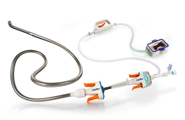
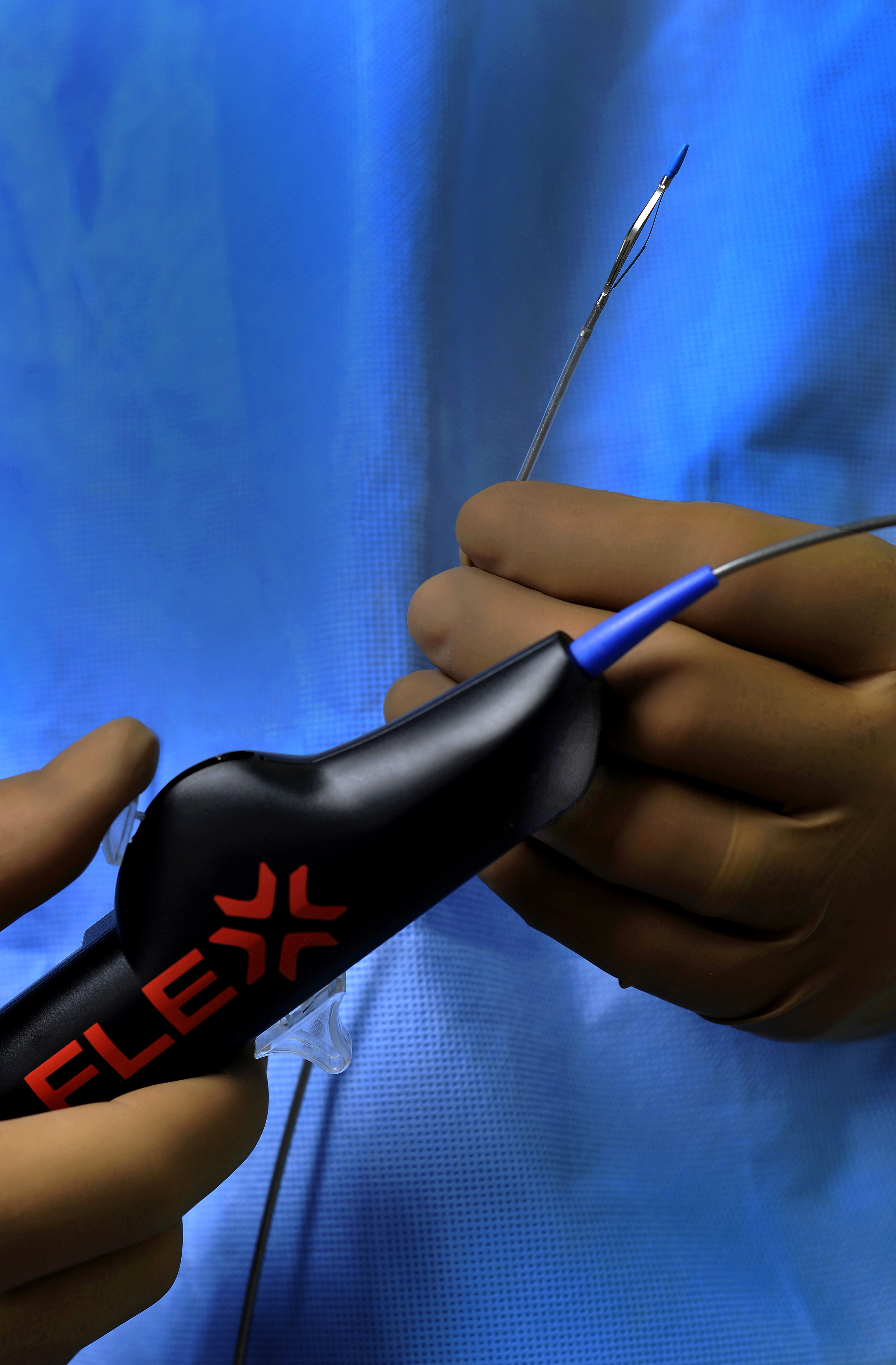
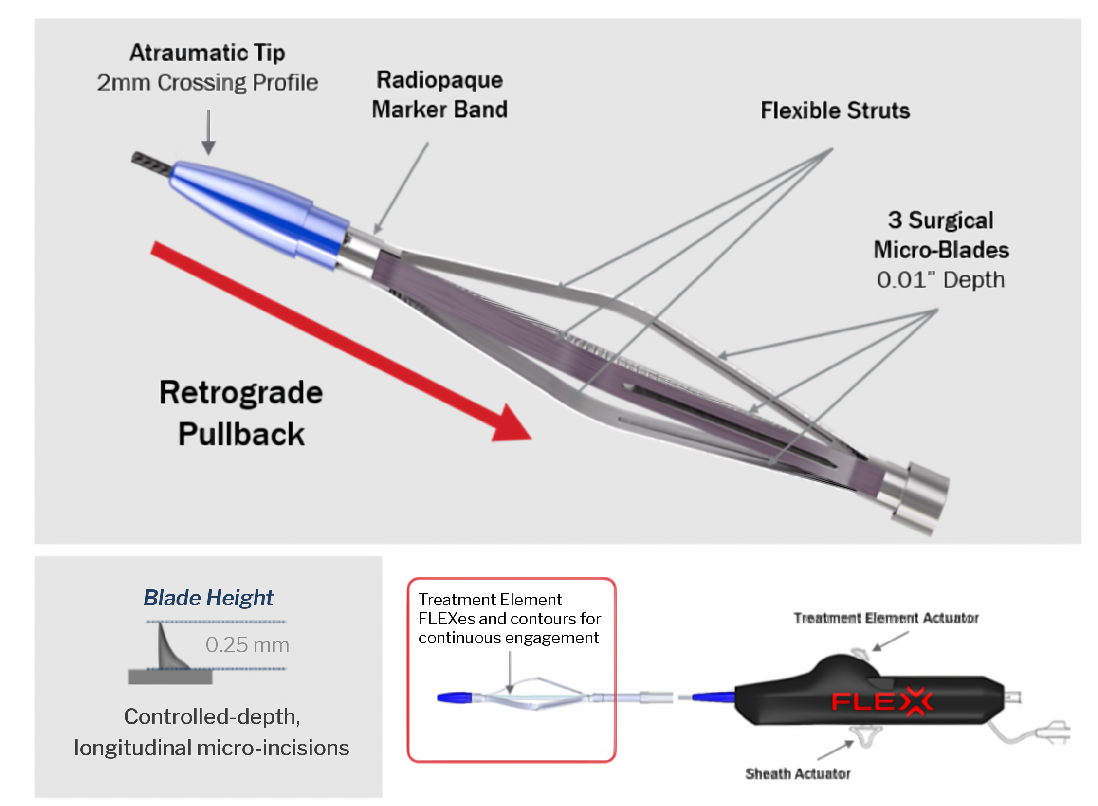
The FLEX Vessel Prep (FLEX VP) System (Flex Vascular) (Figures 1 and 2) represents a different concept of lesion preparation. Rather than relying primarily on high radial balloon pressure or tissue removal, FLEX VP uses controlled longitudinal microincisions to modify plaque and increase vessel compliance before definitive angioplasty or DCB treatment.
The growing clinical evidence suggests that this approach may reduce uncontrolled vessel injury, facilitate luminal expansion, reduce the requirement for extensive stenting, and potentially optimize the vascular environment for subsequent antiproliferative therapy.
The Rationale for Vessel Preparation
Balloon angioplasty works by exerting radial force against an obstructive lesion. In complex atherosclerotic disease, however, the force required to overcome plaque resistance may be substantial. Calcification further increases resistance to vessel expansion and may lead to incomplete balloon expansion or require higher inflation pressures.
The consequences are clinically relevant. Balloon expansion can produce uncontrolled fractures within plaque and the arterial wall. Depending on lesion morphology and the depth of injury, this may result in dissection, elastic recoil, residual stenosis and, ultimately, bailout stenting.
These effects are particularly problematic when DCB therapy is intended. The fundamental objective of DCB treatment is to deliver an antiproliferative agent to the vessel while avoiding a permanent metallic implant. The quality of lesion preparation therefore influences not only the immediate angiographic result but potentially drug transfer and long-term durability.
An ideal vessel preparation strategy should increase vessel compliance, facilitate luminal expansion, minimize uncontrolled dissection and barotrauma, reduce significant residual stenosis, facilitate effective DCB therapy, and minimize the requirement for permanent scaffolding. FLEX VP was developed specifically around these principles.
FLEX Vessel Prep: Mechanism of Action
The FLEX VP System is an over-the-wire, non-balloon vessel preparation catheter designed to create controlled-depth, longitudinal microincisions along the target lesion.
The device incorporates 3 flexible struts with microsurgical blades positioned on their surfaces. According to the device description, the blades have a depth of approximately 0.25 mm. The self-sizing treatment element adapts to the vessel, and controlled microincisions are produced during retrograde pullback through the lesion.
This kinetic mechanism distinguishes FLEX VP from technologies in which blades, wires, or scoring elements are pressed radially into plaque by balloon inflation. The concept has been described as kinetic endovascular microincision creation. Instead of simply applying focal radial force, the device draws microsurgical elements longitudinally through the lesion. Multiple passes can generate circumferentially distributed longitudinal microincisions.
The proposed biomechanical consequence is a reduction in circumferential plaque resistance. By releasing this resistance before balloon angioplasty, subsequent balloon expansion may occur at lower mechanical stress and with a more controlled distribution of force.
Importantly, FLEX VP is not intended primarily as a debulking technology. It modifies lesion mechanics rather than removing substantial plaque volume. This differentiates the technology conceptually from directional, rotational, or orbital atherectomy.
Acute Clinical Evidence
An important evaluation of FLEX VP was reported by Zeller and colleagues in a multicenter real-world study involving 255 patients treated at 38 centers.1
This was a challenging femoropopliteal population. Mean lesion length was 133 ± 88 mm, baseline diameter stenosis averaged 92%, and approximately 44% of patients had chronic total occlusions.
One of the most interesting observations was that FLEX VP itself produced measurable luminal improvement before balloon angioplasty. Mean stenosis was reduced by approximately 27% following FLEX treatment alone.
Despite the complexity of the lesions, no flow-limiting dissection, vessel perforation, or embolization was observed following vessel preparation. Minor type A or B dissections occurred in only 5.9% of patients.
Following subsequent conventional or DCB angioplasty, provisional stenting was required in 19.2% of patients, while final residual stenosis averaged approximately 9%.
These findings provide important mechanistic support for the concept of longitudinal microincision vessel preparation. FLEX VP can modify lesion geometry before balloon inflation without the aggressive radial forces normally required to create luminal gain.
Long-Lesion Experience and 12-Month Outcomes
Dexpert and colleagues subsequently evaluated FLEX VP in 65 patients with particularly complex femoropopliteal disease.2
Median lesion length was 196 mm, with lesions extending to 480 mm. Median stenosis was 90%, approximately one-third of lesions were occlusions, and more than three-quarters demonstrated some degree of calcification.
Following vessel preparation and angioplasty, 82.1% of patients demonstrated either low-grade dissection or no flow-limiting dissection. The provisional stent rate was 16.9%, despite the substantial lesion length, and median implanted stent length was only 60 mm.
Clinical durability was encouraging. Freedom from target lesion revascularization (TLR) was 98.4% at 6 months and 93.7% at 12 months. Freedom from major amputation was 100%.
These results are relevant because lesion length and calcification represent 2 important predictors of poorer outcomes after femoropopliteal intervention. The ability to achieve a low bailout-stent burden while maintaining high freedom from TLR therefore supports further investigation of controlled microincision therapy.
BELONG: FLEX Vessel Preparation Before Drug-Coated Balloons
The BELONG study provides particularly important evidence because it specifically evaluates FLEX VP before DCB angioplasty.4
This investigator-initiated prospective study enrolled 41 patients with 43 femoropopliteal lesions. The population again contained substantial lesion complexity: mean lesion length was 118 mm, mean diameter stenosis was 82%, and 27.9% of lesions were occluded.
Calcification was highly prevalent. Calcification was identified in 85.7% of lesions and approximately two-thirds demonstrated grade 3 or 4 calcification according to the Peripheral Arterial Calcification Scoring System.
Following FLEX VP and DCB treatment, 92.7% of patients were free from flow-limiting dissection.
Sixteen patients underwent stenting; importantly, however, only 1 stent was required because of a flow-limiting dissection. The other stents were placed for focal residual stenosis, and median stent length was 40 mm.
At 12 months, freedom from clinically driven (CD) TLR was 97.5% (39/40). Clinical improvement was also substantial: whereas 41.5% of patients were Rutherford class ≥3 at baseline, 95% were Rutherford class ≤1 at 12 months. No amputations were reported.
Duplex-based primary patency was approximately 84% among evaluable patients.
The distinction between overall stenting and bailout stenting for dissection is important. A crude stent rate of 39% could initially appear relatively high. However, the observation that only 1 patient required stenting for flow-limiting dissection suggests that FLEX VP may alter the pattern of vascular injury produced during subsequent balloon treatment.
Why FLEX May Be Particularly Relevant to Drug-Coated Balloon Therapy
The combination of mechanical lesion preparation followed by biological therapy is conceptually attractive.
DCBs depend on effective contact between the drug-coated surface and the vessel wall. Severe calcification, incomplete balloon expansion, and complex plaque architecture can potentially compromise that interaction.
Longitudinal microincisions could theoretically improve DCB therapy through 2 complementary mechanisms. First, increasing vessel compliance may permit more uniform balloon expansion and vessel-wall contact without requiring excessive pressure. Second, controlled microincisions may potentially facilitate penetration or distribution of antiproliferative drug into the treated lesion.
The latter mechanism remains biologically plausible rather than definitively established clinically. Current studies demonstrate favorable outcomes after FLEX plus DCB, but they do not establish that enhanced drug uptake is the causal mechanism. Dedicated pharmacokinetic or tissue-level studies would be required to prove this effect.
Nevertheless, BELONG provides a meaningful clinical signal: in a population dominated by calcified femoropopliteal disease, FLEX followed by DCB achieved 97.5% freedom from CD-TLR at 1 year.
Supporting Evidence From Arteriovenous Access
Recent evidence from dialysis access provides additional support for the concept that lesion preparation may influence the durability of subsequent DCB therapy.
A retrospective analysis evaluated 175 vascular-access interventions treated with plain old balloon angioplasty (POBA) plus DCB, scoring balloon plus DCB, or FLEX VP plus DCB. At 12 months, target-lesion primary patency was 73.6% with FLEX VP + DCB, 40.1% with POBA + DCB, and 41.2% with scoring balloon + DCB. Reinterventions per patient-year were also significantly lower following FLEX VP.5
Although arteriovenous (AV)-access stenosis represents a different disease process and these results cannot simply be extrapolated to femoropopliteal PAD, the study provides an interesting independent clinical observation: the method used to prepare a lesion before DCB treatment may materially influence subsequent durability.
The prospective, multicenter FLEX FIRST AV registry enrolled 130 patients with 176 dialysis-access lesions treated with FLEX VP followed by POBA.6 The peer-reviewed 6-month analysis by Kramer et al reported target-lesion primary patency of 70.7% and secondary access patency of 97.7%.7 No serious adverse events occurred within 30 days, and 1 stent graft was required for an angioplasty-related dissection. These findings support further evaluation of FLEX VP in AV access, but the single-arm design does not establish superiority over balloon angioplasty alone.
Potential Role in Contemporary Lesion Preparation
The modern approach to PAD increasingly requires matching lesion preparation to lesion morphology.
Atherectomy may be useful where plaque removal is desired. IVL provides an effective strategy for disrupting deep and superficial calcium. Specialty balloons provide focal force during balloon expansion.
FLEX VP occupies a somewhat different position. It does not principally debulk plaque or fracture deep calcium. Instead, it aims to modify the mechanical behavior of the lesion through controlled longitudinal microincisions.
This may be particularly attractive in long femoropopliteal lesions where aggressive balloon angioplasty can produce extensive dissections and where implantation of long metallic stents is undesirable.
Potential scenarios therefore include long diffuse superficial femoral artery disease, mixed-morphology plaque, moderately or severely calcified lesions, lesions intended for DCB treatment, selected in-stent restenosis, and situations where a leave-nothing-behind strategy is preferred.
The technology may also have procedural advantages. It does not require a capital equipment platform, and its self-sizing architecture may reduce the number of device diameters required for treatment.
Limitations of the Current Evidence
The available data are encouraging but should be interpreted appropriately.
Most PAD studies remain relatively small, and several are retrospective or single arm. BELONG, although prospective, included only 41 patients and lacked a randomized comparator. Consequently, comparisons with atherectomy, specialty balloons, IVL, or DCB-alone strategies should not be made solely by comparing outcomes across independent studies.
Furthermore, the relationship between microincision creation and enhanced drug delivery remains a hypothesis supported by mechanistic reasoning and favorable clinical observations rather than definitive biological proof.
Large, randomized studies comparing FLEX VP + DCB with conventional predilatation + DCB would therefore be valuable. Such trials should include independent core-laboratory assessment and evaluate acute luminal gain, balloon inflation pressure, dissection severity, bailout stenting, primary patency, CD-TLR, and health-economic outcomes.
Conclusion
Lesion preparation is increasingly recognized as a distinct and important component of contemporary endovascular therapy.
The FLEX Vessel Prep System introduces a differentiated approach based on controlled longitudinal microincisions rather than high-pressure radial force or plaque debulking.
Across the available femoropopliteal studies, FLEX VP has demonstrated acute luminal improvement, a low incidence of severe dissection, relatively limited provisional stenting, and encouraging 12-month freedom from TLR. n
Affiliations and Disclosures
Michael Lichtenberg, MD, FESC, is from Arnsberg Vascular Center, Klinikum Hochsauerland, Arnsberg, Germany.
The author reports no financial relationships or conflicts of interest regarding the content herein.
Manuscript accepted October 10, 2026.
Corresponding author: Michael Lichtenberg, MD, Arnsberg Vascular Center, Stolte Ley 5, 59759 Arnsberg-Hüsten, Germany. Email: klichte@gmx.net
References
1. Zeller T, Lopez L, Pigott JP. Acute outcomes with a novel plaque modification system in real-world femoropopliteal lesions. J Endovasc Ther. 2019;26(3):333-341. doi:10.1177/1526602819849955
2. Dexpert J, Hayoz D, Engelberger RP, Kreiger C, Meyer MR, Périard D. Arterial preparation by longitudinal micro-incisions before balloon angioplasty of the superficial femoral and popliteal artery: acute and 12-month results. J Endovasc Ther. 2022;29(3):420-426. doi:10.1177/15266028211057089
3. Zeller T, Böhme T, Beschorner U, Noory E. Device profile of the FLEX Vessel Prep System for the treatment of peripheral arterial disease: overview of its safety and efficacy. Expert Rev Med Devices. 2022;19(1):25-29. doi:10.1080/17434440.2022.2020092
4. Demierre A, Pedrazzoli K, Hayoz D, Engelberger RP, Périard D. The BEnefit of LONGitudinal micro-incisions prior to paclitaxel-coated balloon angioplasty (BELONG Study) in patients with lower extremity arterial disease: clinical outcomes at 12 months. J Endovasc Ther. 2026;33(3):1485-1492. doi:10.1177/15266028241312346
5. Shahverdyan R, Hentschel DM, Mehta TI. Vessel preparation with FLEX Vessel Prep device prior to paclitaxel-coated angioplasty improves outcomes of hemodialysis vascular access. J Vasc Access. 2026;27(2):616-626. doi:10.1177/11297298251352690
6. FLEX FIRST Registry Research Protocol. ClinicalTrials.gov. 2025. https://clinicaltrials.gov/study/NCT05616104
7. Kramer A, Schultz SR, Altman SD. Kinetic endovascular microincision creation prior to plain balloon angioplasty in real-world hemodialysis patients with vascular access dysfunction. J Vasc Surg. 2025;82(5):1799-1806. doi:10.1016/j.jvs.2025.07.026