When Balloons Become Entrapped: Stepwise Bailout of a Severely Calcified Coronary Lesion
© 2026 HMP Global. All Rights Reserved.
Any views and opinions expressed are those of the author(s) and/or participants and do not necessarily reflect the views, policy, or position of Cath Lab Digest or HMP Global, their employees, and affiliates.
Yassine Kallala, MD; Matthieu Godin, MD; Quentin Landolff, MD
Department of Interventional Cardiology, Clinique Saint-Hilaire, Rouen, France
Consent statement: The authors confirm that informed consent was obtained from the patient for the study and intervention described in the manuscript and for the publication of their data.
The authors can be contacted via Dr. Quentin Landolff at qlandolff@clinique-sainthilaire.fr
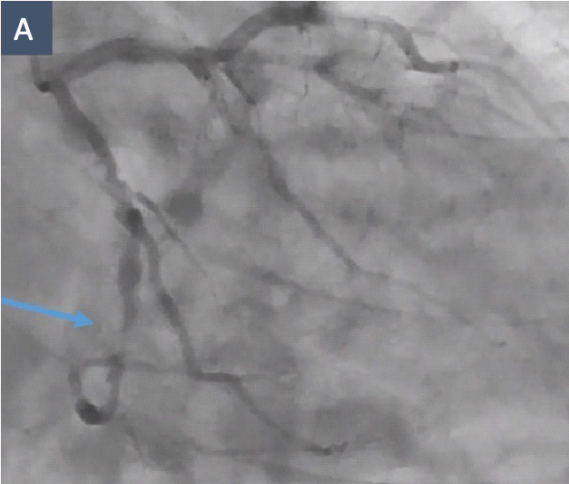
A 71-year-old man with recurrent angina refractory to optimal medical therapy underwent percutaneous coronary intervention for a 22 mm type C, heavily calcified stenosis of a dominant distal left circumflex (LCx) coronary artery (Figure A). The procedure was performed through right distal radial access using a 7 French EBU 3.75 guide catheter.
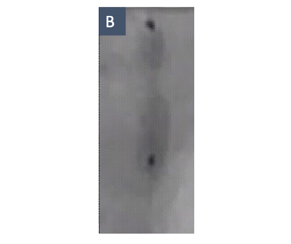
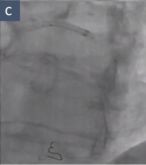
During initial lesion preparation, a 2.5 × 15 mm noncompliant (NC) balloon became entrapped within the calcified nodule (“dogbone” balloon aspect) (Figure B). Attempts at withdrawal resulted in balloon-shaft elongation and rupture (Figure C). Balloon rupture resulted in a type C coronary dissection; however, TIMI-3 flow was maintained, without electrocardiographic changes or hemodynamic compromise. The patient reported mild chest pain that resolved within seconds.
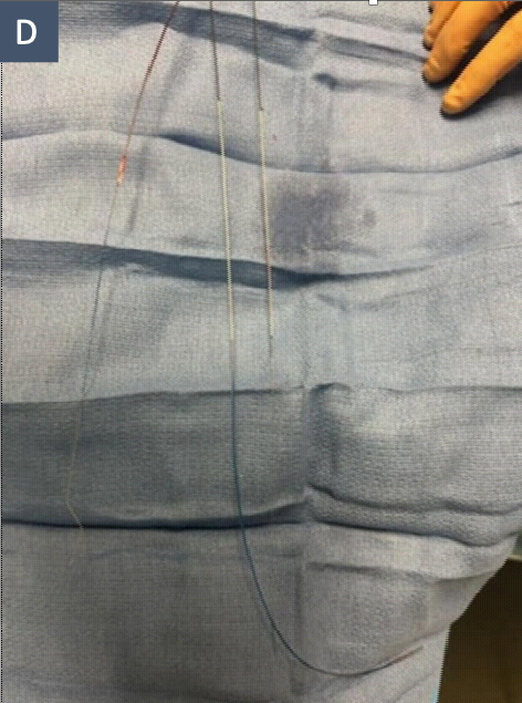
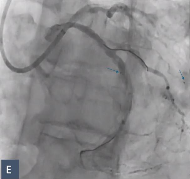
A parallel guidewire was placed, and a 4 mm One Snare endovascular snare system (Merit Medical) was used to retrieve the fractured distal balloon fragment (Figure D). The two radiopaque balloon markers remained entrapped (Figure E): one in a secondary branch and the second adjacent to the calcified nodule. Neither caused flow compromise, and neither was retrieved.
After rewiring with workhorse guidewires, a stepwise calcium-modification strategy was pursued. Because a persistent dogbone deformation remained despite multiple NC balloon inflations, an intravascular lithotripsy (IVL) balloon (Shockwave Medical) was attempted. The IVL balloon did not cross the lesion. Sixty pulses were delivered with the balloon positioned immediately proximal to and in contact with the calcified nodule, an off-label use of the device. The balloon never advanced across or beyond the nodule. Rotational atherectomy (Boston Scientific) was then performed with a 1.75 mm burr, but this proved insufficient. An optical coherence tomography (OCT) catheter (Abbott) and guide-extension catheter could not be advanced in the LCx. After these maneuvers, a 3.5 × 14 mm LithiX Hertz Contact (HC) balloon (Elixir Medical) was advanced with difficulty to the calcified nodule and became entrapped.1,2
Additional percutaneous bailout options were considered. Rewiring with further NC or OPN balloon inflation was thought unlikely to succeed because of the difficulty crossing the resistant calcified lesion alongside the entrapped LithiX HC balloon. Prior attempts with rotational atherectomy and intravascular lithotripsy had not provided adequate lesion modification, and excimer laser was not available. Surgical intervention remained a final option. Given these limitations, ultra-high-pressure inflation of the entrapped LithiX HC balloon was attempted as a final percutaneous bailout strategy.
The rated burst pressure of the LithiX HC balloon is 12 atmospheres (atm), and inflation beyond this pressure is not recommended. Potential risks of ultra-high-pressure inflation include flow-limiting dissection, vessel rupture or perforation with tamponade, and distal embolization of calcific or thrombotic material. Balloon manipulation and multiple inflations at the same site failed to release the balloon. As a bailout maneuver, a single off-label ultra-high-pressure inflation to 28 atm was performed for 30 seconds. The inflation achieved sufficient lesion preparation and vessel expansion to release the entrapped balloon and allow subsequent stent implantation. Because OCT could not cross the lesion, calcium fracture could not be confirmed by intravascular imaging.
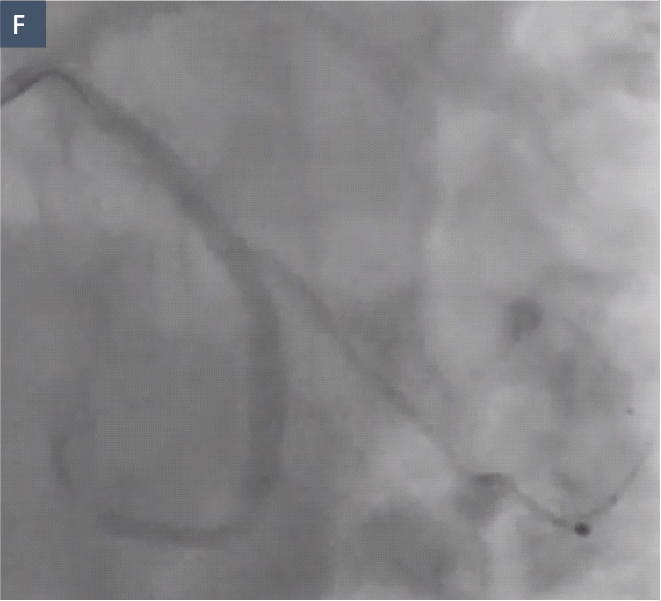
The lesion was stented using two overlapping drug-eluting stents with an excellent final angiographic result and TIMI-3 flow (Figure F). The patient remained asymptomatic and was well at 30-day follow-up.
References
1. Verheye S, Ferdinande B, Paradies V, et al. Hertz contact intravascular lithotripsy for calcified coronary artery disease: the PINNACLE-I trial. EuroIntervention. 2026 Feb 16;22(4):255-264. doi:10.4244/EIJ-D-25-00618
2. Muthusamy TS, Kolanthaivelu J, Choo GH, et al. Hertz contact intravascular lithotripsy for calcified coronary lesions in real-world practice: Primary outcomes of the MY-IVL post-market study. Cardiovasc Revasc Med. 2026 Mar 21:S1553-8389(26)00101-6. doi:10.1016/j.carrev.2026.03.010