Coronary Artery Calcium in PCI: Considerations for Plaque Modification
© 2026 HMP Global. All Rights Reserved.
Any views and opinions expressed are those of the author(s) and/or participants and do not necessarily reflect the views, policy, or position of Cath Lab Digest or HMP Global, their employees, and affiliates.
Jake Rogers, BS, Radiography and ICVT; Professor Richard Merschen, EdS, RT(R)(CV), RCIS
Jefferson College of Health Professions, Philadelphia, Pennsylvania
Disclosures: The authors report no conflicts of interest regarding the content herein.
The authors can be contacted via Richard Merschen, EdS, RT(R)(CV), RCIS, at richard.merschen@pennmedicine.upenn.edu.
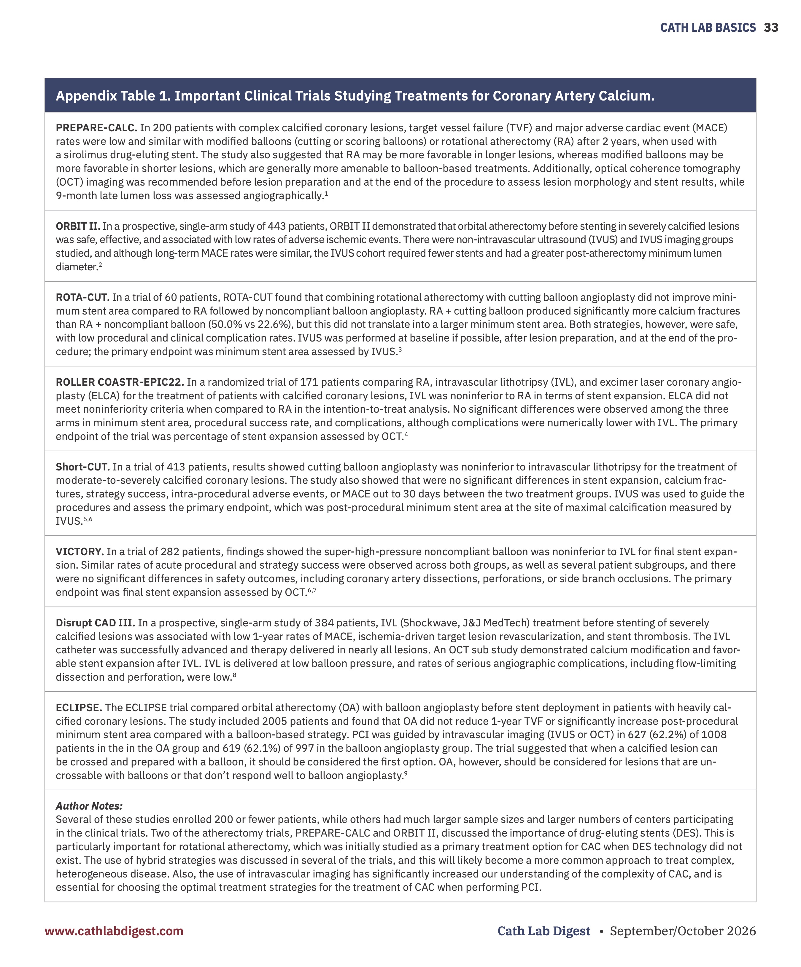
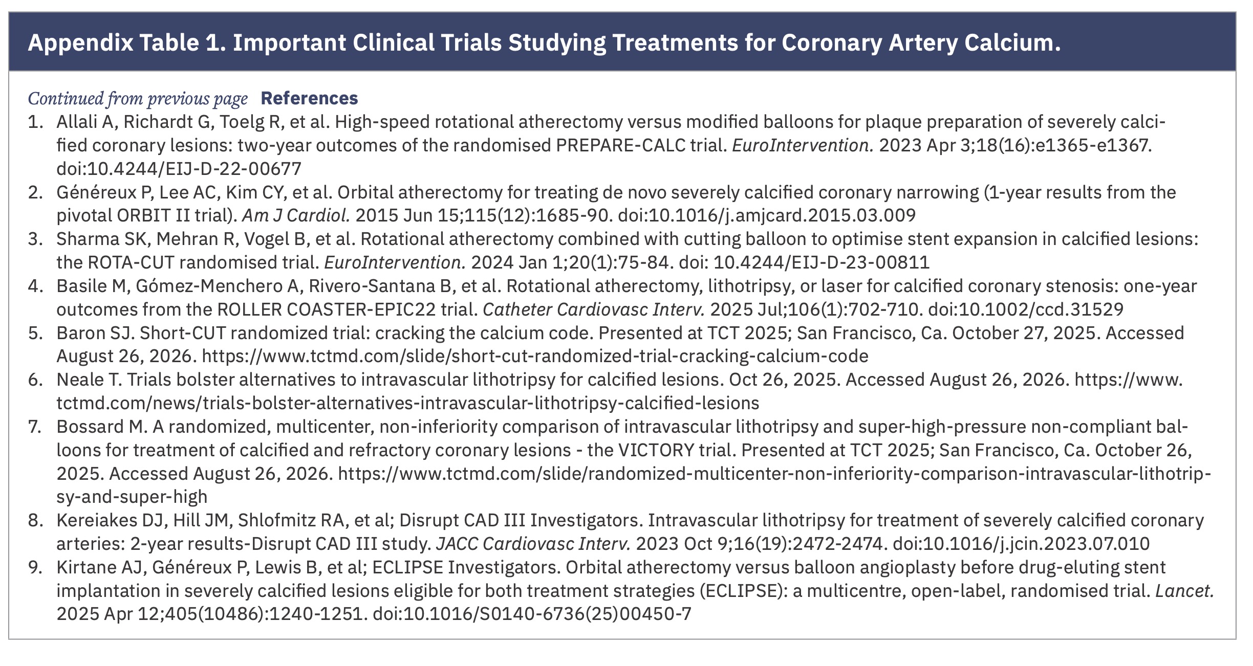
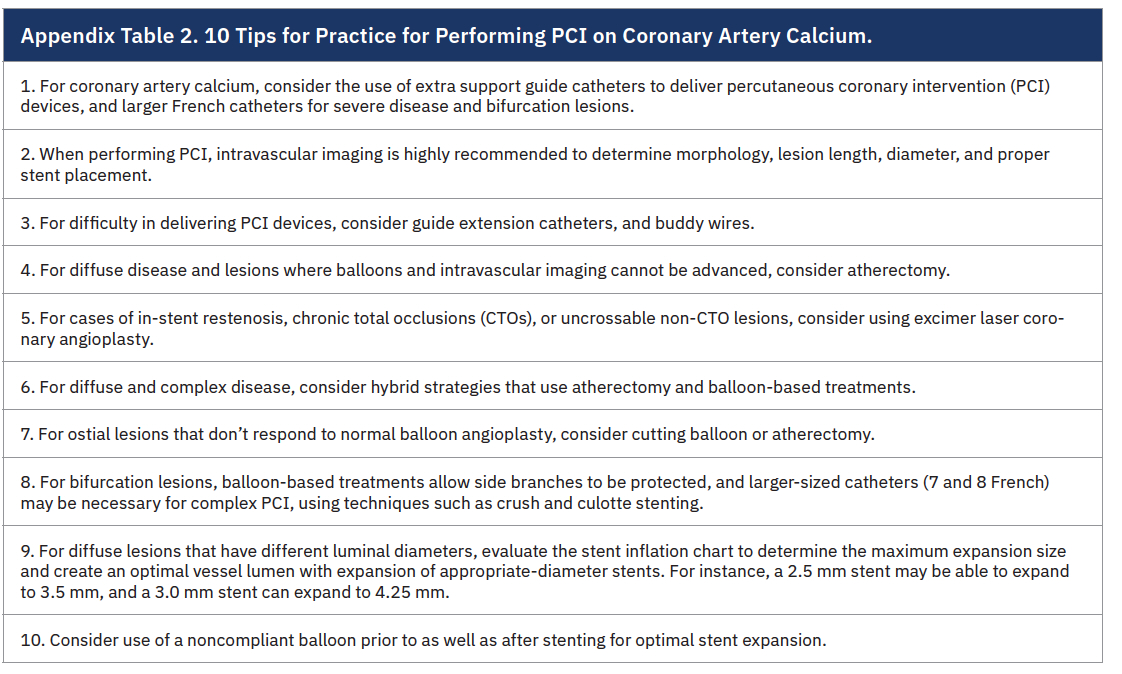
Editor's Note: Appendix Table 1. Important Clinical Trials Studying Treatments for Coronary Artery Calcium and Appendix Table 2. 10 Tips for Practice for Performing PCI on Coronary Artery Calcium are available at the end of the article.
It is estimated that as many as 30% of patients with coronary artery disease (CAD) requiring percutaneous coronary artery intervention (PCI) have lesions with significant coronary artery calcium (CAC).1,2 These numbers are increasing as more people are developing risk factors associated with CAC, including classic cardiovascular risk factors like hypertension, diabetes, tobacco use, obesity, hyperlipidemia and aging.
CAC is also closely linked to chronic kidney disease (CKD), inflammatory processes, and genetic conditions. CKD affects approximately 40 million American adults, and these numbers have more than doubled since 1990.3-5 Patients with CKD also face additional CAC risks because of associated metabolic, mineral, and bone disorders, which are associated with CAC of the media and adventitia.3,4
While CAC begins to develop early in life, it typically becomes detected and clinically significant by age 70. By age 70, more than 70% of adults have some level of CAC.3,6 Men develop CAC about a decade earlier than women, due to estrogen-related protection for women. As the estrogen effect diminishes, women’s CAC rates begin to rapidly accelerate.3,6 Additionally, the population has significantly aged over the past 20 years and the increasing numbers of elderly patients is closely linked to increases in CAC.
Clinical Presentation
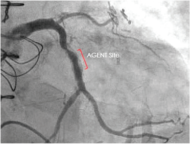
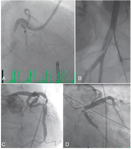
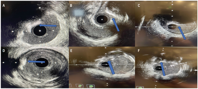
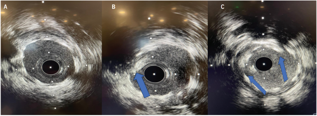
CAC is a complex, diffuse disease process and commonly presents as heterogeneous disease.6-8 It can be superficial or deep, non-obstructive, flow-limiting or completely obstructive, and replace the intima or develop subintimally.7,8 It is not uncommon to see complicated CAC presentations such as eccentric, concentric, flow-limiting, and superficial lesions in the same vessel. Furthermore, CAC may also be complicated by micro calcifications, thrombotic or fibrotic components, extremely dense areas, and layered or concentric sheets (Figure 1).7,8 The vessel lumen may also have different diameters in the same target vessel area, or involve ostia or bifurcations (Figure 2), which can create more challenges when performing PCI.
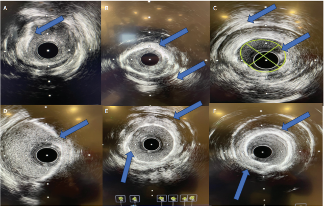
CAC can also be nodular. Calcium nodules (CN) are defined as an eccentric calcium mass with a convex structure within the vessel lumen that may present in as many as 30% of CAC patients (Figure 3).9-12 CNs are usually accompanied by a heavily calcified plaque underneath and acoustic shadowing on IVUS imaging.9-11 In some scenarios, including acute coronary syndromes (ACS), CNs can present as eruptive, unstable lesions with a disrupted fibrous cap and thrombus. CNs may also present as non-eruptive, heavily calcified, stable lesions that are extremely resistant to PCI.9-11 CNs are associated with significantly higher rates of adverse events during PCI, including vessel dissection, perforation, tissue extrusion, or mechanical deployment issues with stents.12-14
Challenges
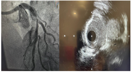
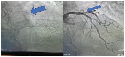
Because of its complexity, CAC can be associated with significant PCI challenges. This includes reduced technical and clinical success rates due to stent damage, stent under deployment and malapposition, and higher stent restenosis rates (Figure 4). Therefore, properly modifying, treating and imaging of CAC is essential to optimizing PCI outcomes. Intravascular imaging is highly recommended to determine the severity of CAC and choose the best strategies, such as atherectomy, balloon-based technologies, and stents, to treat CAC.
CAC Identification on Imaging
CAC is visible on coronary artery tomographic angiography (CCTA) and conventional radiography, as well as fluoroscopy and cine angiography. Coronary computed tomography calcium scoring (CAC scoring), and CCTA are commonly used diagnostic tools for initially evaluating the presence and severity of CAC. CAC scoring, using the Agaston scale, is an important, cost-effective screening tool for assessing the risk of cardiovascular events and determining calcium burden in coronary arteries. However, it cannot differentiate between intimal and medial plaque, nor determine the presence of obstructive disease (Figure 5).15 Calcium scoring only provides a cumulative calcium burden score without identifying plaque composition or vulnerability.15
CCTA is an excellent imaging modality to visualize CAD. However, CCTA is also limited in diagnosing the true severity and complexity of CAC. It can overestimate the severity of disease due to variables such as blooming artifacts, vessel overlap, and smaller plaques in a vessel.16,17 CCTA also requires intravenous contrast, gating techniques to eliminate motion artifacts, and heart rate and rhythm control to produce high-quality images. CCTA is valuable for assessing CAC, but still needs improvements in specificity to quantify CAC severity.16,17
CAC can also be visualized on conventional radiographs like a chest x-ray. In the cath lab, CAC is visualized as a pre contrast injection with fluoroscopy or cine angiography. Angiography alone cannot determine the morphology, complexity, or severity of CAC, because it is a luminogram that does not assess blood flow directly.18,19 When injecting contrast through the vessel lumen, angiography has the potential to overestimate CAC severity because dense calcium can obscure lesion morphology and vessel boundaries. Because of limitations in interpreting CAC with angiography, intravascular imaging is essential to determine the complexity and severity of CAC.
IVUS and OCT
Intravascular ultrasound (IVUS) or optical coherence tomography (OCT) are recommended for PCIs involving CAC. These imaging systems visualize the vessel lumen and wall layers, determine the morphology and severity of CAC and the target vessel revascularization (TVR) area, and help to determine appropriate plaque modification strategies to optimize procedural outcomes. IVUS is a well-established technology using either a mechanically rotating single-element system or phased-array system through a 5 or 6 French catheter. It provides high-quality data that helps determine morphology, diameter, and length of a TVR area. IVUS is also recommended for assessing stent deployment, challenging in CAC. Rotational IVUS, using a pullback sled, can accurately determine lesion length, which also helps optimize stent positioning. Multiple IVUS runs can be performed during a procedure without using iodinated contrast, which is required with OCT, allowing the operator to easily assess the vessel at key procedural intervals, and ensure proper stent expansion and deployment. IVUS is usually more readily available than OCT, another important consideration.
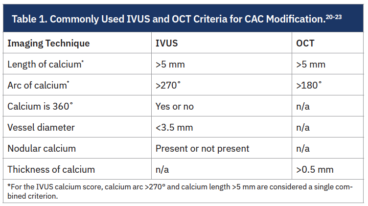
Additionally, IVUS can be performed over a .014-inch guidewire using a monorail catheter and with guide extension catheters. It has a helpful scoring system to assess the severity of CAC and determine if plaque modification is advised. The IVUS calcium score uses four features to assess CAC severity: a superficial calcium arc >270° extending >5 mm, 360° calcium, the presence of a calcified nodule, and vessel diameter <3.5 mm.20 A score of 2 or more suggests a higher risk of stent underexpansion and supports plaque modification.20 Commonly used IVUS and OCT criteria for CAC modification are shown in Table 1. These criteria are relative, and the presence of diffuse flow-limiting disease, severely obstructive calcium, or calcified nodules may also require plaque modification techniques.
Limitations of IVUS include that it can overestimate the lumen area by 10% and that results may be less reproducible compared with OCT.21-23 IVUS is also considered less effective than OCT for assessing deep calcium and stent deployment. Dense calcium can obscure the vessel lumen with acoustic shadowing.22,23 Overall, however, IVUS is easy to set up and use. It provides high-quality information about the TVR area. IVUS can also assess diffuse disease areas within a lesion, provide valuable information about stent deployment, does not use contrast, and reduces radiation exposure by using ultrasound to provide critical lesion data.
OCT uses coherent near-infrared light to visualize the coronary artery lumen and assess CAC. The high resolution of OCT enables better assessment of CAC versus IVUS because OCT visualizes the arterial walls through dense calcium and eliminates acoustic shadowing.23 OCT is also considered to be superior to IVUS in determining the thickness, depth, and eccentricity of calcified lesions. OCT-based imaging is associated with improved post-PCI outcomes because of its superiority in assessing stent apposition23,24 and identifying post-PCI stent complications, including edge dissections, malapposition, and underexpansion. OCT has a “rule of 5” scoring system that is similar to IVUS for interpreting CAC, with a few notable exceptions.21-24 OCT’s main limitation is the need to use iodinated contrast for blood clearance, which is a major consideration with patients who have CKD. OCT generally requires a pre-scripted pullback sequence, whereas IVUS can be manually controlled, making it more versatile in some situations.
Atherectomy
After assessment with intravascular imaging, numerous adjunctive devices are available to treat CAC and prepare the vessel for stenting. This includes atherectomy and balloon-based devices. Atherectomy is an important treatment step for CAC, with three major devices available for use: rotational atherectomy (RA), orbital atherectomy (OA) (Table 2), and laser atherectomy (ELCA). Atherectomy debulks calcium from the lesion and prepares it for further modification and stenting. If a CAC lesion is not crossable with IVUS or balloons, atherectomy devices should be considered as the initial interventional step (Table 3).
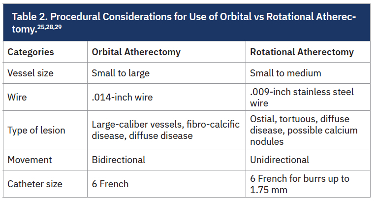
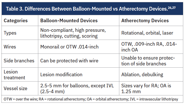
Rotational Atherectomy. RA is a well- established technology that can be used to modify CAC. RA ablates inelastic CAC tissue while maintaining the integrity of elastic tissue, due to the principle of differential cutting.25 RA is effective for ostial lesions, uncrossable lesions, vessels with small-to-medium lumen vessels, tortuous, diffuse disease, and heavily calcified lesions that cannot be dilated or crossed with balloons. It may be beneficial for treating non-eruptive CNs. Additionally, clinical trials are now demonstrating the effectiveness of using RA with either intravascular lithotripsy (IVL) or cutting balloons to further modify CAC before stent placement.26,27
Some important considerations for RA include the need for larger guide catheters for burrs >1.75 mm, which are uncommonly needed. The procedure also requires an .009-inch stainless steel wire, and this wire may not navigate easily through CAC lesions. Additionally, the wire is not optimal for balloon angioplasty and stent delivery, so the procedure may require multiple wire exchanges using an over-the-wire (OTW) catheter. RA can also cause bradycardia in right coronary artery (RCA) lesions. No-reflow phenomenon can occur, especially in cases where there is extreme burr deceleration.
Orbital Atherectomy. Orbital atherectomy ablates CAC and fibro-calcific debris in vessels sized from 2-4 mm using bidirectional sanding, and centrifugal and pulsatile forces. It is a single 1.25 mm device that advances over a tapered .014-inch guidewire. OA is able to treat larger luminal areas than RA by using bidirectional movements and changing the speed of the device.28 It is preferred for larger caliber vessels, but can also be used in ostial disease and smaller lumen vessels. Overall, however, RA is generally considered superior to OA for tight ostial lesions, and smaller caliber and more tortuous lesions.
Data suggests both RA and OA are safe and effective as pre-treatment strategies before stent deployment and have similar overall outcomes. However, OA is associated with lower rates of all-cause mortality, TVR, coronary artery bypass graft (CABG) surgery, and fluoroscopy time.29 OA also provides advantages for setup because it doesn’t require a nitrogen tank or foot pedal to operate. The .014-inch wire also reduces the amount of wire exchanges, as a full intervention can theoretically be completed over the guidewire.
ELCA. Laser atherectomy is not a front-line treatment for de novo CAC lesions. Its best CAC clinical indications are for in-stent restenosis, chronic total occlusions (CTOs) and non-CTO lesions that are truly uncrossable, even with the use of RA or OA.30,31 In these cases, ELCA modifies the plaque and creates a channel that allows for the use of other atherectomy devices and balloons.30,31 However, RA, OA and balloon-based technologies are far more likely to be used for CAC modification in de novo and non-CTO lesions.
Balloons
There are also numerous balloon-based technologies for CAC plaque modification. Balloons modify plaque through a variety of processes such as high-pressure inflations, lithotripsy, and the use of atherotomes and metal scaffolding for cutting into plaque. When specialty balloons can be delivered, there are numerous advantages to balloon-based treatments. All balloon-based options can be performed over an .014-inch wire using either a monorail or OTW system, which allows the entire procedure to be completed with a single wire. Balloon-based technologies also allow for side branch wiring and protection for bifurcation lesions. They can also be used with guide extensions and buddy wires. When a balloon expands in CAC, it helps ensure that the vessel will conform to the 1:1 stent ratio needed for optimal stent deployment. Several studies support a comprehensive approach for CAC treatment that integrates balloons and atherectomy to modify plaque, maximize the vessel lumen, and improve stent deployment success.
Of note, even with advances in the construction of specialty balloons, deliverability of these balloons may be difficult in many patients with CAC. Additionally, balloon-based devices are vulnerable to unsuccessful treatment of an area, and issues such as “dog-boning” or “watermelon seeding” during inflations. Repeated unsuccessful inflations can limit the ability to perform atherectomy during the procedure, and cause dissections, perforations, and other complications. Additionally, many specialty balloons treat small, defined areas, which may limit their effectiveness in treating diffuse CAC. RA and OA should be considered for diffuse disease, along with balloon-based treatment.
Noncompliant (NC) Balloons and High- Pressure Balloons. Since compliant or semi-compliant balloons may not properly expand CAC lesions, NC balloons can be considered as a method to adequately prepare calcified lesions prior to stenting. NC balloons are effective for lesions with superficial, concentric CAC because they have a more predictable diameter and deliver better stent expansion results than compliant or semi-compliant balloons. NC balloons are also less likely to dog bone or watermelon seed. Although NC balloons can be inflated to relatively high pressures (20-24 atmospheres [atm]), they may still be vulnerable to dog-boning or rupture in a heavily calcified vessel.32 There is also increased risk for vascular injuries with high inflations, so high-pressure inflations should be performed carefully. NC balloons are also the preferred balloon for post dilation of stents, especially in CAC patients with suboptimal stent deployment. Therefore, NC balloons are an essential, frontline CAC treatment device.
High-pressure NC balloons (HPB) are double-layered balloons capable of expansion to 35 atm and can be considered when a regular NC balloon fails to properly dilate a CAC lesion. HPBs were recently compared to IVL and scoring balloons, and found to be effective for treating CAC in limited clinical trials.33,34 When using HPBs, it is recommended to initially use a 0.6 to 0.7 ratio, similar to RA, to modify CAC. The vessel can then be expanded to a 1:1 ratio if necessary. Inflations for HPB should be done slowly, using a dedicated inflation device. Any inflation above 20 atm should be increased by five atmospheres every 10-20 seconds.33,34 Deliverability may be more difficult than other devices, due to bulkiness. HPBs may also have little impact on nodular or eccentrically calcified plaques.33-35
Intravascular lithotripsy (IVL). IVL is an important advance in treating CAC. IVL modifies CAC plaques using a balloon catheter that emits acoustic pressure waves, causing calcium micro fractures across a range of calcification arcs.35 IVL is associated with less medial injury than cutting and ultra-high-pressure balloons while effectively inducing calcium fractures, including in lesions with smaller calcification arcs (Table 4).36
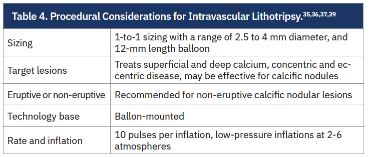
IVL uses low inflation pressure of 2-6 atm because it only needs to make contact with the vessel wall to deliver therapy, minimizing the chances for dissection and perforation. Shockwave (Johnson & Johnson MedTech), for example, has balloons that are 2.5-4 mm in diameter and 12 mm in length, and can deliver 12 rounds of therapy for a max of 120 pulses. The increase in pulses from 80 to 120 in the newer generation of the device improves its ability to treat diffuse disease. IVL has also been studied in calcified nodules and can induce fractures in superficial, deep, and eccentric calcium, broadening its applicability across different calcium morphologies. IVL can also be used in conjunction with atherectomy, especially for diffuse and/or heterogeneous lesions, to optimize outcomes.36,37 Limitations include the available size range (2.5-4 mm for coronary lesions), its relatively short length (12 mm), which can limit its ability to treat diffuse disease, and challenges in deliverability, a challenge for all balloon-based, adjunctive CAC treatments.
Cutting and Scoring Balloons. Cutting balloons are NC balloons with atherotomes, or microsurgical blades, on the surface, that create longitudinal, micro-incisions in CAC with low pressure inflations (6-12 atm).38 Cutting balloons have been found to be superior to compliant and NC balloons for treating CAC, and effective as part of a hybrid strategy using RA. There is limited clinical evidence of non-inferiority to IVL for superficial calcium treatment.39 Overall, cutting balloons, when deliverable, can be effective for ostial lesions, bifurcation lesions, superficial calcium, and fibrocalcific disease.40
Scoring balloons are semi-compliant or sometimes NC balloons, that have external silicone or nitinol wires to create micro incisions in CAC.38,39 Scoring balloons are generally sized up to 75% of the reference vessel diameter and inflated slowly. This allows the balloon to generate focused energy on the area while scoring into the plaque to create micro incisions. Scoring balloons are useful for concentric and eccentric calcification, where focal scoring lines facilitate subsequent stent expansion.39-41 Primary limitations include reduced effectiveness in deep calcification or nodules, where deeper plaque disruption may be required.40,41
No significant differences have been reported between cutting and scoring balloons for complications such as perforation, dissection, slow/no flow, in-hospital death, myocardial infarction, or repeat revascularization.39-41 Therefore, it is operator preference and experience that may guide the decision on whether to use a cutting or scoring balloon.
Conclusion
CAC can pose significant challenges for modification and successfully preparing the vessel for stenting during PCI. Plaque modification can be complicated by heterogenous disease within the target vessel, bifurcations, eruptive and non-eruptive coronary nodules, and deep calcium. CAC is a major challenge for successfully deploying stents and requires a well-planned approach for technical and long-term clinical success. There are numerous adjunctive therapies to treat CAC and optimize outcomes, including atherectomy and balloon-based therapies. Clinical trials have also indicated that hybrid strategies, such as using atherectomy and specialty balloons, may optimize outcomes and better manage complex CAC.
References
1. McInerney A, Hynes SO, Gonzalo N. Calcified coronary artery disease: pathology, prevalence, predictors and impact on outcomes. Interv Cardiol. 2025 Feb 14;20:e02. doi:10.15420/icr.2024.20
2. Rifkin B, Valeri A. Coronaries, calcium, and kidney consequences. Kidney Int Rep. 2024 Aug 13;9(10):2839-2841. doi:10.1016/j.ekir.2024.06.040
3. Madhavan MV, Tarigopula M, Mintz GS, et al. Coronary artery calcification: pathogenesis and prognostic implications. J Am Coll Cardiol. 2014 May 6;63(17):1703-14. doi:10.1016/j.jacc.2014.01.017
4. Sarnak MJ, Amann K, Bangalore S, et al; Conference Participants. Chronic kidney disease and coronary artery disease: JACC state-of-the-art review. J Am Coll Cardiol. 2019 Oct 8;74(14):1823-1838. doi:10.1016/j.jacc.2019.08.1017
5. Cannata-Andía JB, Martín-Carro B, Martín-Vírgala J, et al. Chronic kidney disease-mineral and bone disorders: pathogenesis and management. Calcif Tissue Int. 2021 Apr;108(4):410-422. doi:10.1007/s00223-020-00777-1
6. Gerke O, Lindholt JS, Abdo BH, et al. Prevalence and extent of coronary artery calcification in the middle-aged and elderly population. Eur J Prev Cardiol. 2022 Feb 9;28(18):2048-2055. doi:10.1093/eurjpc/zwab111
7. Riley RF, Patel MP, Abbott JD, et al. SCAI expert consensus statement on the management of calcified coronary lesions. J Soc Cardiovasc Angiogr Interv. 2024 Jan 31;3(2):101259. doi:10.1016/j.jscai.2023.101259
8. Otsuka F, Sakakura K, Yahagi K, et al. Has our understanding of calcification in human coronary atherosclerosis progressed? Arterioscler Thromb Vasc Biol. 2014 Apr;34(4):724-736. doi:10.1161/ATVBAHA.113.302642.
9. Torii S, Sato Y, Otsuka F, et al. Eruptive calcified nodules as a potential mechanism of acute coronary thrombosis and sudden death. J Am Coll Cardiol. 2021 Apr 6;77(13):1599-1611. doi:10.1016/j.jacc.2021.02.016
10. Sato Y, Finn AV, Virmani R. Calcified nodule: A rare but important cause of acute coronary syndrome with worse clinical outcomes. Atherosclerosis. 2021 Feb;318:40-42. doi:10.1016/j.atherosclerosis.2020.12.009
11. Demuyakor A, Hu S, Koniaeva E, et al. Impact of nodular calcification in patients with acute coronary syndrome (ACS) treated with primary percutaneous coronary intervention (PCI). BMC Cardiovasc Disord. 2022 Mar 14;22(1):103. doi:10.1186/s12872-022-02551-7.
12. Fernández-Cordón C, Brilakis ES, García-Gómez M, et al. Calcified nodules in the coronary arteries: systematic review on incidence and percutaneous coronary intervention outcomes. Rev Esp Cardiol (Engl Ed). 2025 Nov;78(11):977-991. English, Spanish. doi:10.1016/j.rec.2025.03.004
13. Ali ZA, Spratt JC, Finn AV, et al. Identification and treatment of calcified nodules in percutaneous coronary intervention. EuroIntervention. 2025 Dec 1;21(23):e1424-e1433. doi:10.4244/EIJ-D-25-00296
14. Hennessey B, Pareek N, Macaya F, et al. Contemporary percutaneous management of coronary calcification: current status and future directions. Open Heart. 2023 Feb;10(1):e002182. doi:10.1136/openhrt-2022-002182
15. Sabouret P, Giamundo DM, Rosencher J, Figliozzi S. Coronary artery calcium scoring in 2026: strengths, limitations, and optimized clinical use. Front Radiol. 2026 Apr 13;6:1822303. doi:10.3389/fradi.2026.1822303
16. Gupta A, Bera K, Kikano E, et al. Coronary artery calcium scoring: current status and future directions. Radiographics. 2022 Jul-Aug;42(4):947-967. doi:10.1148/rg.210122
17. Latina J, Shabani M, Kapoor K, et al. Ultra-high-resolution coronary CT angiography for assessment of patients with severe coronary artery calcification: initial experience. Radiol Cardiothorac Imaging. 2021 Aug 26;3(4):e210053. doi:10.1148/ryct.2021210053
18. Kyriakoulis I, Nanna MG, Rao-Brito M, et al. The additive value of FFR-CT in the evaluation of calcified coronary arteries with coronary CT angiography. Am J Cardiol. 2026 Jan 15;259:74-80. doi:10.1016/j.amjcard.2025.09.024
19. Gurav A, Revaiah PC, Tsai TY, et al. Coronary angiography: a review of the state of the art and the evolution of angiography in cardio therapeutics. Front Cardiovasc Med. 2024 Nov 25;11:1468888. doi:10.3389/fcvm.2024.1468888
20. Eltelbany M, Cilia L, Truesdell A. A how-to guide for using intravascular imaging to determine calcium modification. Cardiac Interventions Today. 2023 July/August;17(4):30-32. https://assets.bmctoday.net/citoday/pdfs/cit0723_F1_Cilia.pdf
21. Nishi T, Imura S, Kitahara H, et al. Head-to-head comparison of quantitative measurements between intravascular imaging systems: An in vitro phantom study. Int J Cardiol Heart Vasc. 2021 Sep 1;36:100867. doi:10.1016/j.ijcha.2021.100867
22. Khan SU, Kleiman NS, Shah AR. Intravascular ultrasound imaging-guided percutaneous coronary intervention: evidence and practical implications. Methodist Debakey Cardiovasc J. 2025 Aug 12;21(4):26-36. doi:10.14797/mdcvj.1611.
23. Kubo T, Akasaka T, Shite J, et al. OCT compared with IVUS in a coronary lesion assessment: the OPUS-CLASS study. JACC Cardiovasc Imaging. 2013 Oct;6(10):1095-1104. doi:10.1016/j.jcmg.2013.04.014
24. d’Entremont MA, Jolly SS. OCT and calcium-you cannot treat what you cannot see. JAMA Cardiol. 2025 Jul 1;10(7):675-677. doi:10.1001/jamacardio.2025.0753
25. Gupta T, Weinreich M, Greenberg M, et al. Rotational atherectomy: a contemporary appraisal. Interv Cardiol. 2019 Nov 18;14(3):182-189. doi:10.15420/icr.2019.17.R1
26. Leone PP, Sartori S, Farhan S, et al. Rotational atherectomy with or without intravascular lithotripsy for patients with heavy coronary artery calcification. Am J Cardiol. 2025 Jul 15;255:41-48. doi:10.1016/j.amjcard.2025.07.002
27. Altobaishat O, Abouzid M, Tanashat M, et al. Rotational atherectomy with cutting balloon before stenting in severely calcified coronary lesions: a meta-analysis. Future Cardiol. 2024 Dec-Dec;20(15-16):859-870. doi:10.1080/14796678.2024.2440220
28. Vidovich MI, Latif F. Rotational vs. orbital atherectomy: how to choose? SCAI.org. August 21, 2020. Accessed August 25, 2026. https://www.scai.org/quality-improvement-tools/qi-tips/rotational-vs-orbital-atherectomy-how-choose
29. Altobaishat O, Abuelazm M, Amin AM, et al. Rotational atherectomy versus orbital atherectomy for the treatment of calcified coronary lesions: a systematic review and meta-analysis of 81,873 patients. Eur Heart J. 2024 Oct;45(Suppl 1):ehae666.2365. https://doi.org/10.1093/eurheartj/ehae666.2365
30. Yeung JYK, Chiang M. Current role of excimer laser coronary angioplasty atherectomy in calcified coronary artery disease management. JACC Case Rep. 2025 Mar 5;30(5):103025. doi:10.1016/j.jaccas.2024.103025
31. Golino L, Caiazzo G, Calabrò P, et al. Excimer laser technology in percutaneous coronary interventions: Cardiovascular laser society’s position paper. Int J Cardiol. 2022 Mar 1;350:19-26. doi:10.1016/j.ijcard.2021.12.054
32. Maffey MW, Bagur R. Dedicated balloon techniques for coronary calcium modification. Interv Cardiol. 2024 Aug 15;19:e13. doi:10.15420/icr.2024.06
33. Super-high-pressure non-compliant balloons for treatment of calcified coronary lesions noninferior to intravascular lithotripsy: results from VICTORY announced at TCT 2025. CRF.org. October 26, 2025. Accessed June 24, 2026. https://www.crf.org/crf/news-and-events/news/news/3978-super-high-pressure-non-compliant-balloons-for-treatment-of-calcified-coronary-lesions-noninferior-to-intravascular-lithotripsy
34. Rheude T, Rai H, Richardt G, et al. Super high-pressure balloon versus scoring balloon to prepare severely calcified coronary lesions: the ISAR-CALC randomised trial. EuroIntervention. 2021 Aug 27;17(6):481-488. doi:10.4244/EIJ-D-20-01000
35. Kumar S, Trenschel R, Mishra T, et al. Cracking coronary calcium with intravascular lithotripsy: a review. Am J Cardiol. 2026 May 15;267:120-133. doi:10.1016/j.amjcard.2026.02.032
36. Sekimoto T, Fujiyoshi K, Kawakami R, et al. Comparison of vascular injury from intravascular lithotripsy, cutting, or ultra-high-pressure balloons during coronary calcium modification. JACC Cardiovasc Interv. 2025 Sep 8;18(17):2093-2104. doi:10.1016/j.jcin.2025.06.035.
37. Dash D, Bortnick AE. Shocking the rock with coronary intravascular lithotripsy in contemporary practice. American College of Cardiology. February 3, 2026. Accessed August 25, 2026. https://www.acc.org/latest-in-cardiology/articles/2026/02/03/11/30/shocking-the-rock-with-coronary-intravascular-lithotripsy
38. Mangieri A, Nerla R, Castriota F, et al. Cutting balloon to optimize predilation for stent implantation: The COPS randomized trial. Catheter Cardiovasc Interv. 2023 Mar;101(4):798-805. doi:10.1002/ccd.30603.
39. Cader A. Randomised comparison of intravascular lithotripsy vs. cutting balloon treatment in calcified coronary artery disease – The Short-CUT trial. PCRonline.com. October 27, 2025. Accessed June 24, 2026. https://www.pcronline.com/News/Congress-coverages/TCT/2025/Short-CUT-intravascular-lithotripsy-vs.-cutting-balloon-angioplasty-in-calcified-CAD
40. Agarwal P, Mahajan S. Cutting balloon: device, current indication, and usage. Journal of Current Cardiology. 2024 Sept-Dec;2(3):134-139. doi:10.4103/JCC.JCC_22_24
41. Colletti G, Ruzsa Z, Gach O, et al. Calcium modifying dedicated balloons: a contemporary review. Front Cardiovasc Med. 2026 Apr 10;13:1700877. doi:10.3389/fcvm.2026.1700877