Cath Lab Crisis Playbook: A Brief Discussion of What to Do First, Who to Call, and How to Communicate Under Pressure
© 2026 HMP Global. All Rights Reserved.
Any views and opinions expressed are those of the author(s) and/or participants and do not necessarily reflect the views, policy, or position of Cath Lab Digest or HMP Global, their employees, and affiliates.
Morton J. Kern, MD, MSCAI, FACC, FAHA
Clinical Editor; Interventional Cardiologist, Long Beach VA Medical Center, Long Beach, California; Professor of Medicine, University of California, Irvine Medical Center, Orange, California
Disclosures: Dr. Morton Kern reports he is a consultant for Abiomed, Abbott Vascular, Philips, ACIST Medical, and Opsens Inc.
Dr. Kern can be contacted at mortonkern2007@gmail.com
On X @MortonKern
One of the most challenging aspects of invasive cardiology is the ability of the operator and his/her team to efficiently manage a crisis in the lab. Since it’s still early in the academic year with new fellows, I was asked how we think about complications that come up during a procedure. To this end, I thought a ‘playbook’ might organize our thinking. Below is my attempt to provide a highly abbreviated playbook that describes an immediate action plan for the four most-critical cardiac catheterization lab complications: no reflow, perforation, acute closure, and access complications, the steps to begin management, and who to call and what critical communications will help the team members at the start of a crisis.
I recognize that this discussion of complications is incomplete in both context and actions, but it is intended to assist the early- career interventionalist and interventional cardiology fellows formulate an effective plan quickly. During an interventional procedure, it is always critical to plan and build one’s own cath lab crisis playbook. A full discussion of complication management is available from several sources, like the Interventional Cardiac Cath Handbook, which deals with these problems in more detail.1 In addition, Dr. Manos Brilakis of the Minneapolis Heart Institute has produced a remarkable online series of management algorithms for interventional cardiology complications.2,3
Four Vignettes and Complications
A 56-year-old man had 16 hours of anterior chest pain. The electrocardiogram (ECG) shows lateral Q waves and significant inferior lead ST depressions. The angiogram shows a near-total occlusion of the mid right coronary artery (RCA). Troponins were marginally elevated. He was a smoker and was treated for hypertension and hypercholesterolemia, but not diabetes mellitus. His family history was negative for coronary artery disease.
Following the first balloon inflation, ST elevation, chest pain, and drop in blood pressure occurred. Angiography now shows TIMI-1 flow (i.e., no flow). The operator quickly reviews the causes of no reflow. His/her mental playbook should (1) list the steps to a solution, i.e., what to do first, and (2) the need for advice/support from colleagues and/or team members, i.e., who to call.
1. Event: Coronary No-Reflow
Phenomenon
The complication is the no-reflow (NRF) phenomenon, defined as an abrupt reduction in angiographically visualized coronary flow, TIMI grade ≤2 despite vessel patency. Vessel obstructions caused by thrombus/atherosclerosis/dissection/spasm should be treated before calling it no reflow.3 The incidence of no reflow ranges from 12-25%. Large trials such as PAMI and CADILLAC showed 4-7% of treated lesions had TIMI flow ≤2. However, higher rates have been noted when using measurements of microvascular flow other than angiography. While uncommon, NRF more often occurs during primary percutaneous coronary intervention (PCI) for ST-elevation myocardial infarction, usually in the right coronary with a proximal 100% thrombotic occlusion.
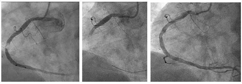
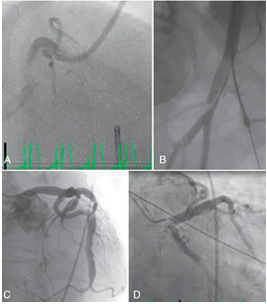
NRF may also occur during saphenous vein graft interventions, rotational atherectomy, or intracoronary air embolus. Clinically, NRF is associated with new or persistent ST elevation, chest pain, and decreased left ventricular function with hypotension. NRF usually resolves in <10-15 minutes. Figure 1 shows the cine frames of the patient before and after management of his no-reflow event.
What to Do First:
1. Stop advancing equipment.
2. Check patient’s blood pressure and ECG for ST elevation.
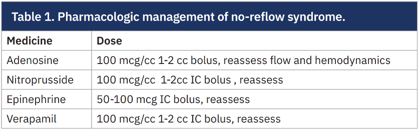
3. If there is no visible mechanical obstruction, administer intracoronary vasodilators (adenosine, nitroprusside, epinephrine, or verapamil [Table 1]). Microcatheters can be helpful in delivering medications to the distal vessel.
4. If the patient becomes hypotensive, begin with fluid resuscitation and vasopressors as needed. Consider an intra-aortic balloon pump (IABP).
5. If no reflow is due to thrombus, manual or mechanical aspiration is appropriate.
6. Additional anticoagulation with more heparin and/or glycoprotein receptor IIb/IIIa inhibitors should be started. Recheck activated clotting time (ACT) levels.
7. NRF that may be due to vasospasm responds to intracoronary (IC) nitroglycerin (NTG) (100 mcg/cc). IC NTG is not effective for the no-reflow phenomenon due to distal microembolization.
Who to Call:
1. Notify a partner or senior interventional cardiologist, especially if you need technical or mechanical circulatory support with IABP/Impella (Abiomed).
2. Call Anesthesia if hemodynamic instability worsens.
Critical Team Communications:
State clearly: “We have a no-reflow situation. Prepare intracoronary [e.g., adenosine, 100mcg for bolus]. Watch the blood pressure.”
2. Event: Coronary Perforation
An 82-year-old man with progressive angina, resting ECG changes with pain, multiple risk factors, and visually calcified mildly tortuous 85% proximal left anterior descending (LAD) on angiography requires revascularization. The operators dilate the calcified lesion with a 3.5 x 15 mm balloon. The next image shows contrast extravasation from the mid part of the lesion.
The playbook scenarios that can lead to coronary perforation include use of a large- diameter balloon or stent in the highly calcified vessel segment, rotational or laser atherectomy, and iatrogenic dissection related to chronic total occlusion PCI. Perforations are graded (Ellis Classifications) by the degree of contrast media stagnating or freely flowing outside the vessel and into the pericardium.
What to Do First:
1. Maintain balloon or wire position.
2. Inflate a balloon at the presumed perforation site (1:1 sizing to block flow) for 1-2 minutes as tolerated.
3. If extravasation is still noted after 2 minutes, re-inflate the balloon for at least 5 to 10 minutes.
4. Reverse heparin anticoagulation with protamine.
5. Obtain urgent echocardiogram to check pericardial tamponade.
6. If cardiac tamponade is diagnosed, start fluid resuscitation, followed by emergency pericardiocentesis.
Who to Call:
1. Echocardiography technologist.
2. Cath lab runner (to grab covered stents and pericardiocentesis kits).
3. Cardiac Surgery team (stat standby).
Critical Team Communications:
State clearly: “We have a perforation in the [vessel name]. The coronary balloon is up. Please get the echo machine in the room. Notify Cardiac Surgery and bring the covered stents and pericardiocentesis tray, please.”
Technical note: A main vessel perforation may be treated by prolonged balloon inflation or covered stent implantation when there is unremitting brisk contrast exiting into the pericardial space. Distal artery perforation and collateral vessel perforation may require embolization of the small branch by coiling or fat embolization.
3. Event: Acute Vessel Closure
Following stenting of a dominant circumflex vessel at a bifurcation with a very large obtuse marginal (OM) branch, the OM1 branch closes. The sudden occlusion of the OM1 branch artery is associated with chest pain and ST changes.
What to Do First:
1. Pass a wire into the occluded branch vessel. Maintain wire position across the main branch.
2. Dilate the occluded branch.
3. Administer intracoronary nitroglycerin to rule out severe spasm.
4. Prepare for deployment of a stent into the OM1.
5. Use intravascular imaging to define the precise cause of the abrupt occlusion (most likely the cause is dissection or tissue shift with thrombus).
Who to Call:
1. A colleague or senior interventionalist, in case help to place a mechanical circulatory support device like Impella may be needed.
2. Notify Cardiac Surgery, especially if very proximal LAD/left main dissection is suspected and difficulty is encountered in reestablishing flow.
Critical Team Communications:
State clearly: “The [name] vessel has closed. I will need another guidewire. Prepare another balloon for inflation. Please pull a [stent size/type] for us.”
4. Event: Vascular Access Complications
A 79-year-old man receiving hemodialysis via left arm fistula reports exertional chest pain and undergoes coronary angiography from the right femoral artery. At the end of the procedure, the patient complains of lower back pain. His blood pressure is now 90/60 mmHg, down from an entry level of 130/80 mmHg. A retroperitoneal hematoma (RPH) is suspected.
Serious vascular access problems are rare but include femoral dissection with occlusion or bleeding, or RPH. For radial access, the most common problems are acute radial occlusion due to dissection, thrombosis, or spasm. Other minor complications like small hematomas or pseudoaneurysms can often be managed outside the lab. The only acute life-threatening access-related complication needing immediate intervention will be uncontrolled retroperitoneal bleeding (RPH).
What to Do First:
1. Recognize signs/symptoms of RPH (recall back pain, leg pain, hypotension, vagal reactions). Fluoroscopy of the bladder may reveal compression from the RPH.
2. Repeat femoral angiography for a correctable cause of RPH. If iliac artery dissection/perforation is the cause of RPH, perform emergency balloon tamponade of the femoral bleeding site. Contralateral femoral access may be required.
3. Start intravenous (IV) fluids and call for crossmatched blood.
Who to Call:
1. Vascular Surgery team (if pulsatile mass expands or patient remains unstable).
2. Ultrasound technologist for bedside imaging.
3. Cath lab runner for peripheral covered large vascular stent, if needed.
Critical Team Communication:
State clearly: “The patient is complaining of groin and back pain, possible RPH. He is hypotensive. Let’s apply manual pressure at the puncture site. Open the IV fluids and send blood for crossmatch. Please call Vascular Surgery. Prepare for contralateral femoral access.”
Notes on Crisis Communications
In the cath lab, under extreme stress, clear team communications are essential to prevent chaos. Several techniques for critical team communications duplicate what should be routine practice in the cath lab. The team should always use closed-loop communications. When giving an order, name the person to perform the task needed. The person acknowledges the order and repeats the order back:
Operator: “John, give 50 mg IV protamine.”
John: “Copy that, giving 50 mg protamine…protamine given.”
Operator: “Thank you.”
When a complication occurs, declare a state of emergency so all know to get in the game now. Calmly and clearly state the complication aloud so the entire room shifts from secondary tasks and focuses on the immediate problem. Assign one voice as the captain of the ship. Only the primary operator or a designated lead speaks to external teams (Surgery, Anesthesia) to avoid conflicting messages.
The Bottom Line
Over the years, Cath Lab Digest has addressed many of these issues on complications, critical communications, cardiopulmonary resuscitation in the lab, and other emergencies (Table 2, Figure 2). I thought that a quick review of what a cath lab crisis playbook would look like for the early career interventionalist and interventional fellow-in-training would be valuable. Thinking ahead is often the best medicine under crisis conditions.
|
Table 2. Cath Lab Digest articles on complications, communications, and leadership during a cath lab crisis.
|
References
1. Lim MJ, Sorajja P, Kern MJ, eds. The Interventional Cardiac Catheterization Handbook. 5th ed. Elsevier; 2023.
2. Brilakis M. Complications – How to Manage. EuroCTO Club. October 1, 2016. Accessed August 31, 2026. https://www.slideshare.net/slideshow/emmanouil-s-brilakis-complications-how-to-manage/67009373
3. Brilakis M. Managing PCI Complications. December 17, 2025. Accessed August 31, 2026. https://www.youtube.com/watch?v=Jz7yqWXLYWk.
4. Eeckhout E, Kern MJ. The coronary no-reflow phenomenon: a review of mechanisms and therapies. Eur Heart J. 2001 May; 22(9): 729-739. doi:10.1053/euhj.2000.2172