




Henry Ford Health
© 2026 HMP Global. All Rights Reserved.
Any views and opinions expressed are those of the author(s) and/or participants and do not necessarily reflect the views, policy, or position of EP Lab Digest or HMP Global, their employees, and affiliates.
EP LAB DIGEST. 2026;26(11):20-27.
Arfaat Khan, MD
Detroit, Michigan
Electrophysiology (EP) has changed significantly over the years, and so has the way we deliver care. What was once a small part of cardiology is now a highly specialized, fast-moving field centered on complex procedures, advanced technology, and coordinated, team-based care.
At Henry Ford Hospital, the EP program has grown into a regional referral center that handles everything from routine arrhythmia management to high-risk ablations, device therapy, and complex lead extractions. The work is demanding, but it runs on a steady rhythm established by a team that has learned to stay aligned, even when cases become unpredictable.
When was the cardiac EP program started at your institution, and by whom?
EP services at Henry Ford Hospital began in 1967 with the early adoption of cardiac pacing and implantable device therapy, including one of the institution’s first pacemaker implantations.
The modern EP program was formally established in 1998 under Claudio Schuger, MD, transitioning EP from a procedural subset of general cardiology into a dedicated service line within the Heart & Vascular Institute.
The program is currently led by Arfaat Khan, MD, Section Chief of Cardiac EP for the Henry Ford Medical Group and Co-Director of the Heart Rhythm Council for Henry Ford Health. In this role, he oversees clinical operations, advanced procedural expansion, and regional referral development.
What prompted the need for an EP program?
The need for a dedicated EP program was driven by sustained growth in arrhythmia prevalence and increasing procedural complexity that exceeded the scope of general cardiology practice. Three primary drivers shaped program development:
- Rapid expansion in atrial fibrillation (AF) incidence requiring catheter-based rhythm control strategies
- Increasing ventricular tachycardia (VT) referrals, particularly scar-mediated and post-infarct VT
- Growth in implantable cardiac device therapy requiring long-term management, upgrades, and extraction capability
Over time, Henry Ford Hospital evolved into a regional referral center for complex EP care, receiving high-acuity cases from across Michigan, northern Ohio, and cross-border referrals from Canada. A sizable portion of volume now reflects tertiary referrals for complex ablation, device complications, and extraction cases that require hybrid operating room (OR) capability.
What is the size of your EP lab facility? Has it expanded or will it soon?
The EP program currently operates across a hybrid procedural infrastructure consisting of:
- 2 dedicated EP laboratories
- 1 shared catheterization laboratory utilized for EP procedures approximately 3 days per week
- 1 hybrid OR used on a scheduled and emergent basis, particularly for lead extraction and complex ventricular arrhythmia procedures
Operationally, this functions as a dynamic 3-room EP system, with capacity flexing based on case complexity and resource allocation.
Construction is underway on a $3.2 billion expansion of Henry Ford Hospital. The state-of-the-art building, expected to open in 2029, will include vastly expanded surgical facilities and a new patient tower. This hospital will add 4 dedicated EP laboratories designed to centralize EP services, reduce dependency on shared cardiac catheterization lab space, and increase capacity for advanced ablation, device therapy, and extraction procedures.
Does your institution offer EP-related procedures in an ambulatory surgical center (ASC)?
The EP program does not operate within a standalone ASC model. Instead, EP services are delivered through a hospital-based, outpatient-first structure.
A considerable proportion of procedures are performed in the outpatient setting, with structured pathways supporting same-day discharge (SDD) when clinically appropriate.
These include:
- Cardioversion and transesophageal echocardiography (TEE)-guided cardioversion
- Loop recorder implantation
- Device implantations
- Catheter ablation cases in appropriate patients
The system is designed to maintain inpatient-level safety infrastructure while functioning operationally as a high-throughput outpatient EP service.
Who manages your EP lab, and what are their credentials and experiences?
EP and cardiac catheterization lab operations are overseen by Christa Means, RN, BSN, Clinical Practice Director for Cardiac Catheterization and EP Services. She brings 12 years of nursing experience, 7 years in progressive leadership roles, and 3.5 years within Henry Ford Health. Her credentials include RCIS, MSN, MBA, and NE-BC.
She works closely with Ed Muxlow, RN, our EP lab leader. Ed has worked as a nurse in the cath/EP space at Henry Ford Hospital since 2001 and has been in his current EP lab lead role since 2006.
Together, their responsibilities include:
- Daily operational oversight of EP and cath lab workflow
- Staffing models and shift coordination across procedural suites
- Supply chain and inventory governance
- Coordination with anesthesia, cardiothoracic (CT) surgery, and interventional cardiology
- Throughput optimization and procedural readiness planning
What is the number of staff members?
The EP program includes:
- 7 electrophysiologists
- 5 advanced practice providers (APPs)
- Approximately 10 registered nurses assigned to procedural areas
- 1 dedicated EP technologist
- 4 EP fellows in training
- Supporting scheduling, administrative, and coordination staff
This structure supports simultaneous multi-room procedural operation while maintaining academic training, add-on case flexibility, and high-complexity procedural coverage.
What types of procedures are performed?
The EP program provides full-spectrum arrhythmia and device therapy across both routine and complex referral cases.
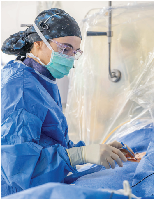
Core ablation services include AF (pulsed field ablation [PFA]), atrial flutter (AFL), atrioventricular nodal reentrant tachycardia, atrioventricular reentrant tachycardia, accessory pathways, premature ventricular complexes (PVCs), and complex VT, including epicardial VT ablation.
Device-based procedures include implantation of permanent pacemakers, implantable cardioverter-defibrillators (ICDs), cardiac resynchronization therapy defibrillators (CRT-Ds), leadless pacemakers, and implantable loop recorders (ILRs). Conduction system pacing (CSP), including left bundle branch area pacing for patients requiring physiologic ventricular activation, has become a routine strategy.
Structural EP services include left atrial appendage occlusion (LAAO) performed in coordination with imaging and anesthesia teams, as well as hybrid AF ablation performed in collaboration with CT surgery for patients with persistent and long-standing persistent AF.
Lead extraction represents a major referral-based service line and includes infected system removal, lead malfunction, venous occlusion, and device system upgrades in structurally complex patients.
How many ablations, device implants, lead extractions, and LAAO procedures are performed each week?
The program maintains consistently high procedural throughput across multiple EP rooms, with variability based on case complexity and hybrid resource utilization.
Under routine conditions, EP rooms collectively support a high weekly ablation volume distributed across AF, VT, and supraventricular tachycardia procedures. Device implantation volume remains steady, driven by both de novo implants and generator replacements, with additional ILR volume integrated into outpatient pathways.
Lead extraction is performed on a structured schedule (typically every other week), with additional urgent cases added for infection or system failure. Because of their extended procedural time and hybrid OR requirements, these cases significantly affect room allocation and overall daily throughput.
LAAO is performed regularly as part of the structural EP workflow, with shared imaging and anesthesia support.
On days with a high volume of VT ablations or lead extractions, the total procedural volume per room decreases due to procedural duration, mapping complexity, and the need for intraprocedural escalation capability. The program performs an average of 30 to 40 cases per week.
What EP equipment is commonly used?
The laboratory operates a multi-platform EP ecosystem designed to support operator preference, case complexity, and redundancy across systems.
Electroanatomic mapping platforms include CARTO (Johnson & Johnson MedTech), EnSite X (Abbott), and the Affera Mapping and Ablation System (Medtronic).
Ablation technologies include PFA systems (FARAPULSE [Boston Scientific]; VARIPULSE [Johnson & Johnson MedTech], and the Sphere-9 Catheter [Medtronic]) as the primary AF ablation modality. Radiofrequency energy (RF) is used selectively for atypical AFLs, VT substrate modification, and lesion completion strategies.
Intracardiac imaging and procedural support are provided using ACUSON SC2000 intracardiac echocardiography (ICE) (Siemens Healthineers), Philips imaging systems, and the CardioLab (GE HealthCare) recording system, with Micropace used for programmed stimulation.
Lead extraction tools include laser systems from Spectranetics, with adjunctive use of systems from Shockwave Medical and the AlphaVac System (AngioDynamics) in select anatomically complex cases.
Vascular access closure is standardized using the MYNX CONTROL Vascular Closure Device (Cordis) and the Perclose (Abbott) system.
What new technologies or techniques have been introduced in your lab, and how have they changed procedures?
Recent innovation has centered on PFA adoption, expansion of CSP, and reduction of fluoroscopy dependency across most procedural types.
The introduction of PFA has significantly altered AF workflow design, shifting ablation strategy from lesion-by-lesion thermal delivery to anatomically standardized energy deployment.
CSP has expanded as a preferred physiologic pacing strategy, particularly for patients requiring ventricular synchrony preservation.
Parallel adoption of multi-platform mapping systems has allowed operator-specific flexibility while maintaining procedural consistency and reproducibility across the lab.
Discuss your program’s use of PFA.
PFA was introduced in August 2024 and has rapidly become the dominant modality for AF ablation. Approximately 95% of our AF ablations are now performed using PFA technology.
RF energy is selectively used in cases of typical or atypical AFL ablations or where lesion reinforcement is required based on operator assessment.
The transition to PFA has reduced procedure variability, improved workflow efficiency, and standardized AF ablation across multiple operators while maintaining consistent acute procedural endpoints.
Discuss your use of hybrid AF ablation.
Hybrid AF ablation is reserved for persistent and long-standing persistent AF cases requiring both epicardial and endocardial intervention.
Procedures are performed in the OR in collaboration with CT surgery. Epicardial ablation is performed first, followed by endocardial mapping and targeted ablation when indicated. Using a portable EnSite X system, our surgeons identify areas of voltage in the epicardial space, allowing for improved localization of AF targets and procedural endpoints.
The program benefits from integrated access to EP labs, cath labs, and OR environments, enabling rapid escalation for complex anatomy or intraprocedural decision-making.
Discuss your use of robotic magnetic navigation.
Stereotaxis robotic navigation is used selectively, primarily for PVC cases or ablations in patients with congenital heart disease that may be considered anatomically complex. Its use is operator-dependent and case-specific rather than routine.
How is inventory managed?
Inventory is managed through Epic Cupid, with a dedicated EP inventory structure integrated into daily operations.
Daily pre-case huddles assess upcoming procedural demand, case complexity, and supply readiness. This allows proactive adjustment of inventory staging prior to first-case start.
High-cost consumables are controlled through structured utilization pathways, with selective opening of devices based on procedural confirmation rather than preemptive setup.
Billing supply audits are performed by dedicated staff to ensure charge accuracy and alignment with procedural documentation.
The system is designed to minimize consignment dependency while maintaining readiness for complex, high-variability EP procedures.
Tell us about your device clinic.
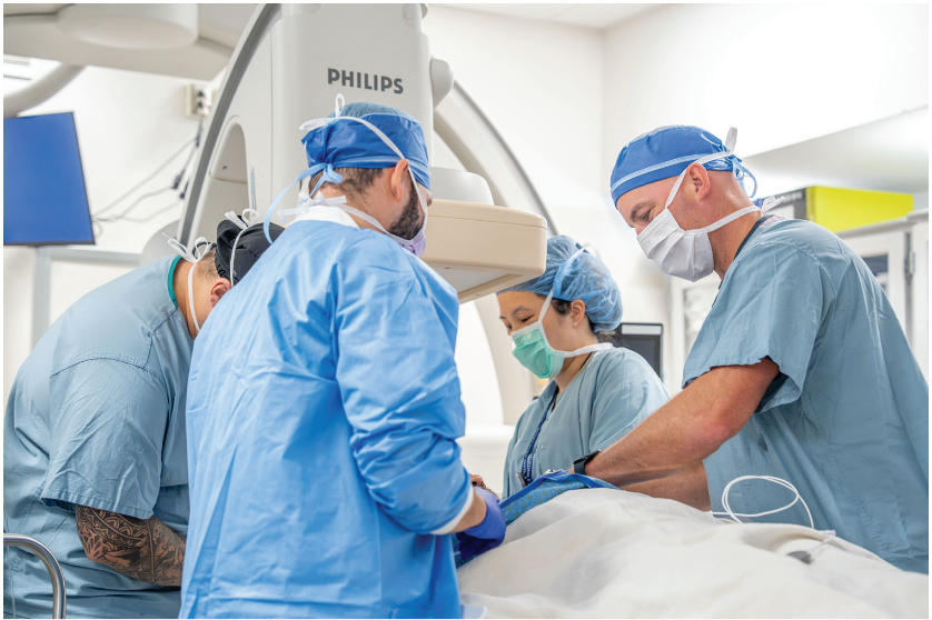
The device clinic functions as an extension of the EP service line, supporting longitudinal management of implanted cardiac devices including pacemakers, ICDs, CRT systems, and ILRs.
Operations include in-person device interrogation, post-implant optimization, generator surveillance, and integration of remote monitoring data into clinical decision-making. We also collaborate with the radiology department to facilitate magnetic resonance imaging (MRI) scans for patients with MRI-conditional and nonconditional devices.
The clinic is structured to manage increasing device complexity and rising remote monitoring volume, with an emphasis on the early detection of arrhythmic events, lead issues, and battery depletion trends.
Device clinic workflows are integrated with EP physicians and APPs to ensure continuity between procedural care and long-term rhythm management.
Discuss your approach to remote monitoring.
Remote monitoring is fully integrated into device and rhythm management workflows and serves as the primary surveillance layer for implanted cardiac devices.
All pacemakers, ICDs, CRT devices, and ILRs are enrolled in remote monitoring when clinically appropriate. Data are reviewed through structured workflows that prioritize high-risk alerts, including AF burden changes, ventricular arrhythmias, lead impedance abnormalities, and device advisories.
Clinical review is supported by device clinic staff and APPs, with escalation pathways directed to electrophysiologists for actionable findings. This model reduces delay in intervention and allows for the early identification of arrhythmia recurrence or device-related complications.
Remote monitoring data are also integrated into longitudinal decision-making for ablation timing, anticoagulation strategy, and device management planning.
What is a typical day like in your EP laboratory?
The EP laboratory operates on a tightly structured daily workflow designed to balance high procedural volume with variable case complexity.
Staff arrival begins at 0630, with one designated early coverage staff member arriving at 0600 to manage callouts, schedule adjustments, and urgent add-on cases. First case start begins at 0730 Monday through Friday, with an 0830 start on Wednesdays to accommodate education, staff meetings, and operational planning.
Case assignment is determined in advance based on procedural type, complexity, and required equipment platforms. Cases are intentionally grouped by similarity to reduce turnover variability, minimize equipment changeover, and improve system efficiency.
Each EP room performs 4 to 6 cases per day under standard conditions. On days involving VT ablation or lead extraction, throughput decreases to 2 to 4 cases due to extended procedural duration, hybrid OR coordination, and increased anesthesia intensity.
Cardiac anesthesia is assigned to all cases and remains dedicated from 0700-1700 with extensions based on procedural length.
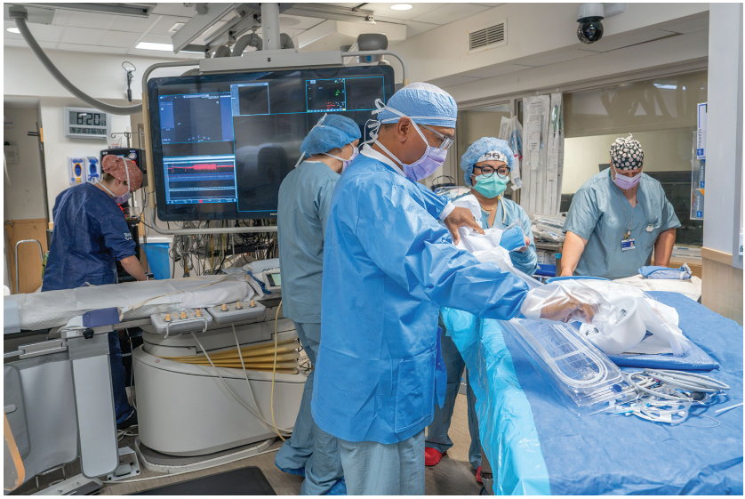
Turnover is executed as a parallel workflow: while one patient transitions to recovery, the next case is actively being prepared, including equipment staging, mapping system setup, and device preparation. One team member accompanies the patient after the procedure while the remaining staff complete room turnover.
Describe your use of vascular closure devices and approach to SDD.
Vascular access closure is standardized across all femoral access cases using closure devices, primarily MYNX and Perclose, or figure-of-8 suturing.
Postprocedural recovery follows a structured pathway:
- 2 hours of flat bed rest
- 1 hour of upright observation
- Ambulation trial prior to discharge, for a total of 3 to 4 hours whenever possible.
This protocol supports early mobilization while maintaining vascular safety standards. SDD is achieved in >90% of eligible outpatient EP cases, with exceptions primarily driven by procedural complexity, travel distance, or patient-specific comorbidity and support considerations.
Has your lab recently undergone a national accrediting inspection?
The EP program operates under multiple layers of institutional and external accreditation, including Corazon cardiovascular accreditation, designation as a Blue Cross Blue Shield Center of Excellence, an ACGME-accredited EP fellowship training program, Intersocietal Accreditation Commission imaging accreditation, and Joint Commission hospital accreditation.
The program also participates in national quality registries, including the National Cardiovascular Data Registry (NCDR) EP device registry and the REAL-AF registry, supporting continuous benchmarking and outcomes tracking against national standards.
How do you ensure timely case starts and patient turnover?
Timely starts are achieved through structured pre-day planning, case sequencing by complexity, and proactive equipment staging prior to first case arrival.
Room turnover is executed as a parallel process with dedicated roles for patient transport, room preparation, and equipment reset. Mapping systems and high-use devices are pre-staged based on predicted case flow to minimize downtime between procedures.
Vendor coordination is completed prior to procedural day to prevent intraday delays or uncertainty about equipment availability.
How does your laboratory schedule call coverage?
The EP laboratory does not operate with a routine staff call coverage requirement.
After-hours EP emergencies are managed by the on-call electrophysiologist, with procedural support scheduled as clinically required. This structure allows continuity of care without maintaining full in-house procedural staffing outside scheduled operational hours.
Do you have flexible or multiple shifts? How do you handle slow periods?
The EP program operates on a 4x10-hour shift model, with designated late coverage teams to accommodate extended procedures and variable case duration.
During lower procedural volume periods, staff are allocated to structured nonprocedural activities including inventory optimization, mandatory education, quality improvement projects, and equipment readiness checks.
This approach maintains operational efficiency while ensuring staff development and system readiness are continuously reinforced, even during lower procedural demand periods.
How are vendor visits managed?
Vendor engagement is structured, controlled, and case-driven rather than open access.
All vendor activity is scheduled in advance through Vendormate Credentialing (GHX), with defined procedural time windows aligned to specific case requirements. Vendors are assigned based on planned procedural needs, particularly for complex ablations, device implants, and extraction cases requiring specialized equipment.
This system reduces intraprocedural disruption, standardizes equipment availability, and ensures vendor presence is purposeful rather than incidental. Urgent or add-on vendor needs are coordinated through a rapid escalation pathway tied to daily case review.
What are the best features of your EP laboratory’s layout or design?
The EP infrastructure is designed as an integrated procedural ecosystem rather than isolated lab spaces.
A defining strength is the close physical adjacency of the EP labs, catheterization labs, and hybrid OR room, allowing rapid transition between EP, structural heart, and surgical environments when procedural complexity escalates.
The presence of an adjoining control room between the EP laboratories enhances real-time communication, coordination of resources, and cross-room procedural awareness.
Preprocedural and postprocedural recovery areas are located within close proximity to the EP suite, significantly reducing patient transport time and enabling rapid turnover and SDD workflows.
This layout supports both high-volume outpatient rhythm management and high-acuity referral-based EP care within the same operational footprint.
We also work closely with our inpatient rounding service. The common control room serves as a central hub where each morning, cases on the inpatient department list are discussed with the rounding physician and EP team. This creates a collegial and enjoyable environment within our service.
What measures has your laboratory implemented to cut or contain costs?
Cost containment is achieved through a structured combination of inventory control, procedural discipline, and supply utilization of governance.
Key strategies include the selective use of reprocessed EP products when clinically appropriate, controlled opening of high-cost consumables only after procedural confirmation, and strict avoidance of unnecessary opening of materials before a case.
Device and lead extraction cases include systematic evaluation for warranty recovery and eligible manufacturer reimbursement pathways when applicable.
All billing supplies are audited by dedicated staff to ensure charge accuracy and alignment with procedural documentation.
Inventory forecasting is integrated into daily operational huddles to match supply readiness with scheduled procedural demand while minimizing excess consignment dependence.
What quality control measures are practiced?
Quality assurance is embedded into daily workflow rather than treated as a separate function.
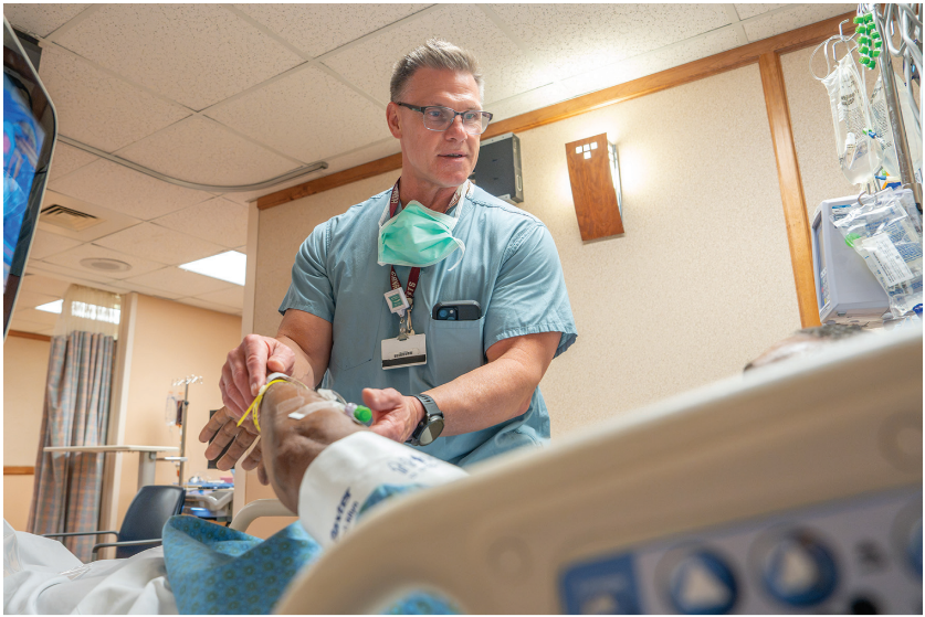
Each lab performs daily equipment and safety checklists prior to first case start, including imaging systems, mapping platforms, defibrillation readiness, and procedural supply verification.
Radiation safety is actively monitored through staff badge tracking and adherence to low-fluoroscopy or zero-fluoroscopy protocols when feasible.
Formal quality governance includes morbidity and mortality review, vascular complication tracking, infection surveillance, and structured escalation review for complex or unexpected procedural outcomes.
Annual competency validation is required for all procedural staff and is supplemented by radiation safety training and emergency response simulations, including open-chest and high-acuity extraction protocols.
Quarterly morbidity and mortality conferences are held to discuss cases that may offer opportunities for learning and improvement.
What works well for onboarding new team members?
Onboarding is structured, competency-driven, and phased over approximately 3 months.
New staff undergo progressive exposure beginning with foundational procedural observation, followed by supervised participation in routine EP cases, and ultimately, advancement to independent role execution once competency benchmarks are met.
Critical care principles are emphasized early in training to ensure staff are prepared for high-acuity EP environments, including VT ablation and lead extraction cases.
Two dedicated educators support both cath and EP services, ensuring standardized competency development and consistent skill acquisition across staff roles.
What continuing education opportunities are provided? How do staff typically maintain and renew credentials?
All procedural staff maintain current Advanced Cardiovascular Life Support certification as a baseline requirement.
Continuing education is delivered through a combination of internal hospital education sessions, structured vendor-led in-services occurring biweekly, and access to hospital-supported educational resources.
Additional education is supported through selective tuition reimbursement programs, although external conference funding is limited.
Skill maintenance is reinforced through annual competency assessments, radiation safety refreshers, and procedure-specific training updates aligned with evolving EP technologies.
Discuss the role of mid-level practitioners in your laboratory.
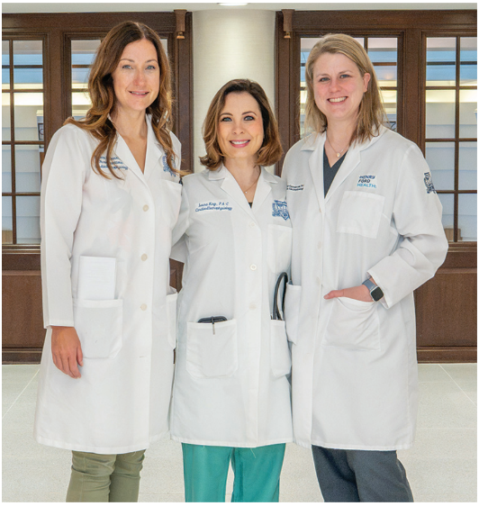
APPs are fully integrated into the EP care continuum and function as core members of both inpatient and outpatient EP operations. Their responsibilities include preprocedural patient evaluation, inpatient rhythm management, post-ablation follow-up, device clinic support, and coordination of remote monitoring workflows.
APP-led AF clinics play a significant role in early patient assessment, enabling rapid access evaluation within 48 to 72 hours of referral and facilitating early rhythm control decision-making. This has led to a tremendous gain in patient access and quality in managing patients with AF.
This structure allows electrophysiologists to focus on procedural complexity while maintaining a high continuity of care across the patient’s lifecycle.
Share a memorable case from your EP laboratory and how it was addressed.
Two of our most memorable cases involved critically ill patients with refractory VT and cardiogenic shock who required mechanical circulatory support. Both patients were extremely ill, and palliative care had been involved with hospice under consideration. Despite their high procedural risk, our multidisciplinary team proceeded with complex VT ablations.
The VT ablations were performed on 11/19 and 11/21, with the cases completed at 1922 and 2132, respectively. These were long and challenging cases requiring close collaboration between EP, anesthesia, heart failure (HF), critical care, lab staff, and our mapping teams. Both ablations were successful, and remarkably, both patients recovered sufficiently to leave the hospital and return home to their families. One of the most rewarding moments was learning that one of the patients was able to spend Thanksgiving at home with his family. These cases highlight what our EP lab can accomplish through advanced technology, multidisciplinary collaboration, and a team willing to take on the highest-risk patients.
Does your lab use a third party for reprocessing or catheter recycling? How has it impacted your lab?
We work with 2 separate reprocessing companies. Eligible EP devices and consumables are selectively reprocessed under institutional guidelines to support cost efficiency while maintaining procedural safety and regulatory compliance.
This approach contributes to cost containment without compromising procedural reliability or patient safety standards.
Discuss your approach to CSP.
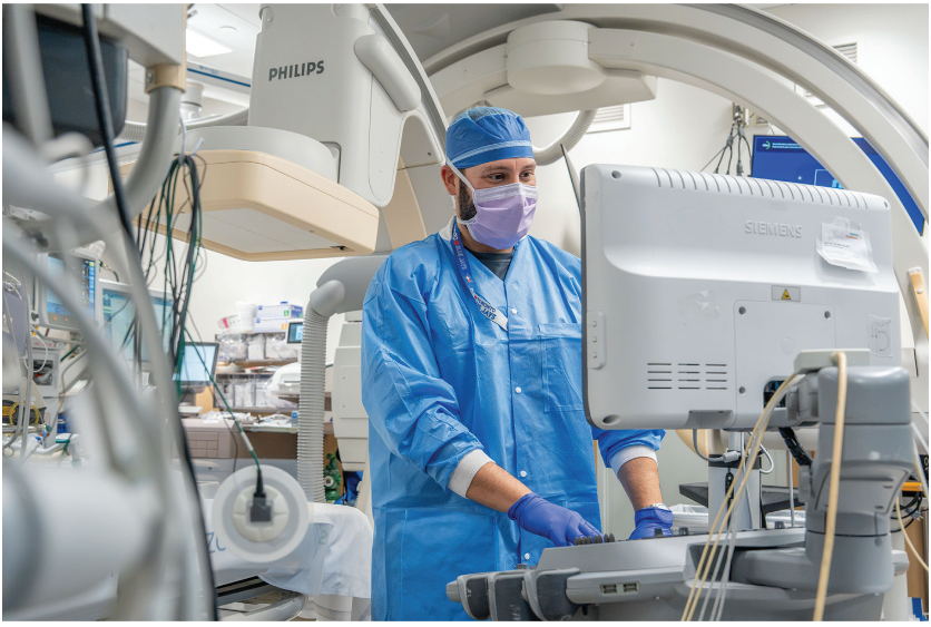
CSP is an established and expanding strategy within the EP program, with increasing use of left bundle branch area pacing as a physiologic alternative to traditional right ventricular pacing.
This approach is used particularly in patients requiring long-term ventricular synchrony preservation, including those with HF risk, pacing-induced cardiomyopathy prevention needs, or CRT optimization challenges.
Implant strategies are guided by real-time electrogram mapping. The program views CSP as a foundational shift in device therapy rather than a niche technique.
Tell us about your primary approach for LAAO.
LAAO is performed through a collaborative, team-based approach involving both the structural heart and cardiac EP program, with joint scrubbing and coordinated imaging and anesthesia support.
Procedures are typically TEE or ICE-guided with a standardized transseptal approach, integrating preprocedural imaging review with CT planning and intraprocedural echocardiographic confirmation of appendage anatomy.
Patient selection is focused on AF patients with elevated bleeding risk or contraindications to long-term anticoagulation, with shared decision-making between the EP and structural heart teams.
The program integrates LAAO into broader AF management pathways rather than treating it as an isolated procedure.
Does your program have a dedicated AF clinic or is it considered an AF Center of Excellence?
The Henry Ford AF Center of Excellence program functions as a formalized, multidisciplinary care model integrating EP, cardiology, CT surgery, primary care, the emergency department, sleep medicine, dietary, and APPs into a unified treatment pathway.
Patients are evaluated through a rapid-access AF clinic model, with assessment typically occurring within 48 to 72 hours of referral. This structure enables early stratification into rhythm control, anticoagulation management, or procedural pathways including ablation or LAAO. AF-specific care pathways have been established to facilitate early referral for AF management and avoidance of hospital admissions.
The program incorporates structured registry participation (including NCDR and REAL-AF) to track outcomes and ensure continuous benchmarking against national standards.
Hybrid AF ablation is available for persistent and long-standing persistent AF, with surgical and catheter-based teams collaborating within a shared procedural framework.
The center is designed to provide longitudinal AF management rather than episodic procedural care, integrating risk factor modification (RFM), device-based monitoring, and early rhythm intervention strategies.
Discuss your approach to lead extraction and management.
Lead extraction is a high-complexity, referral-driven component of the EP program and is performed in a hybrid OR setting under general anesthesia.
The primary technique is laser-assisted transvenous extraction, with adjunct tools used selectively based on lead age, fibrosis burden, and anatomical complexity. Cases include infected systems, lead malfunction, venous occlusion, and device upgrades in structurally constrained anatomy.
Each case is supported by a multidisciplinary team including EP, CT surgery backup, anesthesia, and vascular access support when needed, ensuring immediate escalation capability if required.
The program functions as a regional referral center for complex extraction cases, including patients transferred specifically for complete system removal and re-implant strategy planning.
Extraction procedures are performed both in the EP lab or the hybrid OR based on case complexity and surgical backup requirements. To help facilitate access, we have developed lead dwell time criteria to determine the location of the procedure. Higher risk cases are performed in the hybrid OR, and lower risk cases are performed in the EP lab. This has improved access to these patients.
Discuss your program’s approach to lifestyle RFM for reduction of AF.
The AF program incorporates structured RFM into clinical pathways rather than treating it as adjunct counseling.
Key domains include weight management, hypertension control, sleep apnea identification and referral, alcohol reduction strategies, and physical activity optimization.
These interventions are embedded into APP-led AF clinics and pre-ablation evaluation workflows. Patients are systematically screened for modifiable contributors to AF progression, and intervention plans are initiated in parallel with rhythm management decisions. We work closely with the Henry Ford Health metabolic wellness and sleep medicine program.
This integrated model supports both symptom reduction and long-term recurrence prevention following ablation or medical therapy.
Discuss your approach to AF management in HF.
AF in patients with HF is managed through an aggressive rhythm control strategy. All patients are also evaluated by our advanced HF service.
Catheter ablation is increasingly used earlier in the disease course when appropriate, particularly in patients with reduced ejection fractions, for whom rhythm control may improve functional status, HF trajectory, and mortality.
Device therapy, including CRT and CSP, is integrated when indicated to optimize ventricular synchrony.
Management is coordinated between EP and HF teams to ensure alignment between rhythm strategy and hemodynamic optimization.
Discuss your approach to arrhythmia management in athletes.
High-risk arrhythmias in athletes are referred to us in our outpatient clinic.
Assessment includes rhythm monitoring, genetic testing, advanced imaging, and EP testing when indicated. Common concerns include ventricular ectopy and exercise-induced arrhythmias as well as identifying structural heart disease or genetically determined cardiomyopathies.
Management is individualized based on arrhythmia burden, symptom profile, and risk stratification, with return-to-play decisions guided by objective electrophysiologic findings and clinical stability.
The program prioritizes both safety and preservation of athletic participation when clinically appropriate.
Is your EP lab involved in clinical research?
The EP program is actively involved in clinical research across device therapy, AF management, lead extraction outcomes, and emerging ablation technologies. We have 2 full-time research nurses and 2 assistants. Our team meets quarterly to discuss the industry-sponsored clinical trials in which we participate and, separately, physician-initiated research trials.
We participate in 8 to 10 industry-funded research trials and 5 to 10 physician-led clinical trials.
What is historic about your program or hospital? Has your program or hospital recently experienced any “firsts”?
The institution has a long-standing history in cardiovascular innovation and remains a major referral center for complex cardiac care in the region.
The EP program has achieved multiple procedural and technological firsts within its regional network, particularly in advanced device therapy, complex ablation strategies, and structural EP integration, including:
- First electromagnetic navigation lab in the state using the Stereotaxis system in 2005
- First LAAO in the state using the LARIAT (AtriCure, Inc) system in June 2012
- First subcutaneous ICD implantation in Michigan in December 2013
- First commercial Watchman implantation in the country in 2015
- First use of the dual-energy THERMOCOOL SMARTTOUCH SF Catheter (Johnson & Johnson MedTech) in the Great Lakes region in August 2026.
How does your EP laboratory address radiation safety?
Radiation safety is embedded into procedural design rather than treated as a compliance requirement.
The EP program prioritizes low- and zero-fluoroscopy workflow across sources. This is enabled through widespread use of electroanatomic mapping systems, ICE, and standardized operator techniques that minimize reliance on continuous fluoroscopic guidance.
All staff participate in mandatory radiation safety training and are monitored using personal Landauer dosimetry badges. Exposure trends are reviewed at the departmental level to ensure adherence to ALARA principles and identify opportunities for further reduction.
Radiation reduction is reinforced through technology adoption, workflow standardization, and operator training consistency rather than individual preference variability.
What percentage of cases are done without fluoroscopy? How do you record fluoroscopy times/dosages?
A considerable proportion of ablation procedures are performed with minimal fluoroscopy, with select cases completed entirely without fluoroscopy depending on anatomy and operator approach.
Device implantation workflows increasingly utilize nonfluoroscopic venous access techniques for ultrasound guidance.
Fluoroscopy time and dose metrics are tracked systematically and reviewed as part of ongoing quality improvement initiatives.
Discuss your approach to intravenous (IV) sotalol loading for patients with AF, including effect on hospital length of stay.
We recently received approval for the initiation of IV sotalol across our hospital. Sotalol loading is performed in a monitored inpatient setting with telemetry oversight and structured QT interval surveillance. The protocol is used both for rhythm stabilization and as part of pre-ablation or antiarrhythmic strategy selection in atrial and ventricular arrhythmia patients. Elective outpatients who require rhythm control are assigned to a bed on the morning of initiation and are usually discharged home the following day.
Patients undergo standardized monitoring for bradycardia, QT prolongation, and proarrhythmic risk, with dosing adjustments guided by renal function and electrophysiologic response. This approach supports safe initiation while minimizing outpatient initiation risk in higher-complexity patients.
Discuss your program’s approach to initial treatment and management, including referrals, for patients with postural orthostatic tachycardia syndrome (POTS) and long COVID.
We have seen a tremendous increase in referrals for the evaluation of autonomic dysfunction over the past few years. Patients with suspected POTS or post-viral autonomic dysfunction are evaluated using a structured framework for assessing arrhythmia and autonomic symptoms.
The initial evaluation focuses on excluding primary arrhythmias, structural heart disease, and secondary causes of tachycardia. Management is symptom-directed and includes heart rate control strategies, volume optimization, and referral to multidisciplinary services when appropriate.
EP input is used primarily to distinguish rhythm-based pathology from autonomic dysfunction requiring noninvasive management.
What dominant trends do you see in EP?
Several key trends are shaping EP practice within the program:
- Rapid adoption of PFA as a primary modality for AF
- Focus on workflow efficiencies to enhance throughput and access in the procedural platform
- Expansion of CSP as a physiologic alternative to traditional ventricular pacing
- Increasing procedural complexity driven by higher-risk referral populations
- Growth in lead extraction demand due to device longevity and infection burden
- Integration of hybrid surgical-catheter approaches for advanced arrhythmia management
- Increase utilization of allied health professionals to lead AF clinics for improved access for patients in the outpatient setting
- Michigan recently passed a rule allowing AF ablations to be performed at sites without CT surgery backup. This has enabled a significant shift of routine AF ablations to other sites, allowing patients to receive care closer to home while improving access to complex ablation procedures at our Detroit EP lab.
How do you incorporate digital health, wearable technologies, and artificial intelligence into your treatment strategies? Have you seen an increase in their use among patients, and what benefits or challenges have you encountered? Have these technologies improved patient care or outcomes?
Digital health tools are increasingly integrated into rhythm management pathways, particularly for AF detection, symptom correlation, and postprocedural monitoring.
Wearable devices contribute to earlier identification of arrhythmia burden and support decision-making regarding anticoagulation, ablation timing, and recurrence monitoring. Data from consumer and clinical wearables are selectively incorporated into clinical evaluation when corroborated by medical-grade monitoring systems.
The primary benefit is earlier detection of rhythm abnormalities, although variability in data quality remains a challenge that requires clinician interpretation.
We also participate in clinical research studies that use wearables to determine the need for anti-coagulation in patients with a low burden of AF.
Describe your city or general regional area. How is it unique?
The program serves a large and diverse catchment area spanning urban, suburban, and cross-border populations.
This geographic distribution creates a broad referral base, including patients with advanced structural heart disease, complex arrhythmias, and high device burden who require tertiary-level EP care.
The regional population mix contributes to high procedural diversity and supports the development of advanced subspecialty EP capabilities.
What specific challenges does your hospital face given its unique geographic service area?
The need for a highly structured, high-capacity EP system is reinforced by several key regional challenges, including:
- High prevalence of cardiovascular disease and comorbid conditions such as obesity, diabetes, and HF
- Large geographic catchment area leading to delayed presentation in some patients
- Cross-border care coordination requirements for Canadian referrals
- High procedural complexity due to late-stage referrals and device burden
- Resource balancing between high-volume outpatient care and complex inpatient EP cases
What is special about your EP laboratory and staff?
The EP program is defined by a combination of procedural breadth, clinical complexity, adaptability, and a deeply integrated team culture.
Our lab supports the full spectrum of EP care, from high-volume AF ablation to complex VT ablation, lead extraction, and advanced device procedures. As the field of EP has rapidly evolved, our team has consistently adapted, embracing new technologies and techniques while expanding our ability to care for increasingly complex patients.
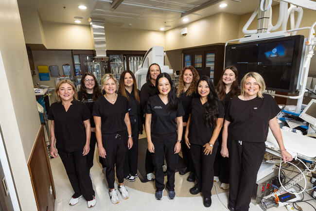
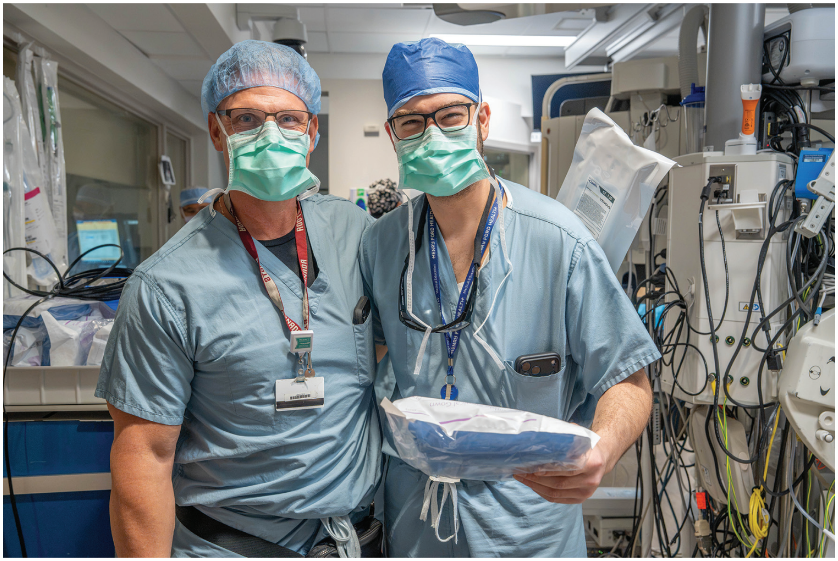
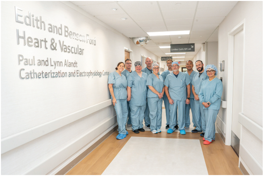
What truly distinguishes our EP lab, however, is not a particular technology or procedure; it is our people and our culture. We have developed a remarkably collegial, family-like atmosphere, with many of our lab staff having worked together for more than 10 years. That longevity has created an exceptional level of trust, familiarity, and teamwork.
There is tremendous mutual respect among our nurses, technologists, APPs, physicians, and support staff. Despite the complexity and intensity of the work we do, the atmosphere in our lab is friendly, supportive, and genuinely enjoyable. We work hard, but we also have fun together and take pride in each other’s successes.
Our greatest strength is that we function as a family. That sense of trust, respect, and camaraderie allows us to work seamlessly together, particularly when caring for our most challenging patients, and is what makes our EP lab a truly special place to work.
Conclusion
EP today is about more than procedures. It is about consistency, teamwork, and being able to manage complexity without losing sight of the details.
What stands out about this program is not any single technology or case type—it is how the lab, staff, and physicians work together to keep a high-volume, high-acuity service running safely and efficiently every day. Over time, that kind of coordination becomes second in nature, especially in a setting where no 2 cases are the same.
The work is fast-paced and technical, and it requires constant adaptation. At its core, however, it is still about caring for patients with rhythm disorders—and doing it well every time.