Optimal Timing for Minor Amputation After Revascularization in Ischemic Diabetic Foot: A Case-Based Review
Abstract
Objective: To assess the evidence on the optimal timing of minor amputation following lower-extremity revascularization in patients with diabetic ischemic foot. A clinical case is presented to illustrate the relevant considerations. Methods: A clinical case involving a 62-year-old man with chronic limb-threatening ischemia is presented and analyzed in the context of the available literature on the optimal timing of minor amputation after revascularization, with emphasis on the potential effects of delayed amputation on tissue perfusion and wound healing. Results: Successful femoropopliteal and tibial angioplasty was followed by a transmetatarsal amputation of the first and second toes 12 days later. Healing progressed favorably, with complete consolidation observed at 25-day follow-up. This case highlights the need to balance infection control with sufficient reperfusion time prior to amputation to optimize healing outcomes. Conclusion: Current evidence does not establish a consensus on the ideal interval between revascularization and minor amputation. Available data suggest that, in select patients, delayed amputation post-revascularization may promote wound healing and improve clinical outcomes.
J CRIT LIMB ISCHEM 2026:6(3):E87-E91. doi: 10.25270/jcli/CLIG-2600005
Key words: diabetic foot, chronic limb-threatening ischemia, revascularization, minor amputation, peripheral arterial disease
Peripheral arterial disease (PAD) affects up to 50% of patients with diabetes-related foot ulcers. This condition is associated with an increased risk of delayed or nonhealing wounds, gangrene, and amputation, as well as high rates of cardiovascular disease and mortality.1,2
Multiple factors can contribute to delayed or failed healing of diabetic foot ulcers (DFUs), including infection, wound size and depth, excessive pressure at the lesion site, and suboptimal local wound care. However, PAD remains the most important determinant of outcome.2
The available literature shows that early revascularization within the first 8 weeks of ulcer onset is associated with a greater likelihood of healing and a reduced risk of limb loss in DFUs.3,4 Among people with diabetes, the lifetime risk of developing a DFU is 19% to 34%; of those who develop a DFU, 4.2% to 9.6% require amputation.
Several studies have aimed to determine the most effective type of revascularization, comparing bypass surgery with endovascular procedures. Results indicate that when a suitable venous conduit for bypass is available, this technique yields better outcomes in terms of limb salvage and mortality than endovascular revascularization.5
There is currently no strong guideline recommendation regarding the optimal timing of minor amputation after revascularization.
The purpose of this article is to review the current literature using a clinical case to determine the optimal timing for performing amputation in patients with DFUs and established PAD.
Methods
A narrative literature review was performed using PubMed and SciELO. Search terms included “minor amputation”, “revascularization”, “diabetic foot”, “critical limb ischemia”, and “chronic limb-threatening ischemia”, used individually and in combination. Particular attention was given to publications evaluating the timing of minor amputation after lower-extremity revascularization and to studies assessing the evolution of tissue perfusion after revascularization, including changes in microcirculatory parameters and tissue oxygenation.
Case Report
We present the case of a 62-year-old man with a history of former tobacco use, arterial hypertension, end-stage kidney disease requiring hemodialysis, type 2 diabetes mellitus with good metabolic control, and dyslipidemia.
His vascular history was notable for a right fifth-toe transmetatarsal amputation. Because of poor wound healing, 1 month later he underwent an anterior femorotibial bypass using a reverse great saphenous vein graft. One year later, stenosis of the proximal and distal anastomoses required angioplasty, and amputation of the distal phalanx of the first toe and proximal phalanx of the second toe was performed.
Three months later the patient presented to the emergency department with right foot pain and was diagnosed with thrombosis of the anterior femorotibial bypass graft. He reported progressive worsening of intermittent claudication, now occurring at very short walking distances, as well as rest pain.
Physical examination
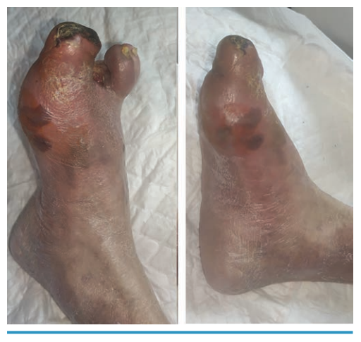
Lower limbs:
- Right leg: Grade 3 femoral pulse. No palpable bypass pulse. Necrosis at the first toe amputation site, with an erythematous-cyanotic area over the first metatarsal head (Figure 1).
- Right leg ankle-brachial index: 0.38 with low amplitude monophasic waveform in dorsalis pedis and posterior tibial arteries. Ankle collapse at 50 mm Hg.
The patient was diagnosed with a diabetic foot ulcer: TEXAS IC, PEDIS 1-2, W1I3fI0 on the right lower limb and Rutherford Category 5 on the right lower limb.
Summary of evolution
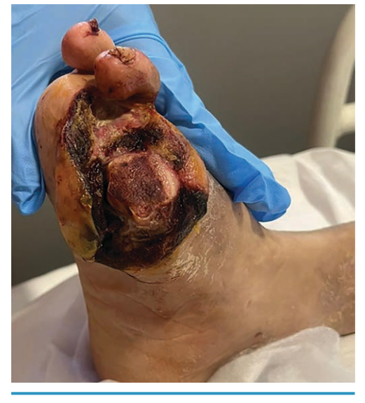
The patient was initially treated with systemic anticoagulation and analgesic therapy during hospitalization, which resulted in improved pain control. His lesions worsened with increased inflammation and intensified pain (TEXAS IIID, PEDIS 3, W1I3fI2).
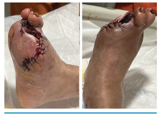
Endovascular angioplasty of the superficial femoral, popliteal, and anterior tibial arteries was performed via a contralateral approach. A transmetatarsal debridement/amputation of the first and second toes was performed because of deep infection. Cultures grew Staphylococcus aureus and Stenotrophomonas maltophilia (Figure 2).
Post-revascularization, the wound bed showed improved viability and decreased inflammation, allowing for proximal reamputation of the first and second toes with near-complete closure 12 days later (Figure 3).
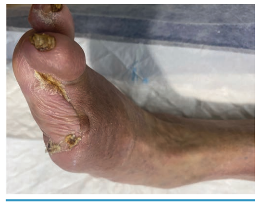
The patient was discharged 16 days after revascularization, continuing oral antibiotics for 1 additional week after 4 weeks of intravenous antibiotics. At follow-up 25 days after the final amputation, the patient reported intermediate-distance claudication, no rest pain, and excellent healing with no dehiscence. Antibiotics had been discontinued (Figure 4).
Discussion
Chronic limb-threatening ischemia (CLTI) associated with diabetic foot infection represents one of the most challenging scenarios in vascular surgery. Successful limb salvage requires adequate control of infection, restoration of arterial perfusion, and appropriate surgical management of nonviable tissue. While current guidelines emphasize the importance of revascularization in patients with ischemic tissue loss, the optimal timing of minor amputation following revascularization remains poorly defined.
Early restoration of arterial inflow has consistently been associated with improved limb salvage outcomes. Sheahan et al demonstrated that delays in revascularization were associated with significantly higher limb loss rates in patients undergoing minor amputations. In their cohort of 670 patients, earlier revascularization was associated with a 76% reduction in nonhealing amputations.6 These findings highlight the importance of restoring blood flow as a first step in the management of ischemic diabetic foot lesions.
Two current international guidelines recommend revascularization before definitive amputation in patients with CLTI when infection is not immediately limb- or life-threatening.7,8 However, neither guideline provides clear recommendations regarding the optimal interval between successful revascularization and subsequent surgical procedures such as minor amputations or definitive debridement.
The limited available evidence suggests that the timing of surgical intervention after revascularization may influence healing outcomes. Tanda et al reported an increased risk of reamputation when minor amputations were performed within 14 days after revascularization.9 However, the study excluded patients with infected diabetic foot lesions, limiting the applicability of these findings in routine clinical practice, where infection is frequently present. Earlier work by Albrektsen et al suggested that delaying debridement for several days following revascularization may improve wound healing even in the presence of necrotic tissue.10
One possible explanation for these observations is the progressive improvement in tissue perfusion that occurs after restoration of arterial inflow. Although macrovascular flow may be restored immediately after revascularization, microcirculatory perfusion and tissue oxygenation often improve gradually. Several studies have demonstrated that parameters such as transcutaneous partial pressure of oxygen (TcPO₂) increase over time after successful revascularization. TcPO₂ values above 25 to 30 mm Hg are generally associated with improved wound healing, while higher thresholds around 40 mm Hg may provide even greater predictive value.11
Evidence from endovascular revascularization studies indicates that recovery of tissue oxygenation is a progressive process that may extend over several weeks after restoration of arterial patency. During angioplasty, TcPO₂ may decrease transiently and often remains close to baseline values at the end of the procedure. Only modest increases are generally observed within the first 24 to 48 hours, whereas the most substantial improvements occur during the following weeks.12,13 These observations are clinically relevant because they suggest that the microcirculatory response to revascularization lags behind the immediate technical success of the procedure.
The full benefit of revascularization at the tissue level may not be apparent in the early postoperative period, supporting the hypothesis that delaying definitive minor amputation could allow time for further improvement in tissue perfusion and wound-healing potential. Other methods may help assess the adequacy of tissue perfusion after revascularization:
Skin perfusion pressure (SPP) has been associated with wound healing, with values above 40 mm Hg generally considered favorable, and some studies suggest that early postoperative SPP measurements may predict healing outcomes more accurately than TcPO₂.
Toe pressure and the toe-brachial index have also demonstrated prognostic value following revascularization; however, their applicability may be limited in patients with previous toe amputations, extensive forefoot tissue loss, or advanced arterial calcification.14,15
Pedal acceleration time has emerged as a useful noninvasive marker of arterial inflow and technical success after revascularization. It also has been associated with limb salvage outcomes.16
Unlike most hemodynamic tests, both TcPO₂ and SPP provide information regarding tissue-level perfusion and microcirculatory recovery, which may be particularly relevant when determining whether sufficient perfusion has been achieved where minor amputation healing must occur.
The progressive improvement in TcPO₂ observed after successful endovascular revascularization suggests that restoration of tissue perfusion is not an immediate event but rather a dynamic process that may continue over several weeks. This observation may be especially relevant when considering angiosome- and woundosome-guided revascularization strategies. Several studies have shown that direct revascularization of the artery supplying the wound area is associated with faster wound healing, improved limb salvage, and lower amputation rates compared with indirect revascularization.17 More recent evidence suggests that indirect revascularization through well-developed collateral circulation may achieve outcomes comparable to direct revascularization, whereas indirect revascularization without effective collateral supply is consistently associated with poorer results.18
From a clinical perspective, these findings suggest that the optimal interval between revascularization and minor amputation may not be the same for all patients. Direct reperfusion of the wound area could potentially allow earlier recovery of tissue perfusion, whereas patients dependent on collateral-mediated flow may require more time to achieve similar physiological conditions. Doyle et al suggest that delaying minor amputations for 15 to 60 days following endovascular intervention may improve healing outcomes in select patients without uncontrolled infection.19
Conclusion
The optimal timing of minor amputation after lower limb revascularization remains uncertain. Available evidence suggests that delayed amputation may improve healing in select patients, but current data are limited by retrospective designs and heterogeneous population. Clinical decisions should therefore be individualized, balancing infection control, tissue viability, and perfusion recovery. Prospective multicenter studies are needed to establish evidence-based recommendations.
Affiliations and Disclosures
Álvaro Moreno, Javier Río Gómez, Elena Menéndez Sánchez, Elena Martín Morales, and Fernando García Boyano are from the Department of Angiology and Vascular Surgery, General Hospital University Gregorio Marañón, Madrid, Spain; Aroa Tardáguila-García and José Luis Lázaro Martínez are from the Diabetic Foot Unit, University Podiatry Clinic, Faculty of Nursing, Physiotherapy and Podiatry, Complutense University of Madrid, Spain.
The authors declare that there is no conflict of interest with respect to the research, authorship, and/or publication of this article.
Written informed consent for publication of this case and any accompanying images was obtained from the patient, and a copy of the consent form is available as a supplementary document.
Manuscript accepted June 16, 2026.
Address for correspondence: Álvaro Moreno, Department of Angiology and Vascular Surgery, General Hospital University Gregorio Marañón, C. del Dr. Esquerdo, 46, Retiro, 28007 Madrid, Spain. E-mail: alvaromocu@yahoo.es
References
1. Leibson CL, Ransom JE, Olson W, Zimmerman BR, O’Fallon WM, Palumbo PJ. Peripheral arterial disease, diabetes, and mortality. Diabetes Care. 2004;27(12):2843-2849. doi:10.2337/diacare.27.12.2843
2. Armstrong DG, Boulton AJM, Bus SA. Diabetic foot ulcers and their recurrence. N Engl J Med. 2017;376(24):2367-2375. doi:10.1056/NEJMra1615439
3. Elgzyri T, Larsson J, Nyberg P, Thörne J, Eriksson K, Apelqvist J. Early revascularization after admittance to a diabetic foot center affects the healing probability of ischemic foot ulcer in patients with diabetes. Eur J Vasc Endovasc Surg. 2014;48(4):440-446. doi:10.1016/j.ejvs.2014.06.041
4. Forsythe RO, Apelqvist J, Boyko EJ, et al. Effectiveness of revascularisation of the ulcerated foot in patients with diabetes and peripheral artery disease: a systematic review. Diabetes Metab Res Rev. 2020;36(Suppl 1):e3279. doi:10.1002/dmrr.3279
5. Farber A, Menard MT, Conte MS, et al; BEST-CLI Investigators. Surgery or endovascular therapy for chronic limb-threatening ischemia. N Engl J Med. 2022;387(25):2305-2316. doi:10.1056/NEJMoa2207899
6. Sheahan MG, Hamdan AD, Veraldi JR, et al. Lower extremity minor amputations: the roles of diabetes mellitus and timing of revascularization. J Vasc Surg. 2005;42(3):476-480. doi:10.1016/j.jvs.2005.05.003
7. Conte MS, Bradbury AW, Kolh P, et al; GVG Writing Group for the Joint Guidelines of the Society for Vascular Surgery (SVS), European Society for Vascular Surgery (ESVS), and World Federation of Vascular Societies (WFVS). Global vascular guidelines on the management of chronic limb-threatening ischemia. Eur J Vasc Endovasc Surg. 2019;58(1S):S1-S109.e33. doi:10.1016/j.ejvs.2019.05.006
8. Schaper NC, van Netten JJ, Apelqvist J, Bus SA, Hinchliffe RJ, Lipsky BA. IWGDF practical guidelines on the prevention and management of diabetes-related foot disease. Diabetes Metab Res Rev. 2023;39(Suppl 1):e3645. doi:10.1002/dmrr.3657
9. Tanda E, Ruiu G, Casula M, et al. Minor amputation after revascularization in chronic limb-threatening ischemia: what is the optimal timing? Vascular. 2024;32(6):1267-1275. doi:10.1177/17085381231214819
10. Albrektsen SB, Henriksen BM, Holstein PE. Minor amputations on the feet after revascularization for gangrene. A consecutive series of 95 limbs. Acta Orthop Scand. 1997;68(3):291-293. doi:10.3109/17453679708996704
11. Leenstra B, de Kleijn R, Kuppens G, Verhoeven BAN, Hinnen JW, de Borst GJ. Photo-optical transcutaneous oxygen tension measurement is of added value to predict diabetic foot ulcer healing: an observational study. J Clin Med. 2020;9(10):3291. doi:10.3390/jcm9103291
12. Gunnarsson T, Lindgren H, Gottsäter A, Pärsson H. Intraprocedural transcutaneous oxygen pressure and systolic toe pressure measurements during and after endovascular intervention in patients with chronic limb threatening ischaemia. Eur J Vasc Endovasc Surg. 2021;62(4):583-589. doi:10.1016/j.ejvs.2021.06.009
13. Wagner H, Schmitz R, Alfke H, Klose K. Influence of percutaneous transluminal angioplasty on transcutaneous oxygen pressure in patients with peripheral arterial occlusive disease. Radiology. 2003;226(3):791-797. doi:10.1148/radiol.2263011728
14. Jeon BK, Ko Y, Lee S, et al. Elevated skin perfusion pressure 48 hours after endovascular therapy predicts early wound healing in chronic limb-threatening ischemia. Vasc Med. 2026;31(2):165-174. doi:10.1177/1358863X251414636
15. Namgoong S, Yang J, Yoo K, Han S, Rha S, Lee Y. Comparison of perfusion values after percutaneous transluminal angioplasty according to the severity of ischaemia in the diabetic foot. Int Wound J. 2019;16(1):176-182. doi:10.1111/iwj.13008
16. Teso D, Sommerset J, Dally M, Feliciano B, Vea Y, Jones RK. Pedal acceleration time (PAT): a novel predictor of limb salvage. Ann Vasc Surg. 2021;75:189-193. doi:10.1016/j.avsg.2021.02.038
17. Guo Z, Guo J, Wu S, et al. Bayesian network meta-analysis and systematic review of endovascular revascularization strategies for infrapopliteal arteries in chronic limb-threatening ischemia. J Vasc Surg. 2026;83(5):1400-1409.e5. doi:10.1016/j.jvs.2025.12.355
18. Shahat M, Mohamed N,A Reyad A, Abdalmoneim M, Hagag M, Abdelmonem M. Do collaterals matter? Comparing the clinical outcomes of direct revascularization (DR), indirect revascularization-with-collateral (IR-tc), and indirect-without-collateral (IR-wc) in endovascular revascularization of chronic limb-threatening ischemia patients. Ann Vasc Surg. 2026;129:219-230. doi:10.1016/j.avsg.2026.03.050
19. Doyle MD, Hastings G, Dontsi M, Dionisopoulos SB, Kane LA, Pollard JD. The effects of endovascular timing and in-line flow on the success of pedal amputations. J Foot Ankle Surg. 2020;59(5):964-968. doi:10.1053/j.jfas.2020.03.018