Use of Intravascular Ultrasound and Extravascular Ultrasound During Endovascular Therapy
Abstract
During endovascular therapy (EVT) for peripheral artery disease (PAD), intravascular ultrasound (IVUS) and extravascular ultrasound (EVUS) can provide information that is not available from angiography alone, including accurate vessel sizing, plaque morphology, calcification distribution, and assessment of dissections or stent expansion. By complementing angiography, these imaging modalities may improve procedural precision and support appropriate treatment strategies. This narrative review summarizes the practical applications and limitations of IVUS and EVUS in contemporary EVT for PAD.
J CRIT LIMB ISCHEM 2026:6(3):E78-E86. doi: 10.25270/jcli/CLIG-2500003
Key words: intravascular ultrasound, extravascular ultrasound, peripheral artery disease, endovascular therapy
Introduction
Intravascular ultrasound (IVUS) and extravascular ultrasound (EVUS) are increasingly used when undertaking endovascular therapy (EVT) for peripheral artery disease (PAD). These modalities provide information that cannot be obtained by angiography alone, including accurate vessel sizing, plaque morphology, distribution of calcification, and assessment of dissections or stent expansion. By complementing angiographic findings, IVUS and EVUS may improve procedural precision and support more appropriate treatment strategies.
The aim of this article is to provide a narrative review of the practical applications and limitations of IVUS and EVUS in contemporary EVT for PAD. It is not a systematic or scoping review; instead, it summarizes well‑established evidence together with procedural concepts and insights derived from the authors’ clinical experience. References were selected from representative, widely cited publications that are commonly recognized in the field and relevant to daily clinical practice.
Intravascular Ultrasound
Can intravascular ultrasound change the treatment strategy for peripheral artery disease?
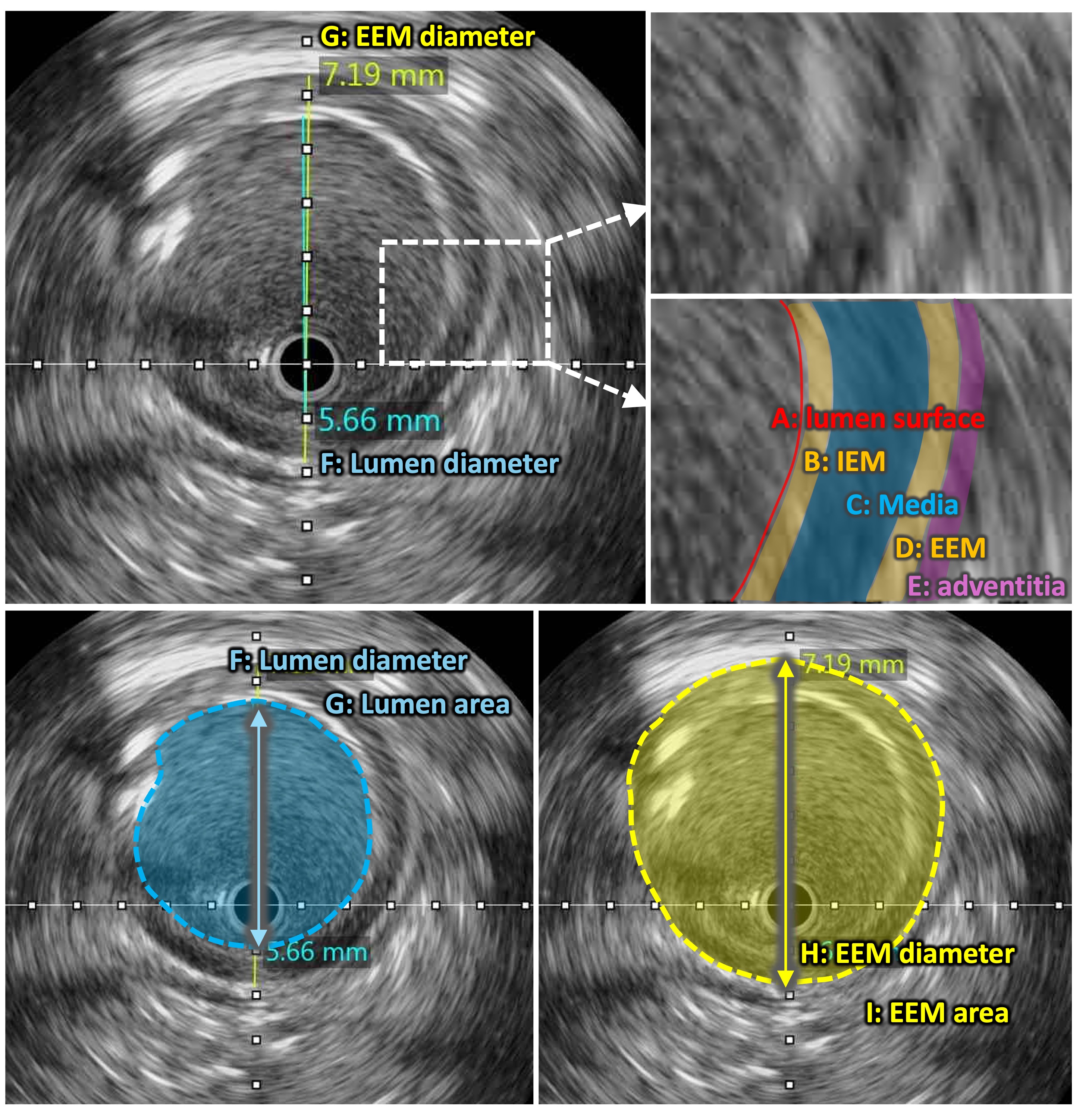
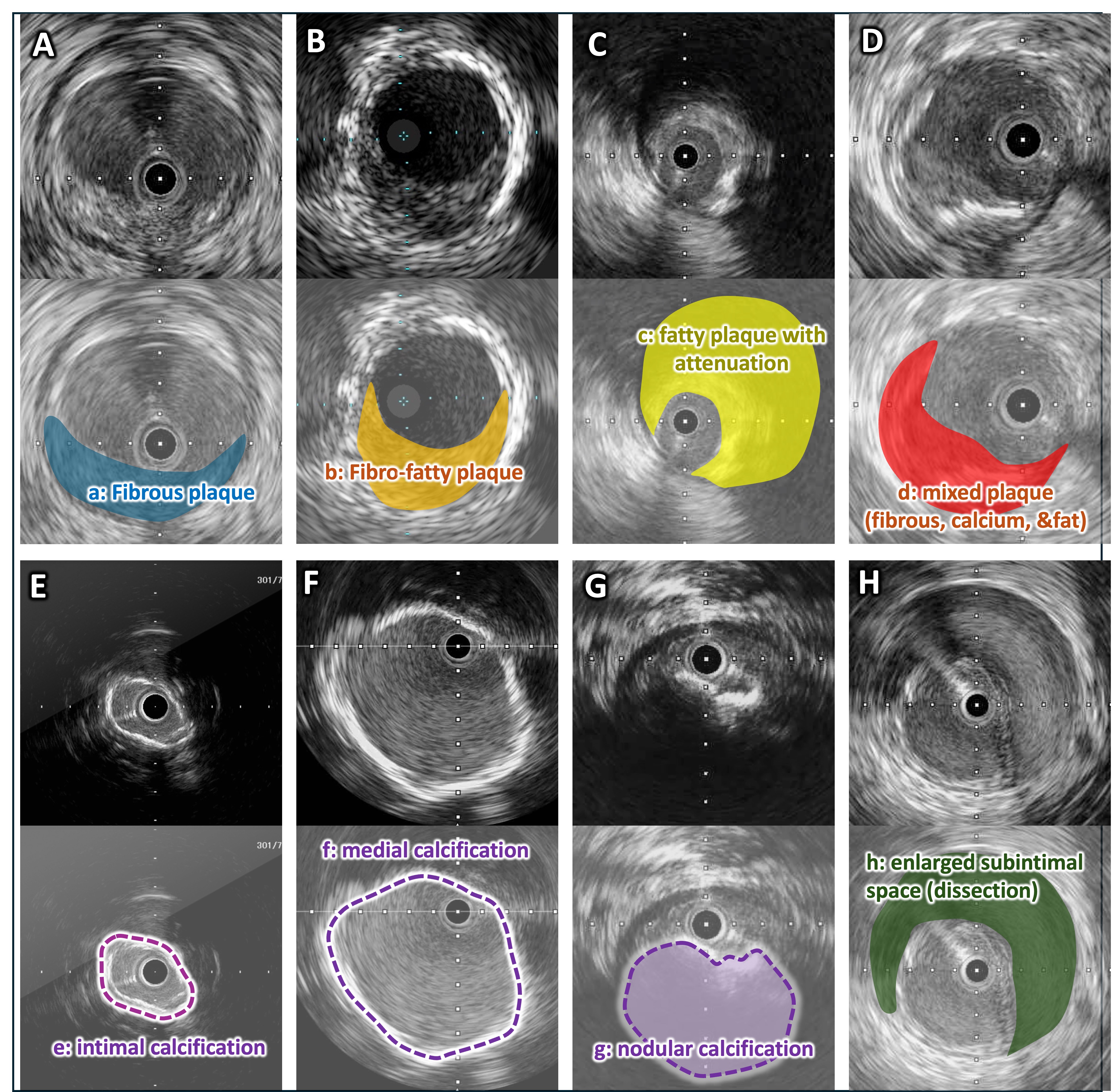
IVUS-guided EVT has received considerable attention as EVT for PAD has evolved. By enabling precise assessment of lesion characteristics (eg, vessel diameter, plaque morphology, and the extent of calcification), IVUS improves understanding of disease pathophysiology and supports procedural planning, device selection, and treatment optimization.1 It also provides important advantages for pre- and post-intervention assessment. IVUS findings help determine the optimal lesion preparation strategy and evaluate its results, including lumen expansion, dissection severity (eg, dissection angle and medial involvement), elastic recoil, and calcified arc fracture—features often difficult to assess using angiography alone. IVUS also guides decisions regarding stent implantation, including identification of appropriate landing zones and optimal stent sizing. After stent placement, it enables evaluation of stent expansion and apposition and helps determine the need for further optimization. Through these mechanisms, IVUS contributes to procedural optimization, improved technical success, and identification of mechanisms of stent failure and post-procedural complications. Definitions of IVUS findings are shown in Figure 1, and typical plaque morphologies are shown in Figure 2.
Interest in IVUS for peripheral interventions has grown rapidly; as of March 2026, more than 40% (264) of 625 IVUS‑related publications indexed in PubMed were published within the preceding 5 years, reflecting increasing clinical adoption and research activity.
Intravascular ultrasound-guided endovascular therapy for femoropopliteal artery lesions
Historically, percutaneous transluminal angioplasty was the primary treatment for femoropopliteal artery (FPA) lesions; however, elastic recoil and flow‑limiting dissections limited durability. Subsequent advances driven by the establishment of new techniques and devices, such as bare‑metal stents, drug‑coated balloons (DCB), drug‑eluting stents (DES), interwoven stents (IWS), and stent‑grafts (SG) have marked the transition toward contemporary antirestenotic strategies. Alongside these developments, IVUS technology has advanced as a tool for acquiring more detailed and precise information about target lesions.
IVUS generates cross‑sectional images of the vessel lumen by emitting ultrasound waves from a transducer mounted on a catheter. IVUS provides information beyond angiography, including accurate vessel diameter measurement and detailed assessment of plaque characteristics such as the distribution and severity of calcification. These insights allow more comprehensive lesion evaluation and may contribute to improved procedural quality and clinical outcomes.
Accumulating evidence supports a beneficial role of IVUS guidance in FPA interventions. The ZEPHYR study (2015) was the first to demonstrate an association between IVUS‑derived parameters and clinical outcomes; a minimum stent area (MSA) ≤12 mm² predicted restenosis at 12 months and helped establish the contemporary concept of lesion preparation.2 Subsequent retrospective and prospective studies have suggested improved outcomes with IVUS‑guided EVT compared with angiography guidance. In 2022, a randomized controlled trial (RCT) from Australia reported improved freedom from binary restenosis with IVUS guidance for FPA lesions. However, no significant difference was observed in clinically driven target lesion revascularization (CD-TLR) rate.3 A similar pattern was observed at the 3-year follow-up. Freedom from binary restenosis was significantly higher in the IVUS group (48.8% vs 34.7%, P=.011), whereas CD-TLR (66.7% vs 64.9%, P=.697) and major adverse events (40.5% vs 32.9%, P=.397) were comparable between the groups. Notably, the benefit of IVUS with respect to freedom from binary restenosis appeared to be particularly pronounced in the DCB-treated subgroup.4 A Korean RCT also reported improved outcomes with IVUS guidance in a DCB‑based strategy.5 Furthermore, a detailed analysis revealed that the effectiveness of IVUS was evident in complex lesions (TASC C/D), with the IVUS-guided group showing significantly higher rates of both freedom from restenosis and freedom from CD-TLR. However, in simple lesions (TASC A/B), no significant benefit of IVUS was observed.6 Importantly, simply passing an IVUS catheter through a lesion does not itself improve outcomes. The clinical value of IVUS-guided EVT depends on accurate image acquisition and interpretation and on integrating IVUS findings into procedural decision‑making. Further analyses and prospective studies are needed to clarify which IVUS‑driven procedural changes translate into improved outcomes.
The mechanisms by which IVUS guidance improves outcomes are primarily attributed to 2 factors: (1) more accurate vessel sizing and (2) more detailed plaque characterization. These advantages facilitate appropriate lesion preparation strategies and guide rational treatment strategy selection, including DCB, DES, IWS, or SG. A substantial body of evidence supports the role of IVUS in accurate vessel sizing.
Recent studies consistently show that the reference vessel diameter (RVD) assessed by IVUS is larger than that estimated by angiography. When vessel size is defined by the external elastic membrane (EEM) diameter, IVUS studies have shown that (1) IVUS‑based RVD is significantly larger than angiographic RVD; (2) approximately half of patients have a ΔRVD (IVUS‑RVD minus angiographic RVD) ≥1 mm; and (3) discrepancies are particularly pronounced in small vessels, chronic total occlusions (CTO), severe calcifications, and previously stented segments.7
Regarding lesion preparation, substantial data focus on calcification. Earlier studies reported that circumferential calcification reduces the efficacy of DCB therapy,8 suggesting the importance of achieving sufficient luminal expansion and improving drug uptake through plaque modification. Early IVUS-guided EVT studies showed that lesions with calcified arcs >180° often fail to achieve adequate minimum lumen area (MLA) with standard plain old balloon angioplasty, even when bare‑metal stents are used to mitigate recoil.9 In such cases, scoring/cutting balloons, intravascular lithotripsy (IVL), or debulking devices may be considered. More recent data suggest that calcified nodules identified by IVUS are associated with inferior outcomes after DCB therapy.10 Calcified nodules may limit luminal gain with balloon‑based therapy and may increase the risk of dissection or vessel rupture. In these settings, plaque modification with IVL or reduction of calcific burden with debulking devices may be reasonable considerations. In percutaneous coronary intervention (PCI), imaging‑based calcium scoring using IVUS11 or optical coherence tomography12 has been proposed to guide the selection of plaque‑modifying devices; developing an analogous evidence‑based framework may be an important future direction for peripheral IVUS‑guided EVT.
IVUS may inform strategy in other lesion subsets. For example, organized thrombus resistant to balloon compression may prompt consideration of thrombectomy devices, relatively rigid fibrous plaque may favor cutting or debulking devices, and lipid‑rich plaque with marked attenuation may raise concerns about distal embolization, for which distal protection devices could be considered. However, robust evidence supporting these approaches remains limited, and further studies are needed to establish standardized algorithms for IVUS‑guided EVT.
Proposed parameters for intravascular ultrasound-guided endovascular therapy
Detailed evaluation of the target lesion should be performed to guide selection of the definitive treatment strategy, including size and length. IVUS can be used to assess lesion characteristics, RVD, and the presence of dissection, and to quantify luminal dimensions before and after lesion preparation. The principles of IVUS‑based vessel assessment in EVT are analogous to those used in PCI. While lesion-specific vessel preparation is a fundamental concept in EVT, accurate IVUS evaluation of predominant plaque morphology and EEM diameter enables a more precise and reproducible, tailored decision-making strategy for device selection and lesion preparation techniques, with the aim of optimizing procedural outcomes.
Evidence of intravascular ultrasound-guided endovascular therapy
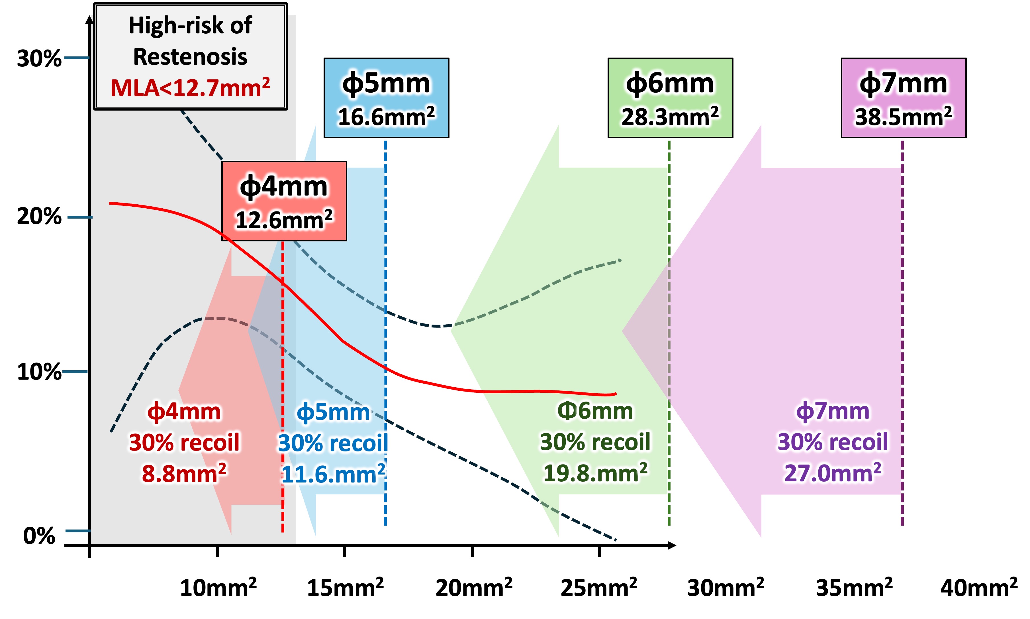
Recent studies suggest that IVUS‑derived parameters can help predict restenosis risk. For example, an MSA <12 mm² after FPA‑DES placement is associated with restenosis risk.2 For fluoropolymer‑based DES, a larger post‑procedural area threshold (eg, 15 mm²) has been proposed in some reports,13,14 emphasizing the need to understand appropriate target MLA/MSA values for each device type. For DCB therapy, retrospective studies have reported an MLA <12.7 mm² as a predictor of restenosis,15 and a subanalysis of a Korean RCT identified an MLA ≥11.6 mm² as a cutoff for 12‑month patency.16 Taken together, achieving an MLA of approximately 11 to 13 mm² after DCB treatment may be a reasonable procedural target to maintain 12‑month patency.
The retrospective multicenter DOLPHINE study reported a dose–response relationship between MLA and restenosis rate.17 The slope of this relationship was steep, between approximately 10.6 and 17.0 mm², suggesting that increasing MLA up to ~17 mm² may improve outcomes, whereas further increases may have less incremental benefit. The theoretical maximum MLA achievable with balloon dilation of 4, 5, 6, and 7 mm is 12.6, 19.6, 28.3, and 38.5 mm², respectively (Figure 3). When accounting for a reported 30% elastic recoil at 15 minutes after balloon angioplasty, these values adjust to 8.8, 11.6, 19.8, and 27.0 mm², respectively.18 Applying these estimates to the reported MLA–restenosis relationship suggests that restenosis rates after treatment with a 4‑mm DCB may be high, supporting the use of at least a 5‑mm DCB when anatomically appropriate. Because the MLA increase between 5‑ and 6‑mm balloons is substantial, IVUS‑based confirmation of EEM diameter and use of the largest feasible balloon (at least 5 mm, preferably 6 mm) may be advisable for lesions treated with 5- to 6‑mm DCBs. In contrast, the MLA increase between 6‑ and 7‑mm balloons may not translate into improved patency and may raise concerns regarding dissection, vessel rupture, and distal embolization. These insights are largely derived from retrospective studies without core‑lab validation and should be confirmed in prospective, core‑lab–adjudicated studies.
1. Application of drug-coated balloon
After successful lesion preparation, DCBs are often selected in appropriate cases. For DCB sizing, a balloon-to-vessel ratio of approximately 1:1 is generally recommended. Kurata et al reported differences between angiography‑based RVD, IVUS‑based minimum lumen diameter, and IVUS‑based EEM diameter,7 with an approximately 2‑mm discrepancy between DCB sizing guided by angiography and sizing guided by IVUS‑EEM. In that report, an IVUS‑EEM–based strategy was associated with lower restenosis rates, suggesting that DCB diameter may be better selected based on EEM diameter.
2. Verification of guidewire passage position
Intraluminal guidewire passage (IP) may be beneficial for maximizing the effectiveness of DCB therapy. IP is associated with procedural conditions that may facilitate favorable outcomes, including (1) the potential to achieve larger acute luminal gain (greater MLA/MSA), (2) a lower likelihood of severe dissection or significant recoil, and (3) more efficient delivery of antirestenotic drugs. In contrast, subintimal guidewire passage (SP) may be less favorable in achieving these procedural goals. Previous studies reported no significant difference in outcomes between intraluminal approach (IA) and subintimal approach (SA) for FPA‑CTO lesions.19 However, many studies classified crossing based on wiring technique rather than actual guidewire position. Several reports demonstrate discrepancies between IA/SA and true guidewire passage assessed by IVUS (IP/SP); Tomoi et al reported such discrepancies in more than one‑third of cases.20 In particular, for vessel preparation devices that require intraluminal wire passage, such as IVL,21 rotational or directional atherectomy,22 and mechanical thrombectomy, accurate assessment of guidewire position by IVUS is critical for procedural decision-making. When subintimal or intramedial wire passage is identified, operators may need to reconsider device selection or modify the treatment strategy, as the use of these devices in an inappropriate vessel layer may increase the risk of complications, including vessel rupture or severe dissection.
Accordingly, IVUS confirmation of guidewire passage is important when evaluating the clinical impact of IP. Mori et al reported that IVUS‑identified IP was protective against restenosis, whereas SP exceeding 14.4% of total CTO length was an independent predictor of restenosis.23 Tsubakimoto et al demonstrated that IVUS‑guided wiring achieved a higher IP rate (91.1% vs 51.3%, P<.001) and a larger post‑procedural MLA (16.4 vs 12.4 mm², P=.044) than conventional wiring techniques.24 Shimada et al suggested that IP may reduce severe dissections detected by IVUS and angiography after balloon angioplasty.25 In addition, a subanalysis of the POPCORN study (a nationwide Japanese registry of DCB treatment) reported higher 12‑month primary patency with IVUS‑confirmed IP than with SP.26 Taken together, particularly when planning a DCB‑based strategy, aiming for IVUS‑verified IP appears reasonable, while recognizing that IP itself does not guarantee optimal lesion preparation or clinical outcomes.
3. Stent deployment
Known target thresholds for post‑stent MSA (eg, 12 and/or 15 mm²)2,13,14 support the importance of adequate lesion preparation before stent placement. In addition, “healthy‑to‑healthy” stent landing is a fundamental concept for stent‑based strategies. Recent reports suggest selecting landing zones with plaque burden <60%27; without IVUS, plaque burden assessment and optimal landing‑zone selection are difficult. IVUS is also useful for detecting edge dissection after stent placement. Accordingly, IVUS guidance may be particularly important for stent‑based strategies in selected cases. The CAPSICUM study identified SP and IVUS use as factors associated with aneurysmal degeneration after FPA‑DES placement.28 Although pathological validation is required, it has been hypothesized that IVUS guidance may lead to selection of larger balloons, resulting in over‑expansion within the subintimal space and injury to the media, potentially contributing to aneurysmal degeneration.
4. Result from recent meta-analysis
Recent meta-analyses have provided additional insights into the role of IVUS in peripheral endovascular interventions. Two large meta-analyses—one from the UK including 246,418 patients and another from the USA including 708,808 patients—demonstrated that IVUS-guided EVT was associated with a lower rate of major amputation compared with angiography-guided EVT.29,30 However, results regarding restenosis and target lesion revascularization were heterogeneous. The UK-based analysis reported better freedom from reintervention in the angiography-guided group, whereas the US-based analysis showed a trend toward improved outcomes with IVUS guidance. Although these findings are difficult to interpret, they suggest that the clinical benefit of IVUS may vary depending on study design and patient population. To establish the true advantage of IVUS, further accumulation of well-designed RCTs and high-quality meta-analyses is warranted.
Limitations of intravascular-guided endovascular therapy
Several limitations should be considered when interpreting the benefits of IVUS-guided EVT. First, IVUS availability depends on insurance coverage and access to consoles and catheters. IVUS is reimbursed in only a limited number of countries (eg, the United States and Japan) and is not readily available worldwide. As in PCI, broader adoption requires substantial investment in equipment and training. In addition, the question of who bears the incremental costs of IVUS—whether covered by insurance or paid out‑of‑pocket—remains a practical challenge. Cost‑effectiveness is also a key issue. Although IVUS guidance may improve acute luminal gain and patency, long‑term cost‑effectiveness remains uncertain. Soga et al reported higher long‑term freedom from TLR with IVUS guidance, suggesting a potential reduction in downstream costs31; however, cost‑effectiveness should be assessed across different health care systems and countries. Finally, IVUS use may increase procedural time, particularly for operators with limited experience in image acquisition and interpretation. In the field of PCI, only approximately 15% of operators report independence in IVUS use and interpretation, according to recent survey data, and in the field of PAD—where IVUS is less routinely utilized—this proportion is expected to be even lower.30 Structured training and clear guidance on key IVUS checkpoints, relevant parameters, and actionable thresholds are therefore necessary to maximize benefit and minimize inefficiency, and to ensure appropriate use of IVUS in clinical practice.
Conclusion for intravascular ultrasound-guided endovascular therapy
IVUS provides comprehensive intraprocedural information for vessel sizing, lesion characterization, and treatment optimization during EVT for PAD. In addition to guiding lesion preparation and selection of the definitive treatment modality, IVUS enables post-intervention assessment of procedural results, including evaluation of lumen expansion, dissection severity, and the need for further optimization. Through these roles, IVUS supports decision-making throughout all stages of the procedure and may contribute to improved technical success and clinical outcomes when its findings are appropriately incorporated into procedural strategies. Although routine IVUS use may be limited by cost, availability, and the need for operator expertise, its use should be considered when feasible, particularly for complex lesions and device‑based strategies.
Extravascular Ultrasound
Role of extravascular ultrasound in endovascular therapy for peripheral artery disease
EVUS has emerged as a useful adjunctive imaging modality during EVT for PAD, particularly in selected cases such as CTO and chronic limb‑threatening ischemia (CLTI). In many centers, EVUS is primarily used for ultrasound‑guided vascular access and procedural support rather than as a routine modality for lesion crossing or device deployment. Utilization varies by operator experience, institutional practice, and procedural complexity.
In EVT for PAD—especially in CLTI—vascular access is most commonly obtained via the common femoral artery (CFA), in contrast to PCI where radial access is frequently used. Patients with PAD often have concomitant atherosclerotic disease in the CFA, including stenosis and calcification, which may increase the risk of access‑site bleeding complications. EVUS guidance allows precise assessment of the puncture site, enabling operators to avoid heavily calcified or plaque‑rich segments and thereby potentially reduce access‑related complications. Careful EVUS assessment of the CFA before puncture is therefore recommended whenever feasible. For infrapopliteal interventions, antegrade ipsilateral CFA access is often preferred to maintain guidewire control, device support, and pushability (except in patients with extreme obesity). Compared with standard retrograde CFA puncture, antegrade access requires a steeper puncture angle near the inguinal ligament and therefore demands careful technique. In such cases, EVUS can help identify the inguinal ligament, CFA, and the bifurcation into the superficial and deep femoral arteries, facilitating safer and more accurate access. Accordingly, EVUS is an important adjunct for femoral access during EVT, particularly in CLTI and complex procedures, while acknowledging variability in use across centers.
Multiple studies have demonstrated that ultrasound‑guided vascular access reduces access‑site complications and improves procedural accuracy compared with landmark‑based techniques.32-36 Patients with CLTI often require multiple endovascular procedures to achieve complete revascularization, with prior studies reporting an average of 1.9 to 2.4 procedures per patient.37 In this population, EVUS‑guided access has been associated with low complication rates; a recent analysis reported an overall complication rate of 1.1% and no tibial access-related complications when ultrasound guidance was used.36
During CTO interventions, maintaining intraluminal guidewire position and safely re‑entering the true lumen are key procedural objectives. Although angiography remains the primary imaging modality for these steps, EVUS can provide complementary real‑time visualization of vascular structures and surrounding anatomy in selected situations. EVUS may also allow visualization of interventional devices within and adjacent to the vessel, allowing operators to adjust strategies based on live imaging feedback.
Extravascular ultrasound assessment of chronic total occlusion morphology
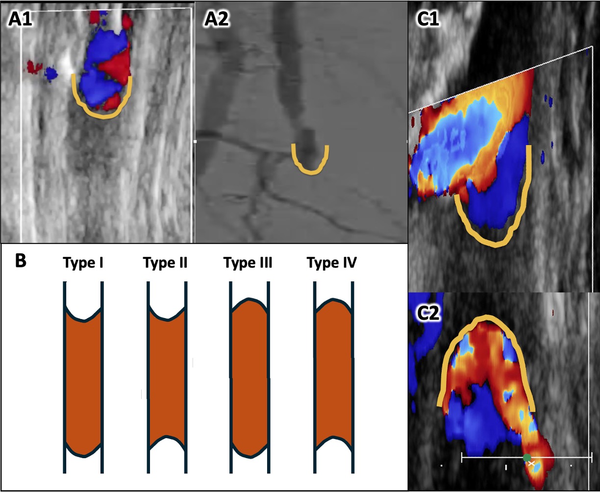
CTOs consist of proximal and distal caps, with lesion composition varying by occlusion length and chronicity. A “hibernating” lumen may exist between the caps in some cases, whereas in others the vessel is completely occluded throughout the segment. EVUS allows direct assessment of CTO cap morphology, which may provide additional information beyond angiography. Based on EVUS findings, CTO caps can be described as concave or convex relative to the direction of approach. This concept underlies the CTO crossing approach based on plaque cap morphology classification (Figure 4).38 Antegrade concave proximal caps (Type I) are generally more amenable to antegrade crossing, whereas lesions with convex or retrograde concave caps (Types II–IV) are associated with increased procedural complexity and a higher likelihood of requiring alternative or retrograde approaches. Representative examples of femoropopliteal CTOs assessed by EVUS are shown in Figure 4.
Extravascular ultrasound-guided chronic total occlusion crossing and device visualization
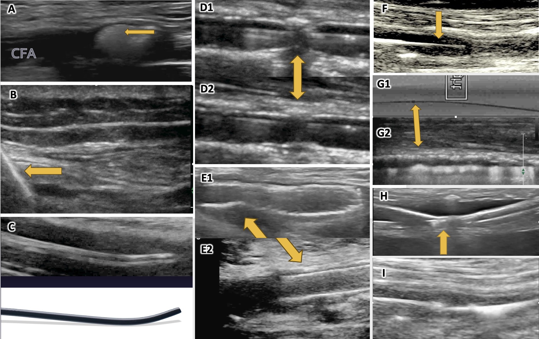
In complex CTO interventions—particularly long or heavily calcified tibial and femoropopliteal lesions—EVUS may assist by providing real‑time visualization of the arterial wall, plaque morphology, and device position. EVUS can help characterize CTO caps and may guide selection of crossing strategies and devices in selected cases. Figure 5 illustrates representative EVUS findings during EVT, including visualization of CTO crossing devices, guidewires, catheters, balloons, stents, and atherectomy systems. Certain CTO crossing and atherectomy devices can generate characteristic ultrasonic phenomena (eg, microcavitation) that may be visible on EVUS and provide real‑time feedback during lesion traversal or plaque modification (Figure 5A and 5I). EVUS can also assist in assessing stent expansion and apposition, helping identify under-expanded segments that may require further optimization (Figure 5E). In distal vessels, particularly the tibiopedal segment, EVUS may be useful when fluoroscopic assessment is limited by tortuosity or overlapping structures. In such settings, EVUS may assist guidewire navigation, balloon sizing, evaluation of lesion response after angioplasty, and confirmation of adequate device deployment.
Practical considerations and limitations of extravascular ultrasound
Despite potential advantages, EVUS is not universally applicable to all EVT procedures. Image quality depends on equipment performance and operator expertise, and severe calcification may cause acoustic shadowing that limits visualization. EVUS-guided procedures may also increase procedural complexity and require dedicated training for accurate interpretation. Accordingly, EVUS should be regarded as a complementary imaging tool rather than a universal solution. Selective use in appropriately chosen cases, such as complex CTOs, CLTI patients requiring ultrasound‑guided access, or situations where fluoroscopic assessment is insufficient, may enhance procedural understanding and safety without replacing standard angiographic guidance.
Conclusions for extravascular ultrasound-guided endovascular therapy
EVUS provides real‑time extravascular visualization of vascular structures and endovascular devices, and it can serve as a valuable adjunct during EVT in selected PAD and CLTI cases. When applied appropriately, EVUS can support vascular access, CTO morphology assessment, device visualization, and procedural decision‑making. However, its use should be tailored to operator experience and procedural context, acknowledging its limitations and complementary role alongside angiography.
Conclusions
IVUS and EVUS complement angiography during EVT for PAD. IVUS primarily supports intraprocedural vessel sizing, lesion characterization, and optimization, whereas EVUS is most commonly used for ultrasound‑guided access and selected complex scenarios. Further studies and standardized training may help clarify optimal indications and workflows for both imaging modalities.
Affiliations and Disclosures
Tatsuya Nakama, MD, is from the Department of Cardiology, Tokyo Bay Medical Center, Urayasu, Japan, and Division of Vascular Surgery, Department of Surgery, The Jikei University School of Medicine, Tokyo, Japan; Fadi Saab, MD, is from the Michigan Outpatient Vascular Institute, Dearborn, Michigan, USA; Mahmoud Abdelghany, MD, is from Mediclinic City Hospital, Dubai, UAE, and Mohammed Bin Rashid University of Medicine and Health Sciences, Dubai, UAE; Imraan Ansaarie, MD, is from Interventional Cardiology and Endovascular Therapies, Ansaarie Cardiac & Endovascular Center of Excellence, St. Augustine, Florida, USA; Mario D’Oria, MD, is from the Division of Vascular and Endovascular Surgery, Department of Clinical Surgical and Health Sciences, University of Trieste, Trieste, Italy; Grigorios Korosoglou, MD, is from the Departments of Cardiology and Vascular Medicine, GRN Hospitals Weinheim and Eberbach, Germany; Masahiko Fujihara, MD, is from the Department of Cardiology, Nozaki Tokushukai Hospital, Osaka, Japan; Pradeep Nair, MD, is from the Cardiovascular Institute of the South, Houma, Louisiana, USA; Bob Tahara, MD, is from Allegheny Vein & Vascular, Bradford, Pennsylvania, USA; Sameh Sayfo, MD, is from the Department of Cardiology, Baylor Scott & White The Heart Hospital, Plano, Texas, USA; and Zola N’Dandu, MD, is from Ascension Sacred Heart, Pensacola, Florida, USA.
Dr Nakama is a consultant for Abbott Vascular, Asahi Intecc, Becton Dickinson, Boston Scientific, Cook Medical, Cordis, Kaneka Medix, NIPRO, and OrbusNeich; Dr Saab is a consultant for Abbott Vascular, Asahi Intecc, and Reflow Medical; Dr Ansaarie is a consultant for Abbott Vascular, Cordis, and Terumo; Dr Korosoglou received speaker honoraria from Philips, Cordis, Boston Scientific, and BARD Peripheral Vascular Inc., and institutional grants from Philips and BARD Peripheral Vascular Inc.; Dr Fujihara is a consultant for Philips, NIPRO, Cordis, Kaneka, and Becton Dickinson; Dr Nair is a consultant for Abbott, Cordis, NIPRO, Philips, Reflow Medical, and Surmodics; Dr Sayfo is a consultant for Philips, Abbott, Medtronic, Boston Scientific, Cordis, AngioDynamics, and Terumo; and Dr N’Dandu is a consultant for Abbott, Becton Dickinson, Boston Scientific, Provisio, LimFlow, Gore, and Inari. The remaining authors report no financial relationships or conflicts of interest regarding the content herein.
Manuscript accepted June 2, 2026.
Address for correspondence: Tatsuya Nakama, MD, Department of Cardiology, Tokyo Bay Medical Center 3-4-32, Todaijima, Urayasu, Chiba 279-0001, Japan. E-mail: tatsuya_nakama@med.miyazaki-u.ac.jp
References
1. Secemsky EA, Parikh SA, Kohi M, et al. Intravascular ultrasound guidance for lower extremity arterial and venous interventions. EuroIntervention. 2022;18(7):598-608. doi:10.4244/EIJ-D-21-00898
2. Iida O, Takahara M, Soga Y, et al; ZEPHYR Investigators. 1-year results of the ZEPHYR registry (Zilver PTX for the femoral artery and proximal popliteal artery): predictors of restenosis. JACC Cardiovasc Interv. 2015;8(8):1105-1112. doi:10.1016/j.jcin.2015.03.022
3. Allan RB, Puckridge PJ, Spark JI, Delany CI. The impact of intravascular ultrasound on femoropopliteal artery endovascular interventions: a randomized controlled trial. JACC Cardiovasc Interv. 2022;15(5):536-546. doi:10.1016/j.jcin.2022.01.001
4. Allan RB, Puckridge PJ, Delaney CL. Three-year outcomes of a randomized controlled trial investigating the use of intravascular ultrasound in femoropopliteal endovascular interventions. J Endovasc Ther. 2025:15266028251398977. doi:10.1177/15266028251398977
5. Ko Y, Lee S, Ahn C, et al; IVUS-DCB Investigators. Intravascular ultrasound-guided drug-coated balloon angioplasty for femoropopliteal artery disease: a clinical trial. Eur Heart J. 2024;45(31):2839-2847. doi:10.1093/eurheartj/ehae372
6. Lee S, Kim T, Lee J, et al; IVUS-DCB Investigators. Intravascular ultrasound-guided vs angiography-guided drug-coated balloon angioplasty in patients with complex femoropopliteal artery disease. JACC Cardiovasc Interv. 2025;18(5):558-569. doi:10.1016/j.jcin.2024.10.052
7. Kurata N, Iida O, Takahara M, et al. Clinical impact of the size of drug-coated balloon therapy on restenosis rate in femoropopliteal lesions. J Endovasc Ther. 2023;30(2):269-280. doi:10.1177/15266028221081082
8. Fanelli F, Cannavale A, Gazzetti M, et al. Calcium burden assessment and impact on drug-eluting balloons in peripheral arterial disease. Cardiovasc Intervent Radiol. 2014;37(4):898-907. doi:10.1007/s00270-014-0904-3
9. Fujihara M, Kozuki A, Tsubakimoto Y, et al. Lumen gain after endovascular therapy in calcified superficial femoral artery occlusive disease assessed by intravascular ultrasound (CODE study). J Endovasc Ther. 2019;26(3):322-330. doi:10.1177/1526602819836095
10. Mori S, Takahara M, Nakama T, et al. Analysis of the impact of nodular calcification on clinical outcome after drug-coated balloon angioplasty for femoropopliteal lesions. CVIR Endovasc. 2025;8(1):65. doi:10.1186/s42155-025-00583-6
11. Zhang M, Matsumura M, Usui E, et al. Intravascular ultrasound-derived calcium score to predict stent expansion in severely calcified lesions. Circ Cardiovasc Interv. 2021;14(10):e010296. doi:10.1161/CIRCINTERVENTIONS.120.010296
12. Fujino A, Mintz GS, Matsumura M, et al. A new optical coherence tomography-based calcium scoring system to predict stent underexpansion. EuroIntervention. 2018;13(18):e2182-e2189. doi:10.4244/EIJ-D-17-00962
13. Yamada T, Tokuda T, Yoshioka N, et al. Predictors of recurrence based on intravascular ultrasound findings after Eluvia placement in symptomatic peripheral arterial disease: a retrospective study. Health Sci Rep. 2023;6(8):e1481. doi:10.1002/hsr2.1481
14. Haraguchi T, Takahara M, Iida O, et al. Impact of postprocedural minimum lumen area on clinical outcome after femoropopliteal drug-eluting stent implantation. Vasc Med. 2023;28(5):412-421. doi:10.1177/1358863X231182742
15. Horie K, Tanaka A, Taguri M, Inoue N. Impact of baseline and postprocedural intravascular ultrasound findings on 1-year primary patency after drug-coated balloon treatment of femoropopliteal lesions. J Endovasc Ther. 2022;29(1):66-75. doi:10.1177/15266028211058683
16. Lee J, Jang JY, Ahn C, et al; IVUS-DCB Investigators. Intravascular ultrasound predictors of 12-month patency loss following drug-coated balloon angioplasty for the femoropopliteal artery. Am J Cardiol. 2025;246:58-64. doi:10.1016/j.amjcard.2025.03.018
17. Horie K, Takahara M, Nakama T, et al. Impact of postoperative lumen gain on the reduction of restenosis risk after endovascular treatment using drug-coated balloon for femoropopliteal lesions assessed by intravascular ultrasound. J Atheroscler Thromb. 2023;30(9):1142-1151. doi:10.5551/jat.63886
18. Baumann F, Fust J, Engelberger RP, et al. Early recoil after balloon angioplasty of tibial artery obstructions in patients with critical limb ischemia. J Endovasc Ther. 2014;21(1):44-51. doi:10.1583/13-4486MR.1
19. Soga Y, Iida O, Suzuki K, et al. Initial and 3‐year results after subintimal versus intraluminal approach for long femoropopliteal occlusion treated with a self‐expandable nitinol stent. J Vasc Surg. 2013;58(6):1547-1555. doi:10.1016/j.jvs.2013.05.107
20. Tomoi Y, Takahara M, Kuramitsu S, et al; IVORY Study Investigators. Subintimal versus intraluminal approach for femoropopliteal chronic total occlusions treated with intravascular ultrasound guidance. J Am Heart Assoc. 2021;10(20):e021903. doi:10.1161/JAHA.121.021903
21. Adams G, Shammas N, Mangalmurti S, et al. Intravascular lithotripsy for treatment of calcified lower extremity arterial stenosis: initial analysis of the Disrupt PAD III study. J Endovasc Ther. 2020;27(3):473-480. doi:10.1177/1526602820914598
22. Kojima S, Tokuda T, Tanaka A, et al; LEADers PAD Investigators. One-year clinical outcomes and predictors of distal embolization after JETSTREAM™ atherectomy for calcified femoropopliteal artery lesions: results from the JET-FORWARD study. Cardiovasc Intervent Radiol. 2025;48(6):760-768. doi:10.1007/s00270-025-04063-1
23. Mori S, Hirano K, Ito Y, et al. Clinical outcomes of the intraluminal approach for long occlusive femoropopliteal lesions assessed by intravascular ultrasound. J Atheroscler Thromb. 2017;24(5):477-486. doi:10.5551/jat.36749
24. Tsubakimoto Y, Isodono K, Fujimoto T, et al. IVUS-guided wiring improves the clinical outcomes of angioplasty for long femoropopliteal CTO compared with the conventional intraluminal approach. J Atheroscler Thromb. 2021;28(4):365-374. doi:10.5551/jat.57166
25. Shimada T, Shima Y, Miura K, et al. Impact of guidewire route on severe dissection after balloon angioplasty for femoropopliteal chronic total occlusion lesions: an intravascular ultrasound analysis. Eur J Vasc Endovasc Surg. 2021;61(5):830-836. doi:10.1016/j.ejvs.2021.01.014
26. Toyoshima T, Takahara M, Iida O, et al; POPCORN Investigators. Intraluminal vs subintimal drug-coated balloon angioplasty for the treatment of femoropopliteal chronic total occlusions. JACC Cardiovasc Interv. 2024;17(5):608-618. doi:10.1016/j.jcin.2023.12.028
27. Kurata N, Iida O, Asai M, et al. Factors influencing in-stent occlusion after femoropopliteal artery stent placement with intravascular ultrasound evaluation. J Vasc Interv Radiol. 2020;31(2):213-220. doi:10.1016/j.jvir.2019.08.014
28. Tsujimura T, Iida O, Takahara M, et al; CAPSICUM Investigators. Clinical impact of intravascular ultrasound-guided fluoropolymer-based drug-eluting stent implantation for femoropopliteal lesions. JACC Cardiovasc Interv. 2022;15(15):1569-1578. doi:10.1016/j.jcin.2022.06.012
29. Sayed MH, Majeed M, Mishrah A, Saha P, Thulasidasan N, Zayed H. Angioplasty combined with intravascular ultrasound vs angioplasty alone in the treatment of peripheral arterial disease. Vasa. 2025;54(5):291-298. doi:10.1024/0301-1526/a001187
30. Tsukagoshi J, Shimoda T, Yokoyama Y, et al. The mid-term effect of intravascular ultrasound on endovascular interventions for lower extremity peripheral arterial disease: a systematic review and meta-analysis. J Vasc Surg. 2024;79(4):963-972.e11. doi:10.1016/j.jvs.2023.08.128
31. Soga Y, Ariyaratne TV, Secemsky E, et al. Intravascular ultrasound guidance during peripheral vascular interventions: long-term clinical outcomes and costs from the Japanese perspective. J Endovasc Ther. 2025;32(3):698-710. doi:10.1177/15266028231182382
32. Shiloh AL, Savel RH, Paulin LM, Eisen LA. Ultrasound-guided catheterization of the radial artery: a systematic review and meta-analysis of randomized controlled trials. Chest. 2011;139(3):524-529. doi:10.1378/chest.10-0919
33. Seto AH, Abu-Fadel MS, Sparling JM, et al. Real-time ultrasound guidance facilitates femoral arterial access and reduces vascular complications: FAUST (Femoral Arterial Access With Ultrasound Trial). JACC Cardiovasc Interv. 2010;3(7):751-758. doi:10.1016/j.jcin.2010.04.015
34. Randolph AG, Cook DJ, Gonzales CA, Pribble CG. Ultrasound guidance for placement of central venous catheters: a meta-analysis of the literature. Crit Care Med. 1996;24(12):2053-2058. doi:10.1097/00003246-199612000-00020
35. Hind D, Calvert N, McWilliams R, et al. Ultrasonic locating devices for central venous cannulation: meta-analysis. BMJ. 2003;327(7411):361. doi:10.1136/bmj.327.7411.361
36. Abu-Fadel MS, Sparling JM, Zacharias SJ, et al. Fluoroscopy vs. traditional guided femoral arterial access and the use of closure devices: a randomized controlled trial. Catheter Cardiovasc Interv. 2009;74(4):533-539. doi:10.1002/ccd.22174
37. Adam DJ, Beard JD, Cleveland T, et al; BASIL Trial Participants. Bypass versus angioplasty in severe ischaemia of the leg (BASIL): multicentre, randomised controlled trial. Lancet. 2005;366(9501):1925-1934. doi:10.1016/S0140-6736(05)67704-5
38. Saab F, Jaff MR, Diaz-Sandoval LJ, et al. Chronic total occlusion crossing approach based on plaque cap morphology: the CTOP classification. J Endovasc Ther. 2018;25(3):284-291. doi:10.1177/1526602818759333