Phototoxic Drug Eruption to Novel Anticancer Agent Pidnarulex
Pidnarulex (CX-5461) is a first-in-class fluoroquinolone derivative used to treat advanced solid tumors with homologous recombination deficiency mutations, including BRCA1, BRCA2, and PALB2, common in breast, ovarian, pancreatic, and prostate cancers.1 The drug stabilizes G-quadruplex DNA, a guanine-rich tertiary DNA structure prevalent in promoters, telomeres, and 5’ untranslated regions that is often transient and easily resolved in normal cells through homologous recombination repair. It also targets topoisomerase II, selectively blocking RNA polymerase Imediated transcription. Thus, tumors with deficient homologous recombination repair mechanisms are unable to resolve the drug-induced DNA damage mediated by Pidnarulex, promoting apopto sis and tumor suppression.
Early clinical trials of Pidnarulex have non-specifically identified phototoxicity as a common adverse effect, using a grading scale based only on eruption duration. Further, phototoxicity has been observed at all dose levels and described as an important adverse effect to patients, although rarely resulting in discontinuation of treatment.1 Despite this report, Pidnarulex-induced phototoxicity, including its clinical course, management, and outcome, has not been formally characterized.1 We report a case of a patient with a phototoxic pustular drug eruption secondary to the novel anticancer agent Pidnarulex for breast cancer therapy.
Case Report
A 69-year-old man with metastatic invasive ductal carcinoma of the left breast with germline BRCA2 mutation presented with a rash 13 days after initiation of a clinical trial for Pidnarulex. Concurrent medications included vitamin and electrolyte supplements, an anticoagulant (rivaroxaban), an antiemetic (prochlorperazine), an analgesic agent (oxycodone), and an antimicrobial (metronidazole). Although metronidazole is rarely associated with photosensitivity, it can be excluded as a cause for this reaction as the patient had tolerated this drug at a stable dose before and throughout the eruption.2 His prior oncologic course included palbociclib, goserelin, and letrozole (March 2020–August 2021); tamoxifen (August–October 2021), discontinued due to bilateral pulmonary emboli; palbociclib with fulvestrant (November 2021– February 2024); and olaparib (March 2024–March 2025), all tolerated without phototoxic reactions. Additional medical and family history were noncontributory.
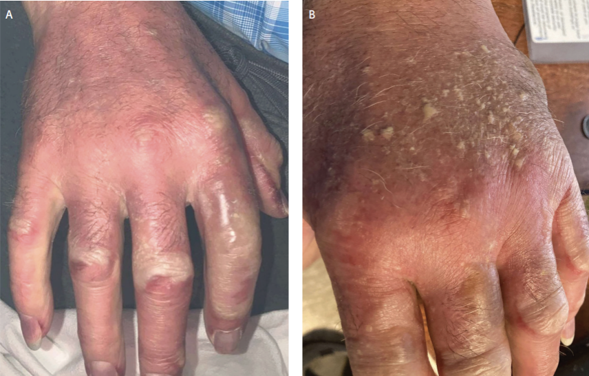
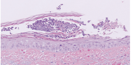
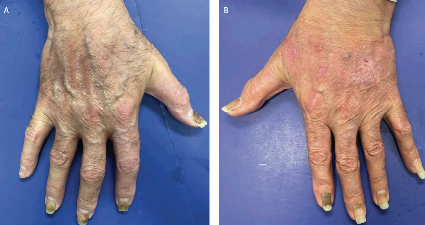
Examination revealed sharply demarcated, erythematous, scaly plaques and flaccid bullae with few pustules on the bilateral dorsal hands, sparing the palms (Figure 1 A–B). Additionally, mild erythema was present on the face and upper chest in a V-shaped distribution. Punch biopsy displayed a subcorneal pustule with superficial acantholysis, supporting the diagnosis of a phototoxic pustular drug eruption (Figure 2).
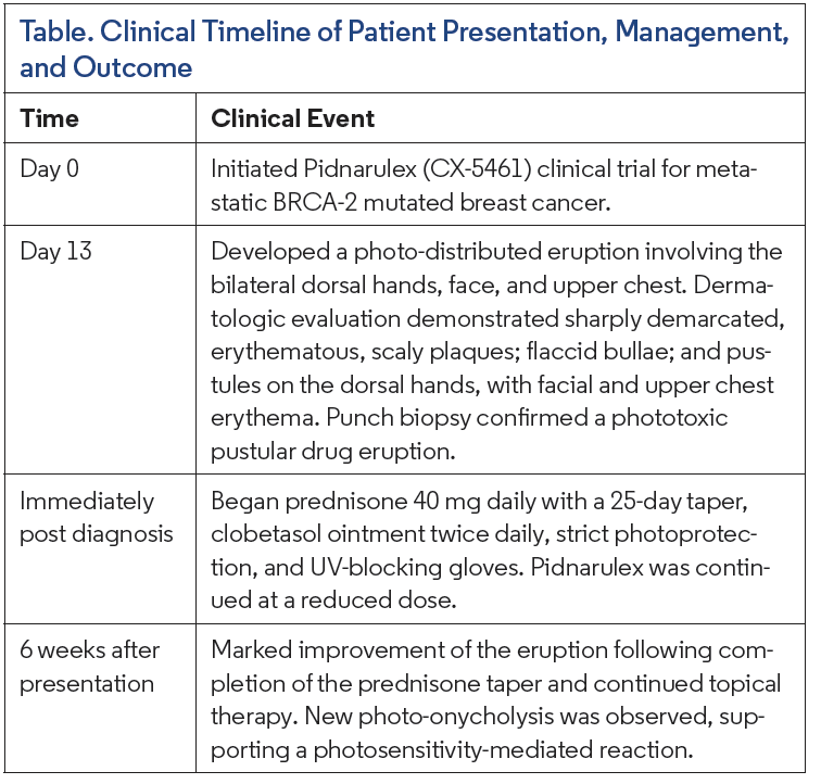 The patient was treated with oral prednisone 40 mg/day with a 25-day taper and clobetasol ointment twice daily. Additionally, he was advised to use UV-blocking gloves and adhere to photoprotective protocols, including sun avoidance and broad-spectrum photoprotection. Given the oncologic importance of maintaining protocol therapy, Pidnarulex was continued with a dose reduction. A timeline of patient presentation, management, and outcome is seen in the Table. At 6-week follow-up, the patient demonstrated marked improvement of the bilateral dorsal hands (Figure 3 A–B). He reported improvement of the eruption after completing the prednisone taper and applying clobetasol with glove occlusion twice daily. He denied any new or changing skin lesions. Photoonycholysis secondary to Pidnarulex therapy was also noted, consistent with the typical 2-week onset of drug-induced photoonycholysis following a photosensitivity reaction.3 This temporal course supports a causal relationship between Pidnarulex therapy and the observed phototoxicity.
Discussion
Fluoroquinolones, the class from which Pidnarulex is derived, confer UV sensitization via amplifying effects of UV-A on the skin, initiate DNA strand breaks, and cause cutaneous reactions manifesting as erythematous, bullous, eczematous, or pustular cutaneous eruptions.4 Fluoroquinolones also have a high affinity for melanin, mediating their toxicity.5 Quarfloxin, a structurally similar anticancer fluoroquinolone-derived G-quadruplex stabilizer, did not demonstrate phototoxic reactions in clinical trials, possibly reflecting differences in melanin-binding affinity.5 Pidnarulex’s additional activity against topoisomerase II may further contribute to this difference, suggesting phototoxicity is not a universal feature among fluoroquinolone-based G-quadruplex agents. Ultimately, this highlights the novel reaction observed with Pidnarulex and the value of prompt dermatologic recognition of this reaction.6
In this patient, the time course after Pidnarulex exposure, lack of other viable triggers, and histopathology support the diagnosis of a phototoxic eruption secondary to Pidnarulex. While punch biopsy was performed, it may not be diagnostically necessary when there is a clear temporal association between Pidnarulex and a phototoxic reaction, notably with photodistribution and improvement upon drug withdrawal or dose adjustment. When obtained, a biopsy can be valuable in excluding alternative diagnoses, including infection. The favorable response in this case suggests that tapered oral corticosteroids combined with potent topical therapy and strict photoprotection provide an effective framework for the management of Pidnarulex-associated phototoxic eruptions.
It is essential to counsel patients on strict photoprotective protocols, including sun avoidance and broad-spectrum photoprotection. Adherence to these photoprotective protocols may prevent this eruption from occurring initially, or upon occurrence may minimize the severity of the reaction and promote recovery. Although not warranted in this case, the immunomodulator hydroxychloroquine is an effective first-line therapy for photosensitive dermatoses, such as polymorphous light eruption and porphyria cutanea tarda, and may be preferred when traditional immunosuppressants are less desirable, including in patients with active malignancy.7 Oral polypodium leucotomos extract, a nonimmunosuppressive photoprotective adjunct, may also be considered for recurrent or severe reactions when further dose reduction or systemic corticosteroids are not an option.8,9
Cutaneous adverse effects observed in patients receiving programmed cell death-1/programmed death ligand-1 therapy are associated with better clinical outcomes, including longer survival time and higher cure rates of the primary malignancy.10 Whether Pidnarulex-associated phototoxicity carries similar prognostic value remains unknown. Additionally, this reaction does not appear to favor a specific treatment cycle, instead representing a doselimiting effect that may occur or recur at any point during therapy.1
Conclusion
As a single-patient report, our findings cannot establish causality or predict phototoxicity risk across the Pidnarulex-treated population. This case highlights the importance of promptly recognizing phototoxic eruptions during Pidnarulex therapy. Early dermatologic intervention with prompt recognition and immunosuppressive treatment may provide a framework for improving this cutaneous reaction. Additionally, thorough patient education on rigorous photoprotection remains critical to both preventing and managing this reaction and optimizing outcomes for patients receiving Pidnarulex.
References
1. Hilton J, Gelmon K, Bedard PL, et al. Results of the phase I CCTG IND.231 trial of CX-5461 in patients with advanced solid tumors enriched for DNA-repair deficiencies. Nat Commun. 2022;13(1):3607. doi:10.1038/s41467-022-31199-2
2. Ngan V. Metronidazole. DermNet. 2005. Accessed September 3, 2026. https://dermnetnz.org/topics/metronidazole
3. Baran R, Juhlin L. Drug-induced photo-onycholysis: three subtypes identified in a study of 15 cases. J Am Acad Dermatol. 1987;17(6):1012-1016. doi:10.1016/S0190-9622(87)70291-6
4. Toipa Lopes R, Gonçalo M. Fluoroquinolones as enhancers of photocarcinogenesis: proposed pathomechanisms. Port J Dermatol Venereol. 2022;80(1):33-41. doi:10.24875/PJD.M22000006
5. Kowalska J, Banach K, Rok J, Beberok A, Rzepka Z, Wrześniok D. Molecular and biochemical basis of fluoroquinolones-induced phototoxicity—the study of antioxidant system in human melanocytes exposed to UV-A radiation. Int J Mol Sci. 2020;21(24):9714. doi:10.3390/ijms21249714
6. Figueiredo J, Mergny JL, Cruz C. G-quadruplex ligands in cancer therapy: progress, challenges, and clinical perspectives. Life Sci. 2024;340:122481. doi:10.1016/j.lfs.2024.122481
7. Morgado-Carrasco D, Ibaceta-Ayala J, Piquero-Casals J. Hydroxychloroquine: an essential drug in dermatology and Its controversial use in COVID-19. Actas Dermosifiliogr. 2022;113(2):T166-T175. doi:10.1016/j.ad.2022.01.012
8. Korman AM, Reynolds KA, Nabhan F, Konda B, Shah MH, Kaffenberger BH. Vandetanib-induced phototoxic drug eruption treated with polypodium leucotomos extract: a case report and review of the literature. J Clin Aesthetic Dermatol. 2019;12(10):35-38.
9. Rodríguez-Luna A, Zamarrón A, Juarranz Á, González S. Clinical applications of polypodium leucotomos (Fernblock): an update. Life (Basel). 2023;13(7):1513. doi:10.3390/life13071513
10. Zhao F, Zhu J, Yu R, et al. Cutaneous adverse events in patients treated with PD-1/PD-L1 checkpoint inhibitors and their association with survival: a systematic review and meta-analysis. Sci Rep. 2022;12(1):20038. doi:10.1038/s41598-022-24286-3