






Symposium on Clinical Interventional Oncology 2026 Abstracts
VASCULAR DISEASE MANAGEMENT. 2026;23(11):E182-E226
Vascular Disease Management is pleased to publish the accepted abstracts from the 2026 Symposium on Clinical Interventional Oncology (CIO).
Percutaneous Sclerotherapy of an Intramuscular Venous Malformation of the Triceps Muscle in an Adolescent Patient
Anirudh N. Maddali, BS; Rajiv Biswal, MD
Purpose: A 16-year-old female presenting with progressive right upper extremity (RUE) pain and swelling limiting movement and physical activity. Patient and family deny any history of trauma to the area. Physical examination showed a tender, palpable fullness in the posterior RUE with no skin changes and reproducible pain with limited flexion and extension of the RUE. Intramuscular vascular malformations of the extremities are uncommon lesions that may present with pain, swelling, and functional limitation. Although surgery has historically been standard treatment, image-guided minimally invasive therapies such as percutaneous sclerotherapy have emerged as effective alternatives, particularly for lesions involving critical muscle groups.
Materials and Methods: Magnetic resonance imaging showed a 2.4 x 3.1 x 5.1 cm predominantly T2 hyperintense, mildly T1 hyperintense, heterogeneous enhancing mass at the mid to distal triceps muscle. Some focal T2 hypointense signal within the mass may represent phleboliths. Procedure: Under ultrasound guidance with a tourniquet applied to the RUE proximal to the lesion, a large venous channel was punctured with a 20-gauge angiocath. A 20-gauge catheter was then advanced into the patient’s venous malformation. Angiogram revealed large filling of the nidus. A total 2-cc mixture of sodium tetradecyl sulfate, ethiodized oil (Lipiodol), and air was then injected. There was good filling of the nidus. There was no filling of the outflow veins.
Results: Post-intervention ultrasound: Targeted evaluation of medial RUE at the level of the triceps muscle was performed. There remains a heterogenous appearance of the triceps musculature, although the extent appears to be decreased. The lesion is less conspicuous and not well delineated to measure on the current study. There are 3 distinct ovoid avascular regions within the musculature measuring 6 x 3 x 5 mm hypoechoic structure involving the lateral triceps muscle, 7 x 9 x 5 mm involving the more medial deep muscle, and 4 x 5 x 3 mm involving the deep muscle more inferiorly with a central echogenic focus, which presumably reflects phleboliths.
Conclusions: Percutaneous sclerotherapy is a safe, effective, and minimally invasive treatment for symptomatic intramuscular venous malformations of the extremities. By avoiding morbidity risks of surgery, sclerotherapy successfully alleviates symptoms while preserving function of surrounding muscle.
Integrating Diagnostics and Image-Guided Procedures in Brain Tumors: Current and Emerging Therapeutic Strategies
Kush Desai, BS; David Monoky, MD; Zahin Alam, BS; Jake Heath, BS; Triston Messer, BS
Purpose: Brain tumors are heterogeneous neoplasms with rising incidence and significant morbidity and mortality. Diagnostic radiology (DR) is central to detection, characterization, treatment planning, and surveillance using advanced modalities (magnetic resonance imaging [MRI], perfusion, MR spectroscopy, positron emission tomography) to define tumor extent and treatment response. Interventional radiology (IR) and image-guided techniques—including IR-adjacent, neurosurgery-led procedures—are increasingly important as minimally invasive options for deep or recurrent tumors, or as promising alternatives to conventional surgeries.
Materials and Methods: A targeted literature review of PubMed, Scopus, and Google Scholar summarized diagnostic and image-guided strategies in glioblastoma (GBM), brain metastases, and meningioma.
Results: In GBM, multiparametric MRI supports grading and surveillance, while emerging therapies include selective embolization, MRI-guided laser interstitial thermal therapy, convection-enhanced delivery, focused ultrasound blood-brain barrier modulation, and intra-arterial chemotherapy. In metastases and meningioma, DR guides diagnosis and operative planning, while neurointerventional approaches include embolization of hypervascular tumors and evolving regional catheter-based therapies. Radiomics and artificial intelligence demonstrate promise for tumor classification and procedural planning but require prospective validation.
Conclusions: Despite significant advances, DR and IR largely function in parallel rather than as an integrated continuum. Emerging theranostic strategies and coordinated imaging–intervention workflows represent a critical opportunity to improve precision and outcomes in neuro-oncologic care. Future efforts should focus on prospective studies evaluating integrated DR–IR pathways and multidisciplinary treatment models.
Cryoablation for Early-Stage Breast Cancer: Emerging Evidence and Future Directions
Christopher Johns, MD; Mina S. Makary, MD
Purpose: To define the role of cryoablation in breast cancer care, including identifying patient and tumor characteristics suitable for cryoablation and procedural technical pearls, and recognizing contraindications. It also aims to summarize recent evidence for breast cryoablation, integration with systemic therapies, and future directions.
Materials and Methods: Five prospective trials and cohort studies evaluating cryoablation for early-stage breast cancer were reviewed. Key studies included ACOSOG Z1072, ICE3, FROST, and ongoing trials such as COOL-IT. Data regarding patient selection criteria, tumor characteristics, procedural techniques, adjuvant therapies, and oncologic outcomes were compiled and analyzed.
Results: Most trials converge on a similar patient profile: older women with small (≤2 cm), unifocal, low- to intermediate-grade HR+/HER2– invasive ductal carcinomas that are node-negative. Exclusion criteria include multifocal disease, extensive ductal carcinoma in situ, or lymphovascular invasion. Optimal technique may include double freeze-thaw cycles, 1 cm or greater ice ball margin, and hydrodissection when lesions are near the chest wall or skin. ICE3 remains the largest prospective study, enrolling women aged 60+ with HR+/HER2– tumors 1.5 cm or less. ICE3 reported a 5-year local recurrence rate of 4.3%, with excellent cosmesis and minimal morbidity. In contrast, the Jean and Bryce 60-patient cohort reported 10% local recurrence at mean 21-month follow-up among nonsurgical patients, reflecting broader inclusion than ICE3. In a recent update from FROST, with criteria similar to ICE3, a 5-year local recurrence rate of 3.6% was reported without serious adverse events. In some cases, cryoablation can be done with endocrine therapy, chemotherapy, and/or radiation. Future directions include integration with systemic immunotherapies and nanoparticles to augment cryoablation's antitumor immune effects in preventing recurrence.
Conclusions: Breast cryoablation is an evolving option demonstrating promising oncologic control, excellent cosmesis, minimal morbidity, and cost-effectiveness for carefully selected patients with early-stage tumors. Prospective randomized studies directly comparing cryoablation with lumpectomy, such as the ongoing COOL-IT trial, are essential to establish oncologic equivalence and guide future clinical adoption.
Emerging Paradigms in Locoregional Treatment of Spinal Metastases: From Palliation to Precision Therapy
Anirudh N. Maddali, BS
Purpose: Spinal metastases are the most frequent site of skeletal spread and a leading cause of cancer-related pain, instability, and neurologic compromise. While surgery and radiotherapy remain foundational to treatment of spinal metastases, advances in interventional radiology (IR) have expanded minimally invasive, image-guided options for palliation and local control. This review aims to synthesize recent developments in embolization, ablation (thermal and nonthermal), image guidance, and combination strategies for spinal metastases, highlighting how evolving IR technologies are transforming patient care.
Materials and Methods: A narrative literature review will be conducted using PubMed and Embase (2015-2025) to identify clinical and technical studies on transarterial embolization, percutaneous ablation, image-guided robotics, and multimodal interventions for spinal metastases. Reported outcomes will include pain response, local tumor control, procedural safety, and innovations in navigation and fusion imaging. Emerging modalities such as irreversible electroporation, pulsed electric field ablation, and magnetic resonance-guided high-intensity focused ultrasound (HIFU) will also be analyzed for feasibility and early oncologic outcomes.
Results: Preoperative embolization reduces intraoperative blood loss by 40% to 60% in hypervascular metastases and decreases transfusion requirements when performed within 48 hours of surgery. Thermal ablation techniques (radiofrequency ablation, microwave ablation, cryoablation) achieve greater than 80% to 90% durable pain relief and 70% to 80% 1-year local control. HIFU offers incisionless palliation with sustained pain reduction and minimal complications. Image-guided robotics and fusion navigation improve accuracy, reduce radiation exposure, and lower needle passes. Combination therapies such as ablation plus cementoplasty or stereotactic body radiotherapy demonstrate synergistic efficacy, with greater than 90% pain response, enhanced vertebral stability, and 1-year local control of 85% to 90% or greater.
Conclusions: Locoregional IR interventions for spinal metastases are evolving toward precision, image-guided, and multimodal care. By integrating embolization, ablation, and robotics, IR now complements surgery and radiotherapy to provide durable palliation and oncologic control. This project will highlight current evidence and address research gaps guiding future spinal oncology practice.
A Practical Right Hepatic Artery Classification for Risk Stratification and Technique Optimization in Yttrium-90
Driss Raissi, MD; Abir Ayoub, MD
Purpose: To describe technical challenges and mitigation strategies for yttrium-90 (Y-90) radioembolization in complex hepatic arterial anatomy and to introduce a practical right hepatic artery (RHA) classification for risk stratification and procedural planning.
Materials and Methods: A 47-year-old man with liver-dominant metastatic cholangiocarcinoma underwent angiographic mapping prior to Y-90 radioembolization. Angiography and cone-beam computed tomography were used to define vascular anatomy, extrahepatic branches, and perfusion territories. A classification framework was applied to categorize RHA branching patterns based on the presence and location of extrahepatic branches and their associated risk of nontarget embolization. A contrast-injectable resin microsphere platform with a split-dose strategy was selected to enable real-time flow assessment and controlled delivery.
Results: Mapping angiography demonstrated a parasitic extrahepatic branch arising from the distal RHA supplying the proximal duodenum, consistent with a high-risk configuration characterized by duodenal collateralization arising near segmental bifurcation. Concurrent high-grade stenosis of the segment V artery reduced distal vascular capacitance and increased reflux risk. Based on this classification, a high-risk strategy was implemented, including distal catheter positioning, staged administration, and continuous fluoroscopic monitoring during contrast-assisted microsphere delivery. Segment V and the remaining right lobe were treated separately with careful titration. Real-time visualization allowed dynamic adjustment of injection parameters in response to altered flow. No reflux or nontarget embolization occurred.
Conclusions: A practical RHA classification based on extrahepatic branching patterns provides a structured framework for identifying high-risk vascular configurations during Y-90 radioembolization. Integration of this classification into procedural planning facilitates tailored delivery strategies, including distal catheterization and real-time monitored injection, which may improve safety in cases with complex anatomy and tumor-induced hemodynamic alteration.
Microwave and Cryoablation for the Management of Recurrent Renal Cell Carcinoma Post Nephrectomy: A Case Report
Karim A. Zaazoue, MBBCH; Brittney Jabot, MPH; Charles A. Ritchie, MD
Purpose: Renal cell carcinoma (RCC) accounts for 90% of malignant renal tumors, and 25% of patients present with metastasis at diagnosis. Additionally, more than 20% of patients with RCC have local recurrence or metastasis after treatment of radical nephrectomy. RCC commonly metastasizes to the lungs, bone, liver, and brain but can metastasize to other organs.
Materials and Methods: We present the case of a 52-year-old patient with a left perinephric nodule identified as metastatic clear cell RCC 2 years post right radical nephrectomy due to stage III RCC.
Results: The mass was first reported as an indeterminate 4-mm enhancing focus in the retroperitoneum along Gerota’s fascia and was hard to biopsy. This was followed up every 3 months and increased in size. The mass was later biopsied, and pathology came back as clear cell RCC. The patient was referred to the interventional radiology department for microwave ablation (MWA) and was disease-free. Magnetic resonance imaging follow-up 18 months after MWA demonstrated a similar 5-mm enhancing nodule adjacent to the ablation site. The patient was referred for cryoablation and on 90-day follow-up remains disease-free.
Conclusions: The case highlights the rare metastasis of RCC to the contralateral retroperitoneal adipose tissue and the successful ablation of RCC metastasis. Metastases to the ipsilateral adipose tissue is relatively common, but metastases to the contralateral adipose tissue is rare, especially after radical nephrectomy.
Microembolic Efficacy Paradox of Sphere Size and Platform Architecture in Sequential Doxorubicin Drug-Eluting Bead Transarterial Chemoembolization
Hussien Abou Ali, MD; Islam Elkherpitawy, MD
Purpose: To evaluate whether microsphere diameter, embolic platform architecture, and cumulative doxorubicin loading influence tumor response and survival outcomes in unresectable hepatocellular carcinoma (HCC) treated with sequential doxorubicin-only in drug-eluting bead transarterial chemoembolization (DEB-TACE).
Materials and Methods: A PRISMA-guided systematic review and pooled exploratory arm-level analysis were performed. Database screening identified 90 studies evaluating DEB-TACE in HCC. Following a screening of 90 databases, 4 studies published between 2012 and 2020 met inclusion criteria. Adults undergoing sequential doxorubicin-only DEB-TACE were eligible, while patients with Barcelona Clinic Liver Cancer Stage D disease, macrovascular invasion, or portal vein thrombosis were excluded. The final dataset included 226 patients across 4 tertiary centers and generated 8 analytical study arms based on microsphere diameter and doxorubicin loading variations. Variables included microsphere size (μm), doxorubicin dose (mg), objective response rate (ORR), complete response (CR), progressive disease, and 1-year overall survival (OS). Pearson correlation coefficients (r) were calculated in JASP at the study-arm level. Missing endpoints were handled using pairwise deletion.
Results: Smaller microspheres demonstrated a moderate negative correlation with ORR (r = -0.427), indicating superior immediate tumor response. Conversely, larger diameters showed a weak positive correlation with progressive-free survival (r = 0.233), suggesting enhanced longitudinal stabilization. Sphere size did not significantly impact CR or 1-year OS. Increased mean doxorubicin dose weakly correlated with diminished ORR and OS; notably, 25-mg low-dose protocols yielded the highest 1-year survival (85.5%). Embozene Tandem cohorts (Boston Scientific) achieved higher ORR than DC Beads (Boston Scientific; 62.89% vs 38.10%) despite lower drug loading, while DC Beads provided superior progression control (50.00% vs 42.18%).
Conclusions: Microsphere diameter and embolic architecture influence tumor response in sequential DEB-TACE for HCC, independent of doxorubicin dose, with smaller calibers improving immediate response and larger ones favoring prolonged progression control. Higher cumulative doxorubicin loading is associated with reduced 1-year OS, underscoring the necessity of preserving hepatic reserve during repeated treatments.
Histotripsy for Liver Tumors: A Review of Clinical Safety and Efficacy
Nick Prus, BS, BA; Jaeson Kim, BA; Michael Tanious, MD
Purpose: Histotripsy is an emerging ablative technology that was granted FDA clearance in 2023 for the treatment of liver tumors. It uses focused ultrasound to destroy tissue via acoustic cavitation, making it a nonthermal therapy that distinguishes it from current ablative techniques. This review aims to summarize recent advances in histotripsy and examine its effectiveness in treating liver tumors.
Materials and Methods: A literature review of studies from 2022 to 2026 was conducted in PubMed, focusing on the safety and efficacy of histotripsy for liver tumors in humans. Sources included clinical trials and retrospective and prospective cohort studies. Preclinical studies and case reports were excluded. The search query generated 73 studies, with 63 being excluded for not meeting inclusion criteria. A total of 10 studies met the inclusion criteria.
Results: Evidence across included studies may suggest that histotripsy is safe and effective for the treatment of primary and metastatic liver tumors. Technical success rates range from 95% to 100% across clinical trials and cohorts. Local tumor control rates of 82.5% to 100% have been reported at 30 days, with 87.5% to 94% reported at 90 days. By comparison, the NOLA registry reports a cumulative local tumor progression of 11.9% at 12 months for microwave ablation of liver tumors with a technical success rate of 98.1% in 615 patients, though direct comparison is limited by study differences. The largest multicenter safety analysis of histotripsy reported an overall complication rate of 5.2% in 230 patients, with only 1.3% classified as Clavien-Dindo grade >II. Most adverse events were minor, including transient pain and skin irritation. Histotripsy has demonstrated efficacy across diverse tumor histologies, including hepatocellular carcinoma and various metastatic histologies.
Conclusions: Current research indicates that histotripsy may provide a safe and effective alternative to traditional hepatic ablative therapies. Its nonthermal mechanisms avoid the heat sink effect common to other ablative technologies, potentially reducing injury to adjacent structures. However, this review is limited by short follow-up duration and a lack of randomized controlled trials, making it difficult to compare directly to established ablative therapies. Continued research and increased follow-up are needed to evaluate the use of histotripsy and determine its place in the current treatment algorithm.
Management of Hypertensive Crisis During Cryoablation of an Adrenocortical Adenoma: Anesthesia-Guided Management
Casey Sun, BS; Carlos Bellido, MD; Robert Heithaus, MD; John Hodgson, MD
Purpose: We present a case of intraoperative hypertensive crisis during cryoablation of cortisol-producing adrenal adenomas. Severe hypertension developed during the second thaw cycle and after probe removal, likely related to thaw-mediated cellular disruption and systemic release of adrenal contents.
Materials and Methods: Computed tomography (CT)-guided subtotal cryoablation of the left adrenal gland was performed using the ICEfx cryoablation system (Boston Scientific). Two IceForce cryoablation needles (Boston Scientific) were advanced into the hyperplastic adrenal nodule under CT fluoroscopic guidance with CT confirmation of positioning. Two freeze-thaw cycles were completed with a freeze (10 minutes), passive thaw (8 minutes), freeze (10 minutes) cycle with intermittent CT demonstrating adequate ice ball coverage. Approximately 80% of the gland was treated based on final CT-estimated ice ball coverage. General endotracheal anesthesia was administered with standard monitors and arterial line placement for continuous blood pressure (BP) monitoring.
Results: Our patient had bilateral functional adrenocortical adenomas with persistent symptoms despite partial adrenalectomy and ketoconazole therapy. He was deemed not to be a further surgical candidate, so cryoablation was performed to target residual benign adrenal nodules. Hemodynamics remained stable until the second thaw cycle and probe removal, when BP acutely rose to 306/126 mm Hg, requiring intravenous administration of hydralazine (Apresoline), nicardipine (Cardene), esmolol (Brevibloc), clevidipine (Cleviprex), and sodium nitroprusside (Nitropress) for BP control. A brief recurrence of hypertension (174/51 mm Hg) resolved without further treatment. The patient was discharged home the following day with improved quality of life, including increased energy, weight loss, and improved morning glucose levels. This case highlights cryoablation as a treatment option for benign functional adrenal tumors while also demonstrating the need for continued monitoring of BP after thaw cycles and probe removal.
Conclusions: Cryoablation is a treatment option for benign functional adrenal tumors. However, BP monitoring should continue after thaw cycles and probe removal, as hemodynamic instability may occur late in the procedure.
Readability, Quality, and Content of Online Patient Education Materials for Yttrium-90 Radioembolization
Milin Patel, BHSc; Thanmayi Parasu, BSc; Moaz M. Choudhary, MD; Patrick D. Sutphin, MD; Manoj K. Kathuria, MD; Sanjeeva P. Kalva, MD
Purpose: To evaluate the readability, quality, and clinical content of online patient education materials for yttrium-90 (Y-90) radioembolization.
Materials and Methods: A descriptive audit of online patient education materials was conducted using Google Chrome Incognito mode. Patient-facing Google search terms related to Y-90 radioembolization were identified and used to screen the first 30 results per query against prespecified eligibility criteria. Readability was assessed using Flesch Reading Ease and 5 validated grade-level indices: Flesch-Kincaid Grade Level, Gunning Fog Index, SMOG Index, Coleman-Liau Index, and Automated Readability Index. Quality and transparency were assessed using the DISCERN instrument and JAMA Benchmark criteria, respectively. Clinical content was evaluated using a structured checklist based on key elements of informed consent.
Results: A total of 54 patient education websites met inclusion criteria. The mean Flesch Reading Ease score was 41.15 ± 14.07, corresponding to difficult readability. The mean readability grade level across the 5 grade-based indices was 11.94 ± 2.09. Individual mean grade levels were 10.29 ± 2.24 for Flesch-Kincaid, 12.53 ± 2.25 for Gunning Fog, 11.57 ± 1.45 for SMOG, 12.46 ± 2.30 for Coleman-Liau, and 12.84 ± 3.04 for Automated Readability. No websites met the recommended 6th grade reading level when averaged across all 5 grade-level indices. Quality, transparency, and clinical content were assessed using DISCERN, JAMA Benchmark criteria, and content scoring.
Conclusions: Online patient education materials for Y-90 radioembolization are written substantially above recommended reading levels, with an average readability level approaching the 12th grade. Given the complexity of Y-90 radioembolization and its role in decision-making for liver malignancies, these findings highlight the need for more accessible, transparent, and clinically complete patient-facing educational resources.
Retrospective Analysis of Oral Oncolytic Recapture Strategies: Identifying Drivers of Prescription Recovery
Jennifer Ghandour, PharmD, CPh
Purpose: To describe the reasons patients receiving oral oncolytic therapy initially left Memorial Specialty Pharmacy (MSP), to characterize the strategies that successfully facilitated the return of those oral oncolytic prescriptions to MSP, and to quantify the financial impact of prescription recapture.
Materials and Methods: This is a single-center, retrospective, descriptive, quality improvement study utilizing de-identified operational data collected at MSP. The study population includes patients whose oral oncolytic prescriptions were initially transferred to an external pharmacy and were subsequently recaptured by MSP during the study period. Recapture reasons were categorized into 5 predefined groups: (1) patient preference for Memorial specialty oncology services, (2) prior authorization approval with successful financial assistance coordination, (3) successful payer appeal following initial prior authorization denial, (4) resolution of a drug backorder or supply shortage, and (5) transition of a limited-distribution drug to open-access availability.
Results: This study is expected to identify the most common drivers of successful oral oncolytic prescription recapture and quantify the relative contribution of each strategy. Financial outcomes stratified by recapture reason, therapeutic class, cancer indication, and individual medication will delineate which strategies yield the greatest clinical and financial return. These findings will inform the development of standardized workflows and proactive monitoring processes to reduce prescription attrition, improve oncology patient retention, and enhance continuity of care at MSP.
Conclusions: This retrospective analysis of existing pharmacy operational data poses no more than minimal risk to subjects. Results will support quality improvement efforts aimed at optimizing oral oncolytic patient retention at a health-system specialty pharmacy and provide a financial framework to justify resource allocation toward recapture initiatives in the oncology patient population.
Experimental and Simulation-Based Validation of Microwave Ablation in Phantom and Ex-Vivo Liver Models
Amira Alouane, B.Eng; Jens Gröbner, PhD
Purpose: Microwave ablation (MWA) is used for local treatment of liver tumors. Reliable prediction of temperature propagation and ablation zone formation is important for procedure planning and technical optimization. This study combined temperature measurements in agar-based tissue-mimicking phantoms and ex-vivo bovine liver with finite-element simulations of microwave-induced heating.
Materials and Methods: Phantom and ex-vivo liver experiments were performed with a microwave ablation system at 50 W. In the phantom, 4 temperature sensors were placed at 20, 40, 60, and 80 mm from the applicator, and ablation durations of 600 and 1200 seconds were investigated. In the liver model, 5 fiber-optic temperature sensors were positioned at 5-mm intervals from the applicator, corresponding to distances of 5, 10, 15, 20, and 25 mm, with ablation durations between 300 and 1800 seconds. Corresponding simulation (COMSOL Multiphysics v 6.4, COMSOL AB) models were generated using the respective antenna geometry, sensor positions, material properties, microwave frequency, input power, initial temperature, and treatment duration.
Results: Temperature increase was highly localized and distance dependent. In the phantom experiments, the nearest sensor reached 49.7 °C after 600 seconds and 79.2 °C after 1200 seconds. At 40 mm, temperatures increased only to 27.9 °C and 32.1 °C, respectively, while more distant sensors remained close to baseline. In ex-vivo liver, the nearest sensor reached 100.2 °C after 300 seconds and 137.0 °C after 1800 seconds; at 10 mm, temperatures increased to 80.2 °C and 109.0 °C, respectively. The simulations reproduced the experimentally observed localized heating and the enlargement of thermal spread with increasing ablation time. In the phantom model, the simulated temperature at the nearest sensor was 50.9 °C after 600 seconds. In the liver model, simulated nearest-sensor temperatures were 115.0 °C after 300 seconds and 141.0 °C after 1800 seconds. Experimental ablation zones and simulated temperature profiles showed similar, drop-shaped morphology; differences in length and half-width were in the range of approximately 0.1 to 0.5 cm.
Conclusions: The experimental and simulation-based approach reproduced essential thermal characteristics of MWA in phantom and ex-vivo liver models. The results support COMSOL-based modeling as a tool for technical validation, procedure optimization, and future simulation-assisted planning of liver tumor ablation.
Closing the Gap: Novel Use of Obsidio Embolization for Persistent Aerodigestive and Bronchopleural Fistulas
Jingjing Chen, MBBS, MPH; Rahmi Oklu, MD; Jonathan D’Cunha, MD; Pedro Reck dos Santos, MD; Kenneth Sakata, MD; Ruth C. McConnell, MD; Merve Ozen, MD
Purpose: Persistent and complex airway and aerodigestive fistulas are associated with high morbidity, prolonged hospitalization, and mortality. Standard therapies, including pleurodesis, endobronchial valves, and surgery, may fail or be unsuitable in high-risk patients. This study aims to evaluate the feasibility and effectiveness of Obsidio gel (Boston Scientific), a shear-thinning embolic agent, in the treatment of refractory bronchopleural, tracheopleural, and esophagopleural fistulas.
Materials and Methods: A retrospective case series of 5 patients with complex, refractory aerodigestive fistulas treated using catheter-directed Obsidio gel embolization, either alone or in combination with adjunctive devices (microvascular plugs and coils). Procedures were performed using standard interventional radiology and interventional pulmonology techniques with microcatheters for selective fistula cannulation and embolic delivery. Fistula types included bronchopleural (n = 2), tracheopleural (n = 1), esophagopleural (n = 1), and combined esophagogastric-bronchial/tracheoesophageal fistulas (n = 1).
Results: Technical success was achieved in all cases with successful delivery of Obsidio gel into the fistulous tracts. Sustained closure was observed in 2 cases (Cases 1 and 5), with long-term imaging confirming resolution. Recurrent fistulas occurred in 2 cases (Cases 2 and 3). Case 3 demonstrated successful retreatment with repeat embolization and adjunctive coil placement. Case 2 had persistent fistula despite multiple interventions, likely due to infection and friable tissue. Temporary closure followed by surgical management occurred in 1 case (Case 4). Outcomes suggest that fistula morphology significantly influenced treatment success. Long, narrow, and irregular tracts demonstrated better durability, while short, wide defects were associated with recurrence or need for adjunctive measures.
Conclusions: Obsidio gel embolization is a promising minimally invasive adjunct for managing complex and refractory aerodigestive fistulas. Treatment success appears strongly dependent on fistula geometry, with optimal outcomes in long, narrow tracts. Hybrid approaches incorporating coils or plugs may improve outcomes in less favorable anatomies. Further prospective studies are needed to establish optimal patient selection, procedural strategies, and long-term efficacy.
Outcomes of Renal Angiomyolipoma Embolization Using Obisdio Conformable Embolic
Merve C. Ozen, MD; Samagra Jain, MD; Berk Mizrak, MD; Pranjal Pawar
Purpose: To evaluate the feasibility, technical success, clinical effectiveness, and safety of embolization of renal angiomyolipomas (AMLs) using a novel shear-thinning nanocomposite hydrogel, Obsidio Conformable Embolic (Boston Scientific) (OCE).
Materials and Methods: A retrospective single-center review was performed of 7 patients who underwent renal AML embolization using OCE between 2023 and 2025. Patient demographics, clinical presentation, laboratory values, tumor characteristics, and procedural details were analyzed. Procedural variables included access site, arteries embolized, microcatheter size, OCE volume, and use of adjunct embolic agents. Post-procedural outcomes included follow-up imaging, tumor size and volume changes, and complications. Technical success was defined as angiographic stasis in the target vessels, while clinical success was defined as measurable tumor volume reduction on 3-month follow-up imaging. All procedures were performed under moderate sedation in an outpatient setting.
Results: Seven female patients (mean age 55.6 years; range 38-73) underwent embolization of 7 AMLs involving 10 renal arterial branches. Most lesions were incidentally discovered (6/7), with primary indications for treatment including tumor size or growth (57.1%), hemorrhage prevention (28.5%), and flank pain (14.2%). Femoral access was used in 6 cases and radial access in 1. OCE was used as a sole agent in 3 procedures and combined with adjunct embolics in 4. The mean pretreatment tumor diameter decreased from 8.8 cm to 6.1 cm, with mean tumor volume decreasing from 359 cm³ to 270 cm³, corresponding to an average reduction of 89 cm³ (24.8%). Mean procedure time was 113.7 minutes. Technical and clinical success rates were both 100% (7/7). No complications, adverse events, or post-procedural hospitalizations were reported.
Conclusions: Embolization of renal AMLs using OCE is a feasible, safe, and effective technique, demonstrating high technical and clinical success with significant tumor volume reduction and no observed complications in this cohort. OCE may serve as a reliable standalone or adjunct embolic agent in the treatment of renal AMLs, though larger studies are warranted to further validate these findings.
Splanchnic Venous Stenting in the Setting of Pancreatic Adenocarcinoma
Ahmad Z. Alkadri, BS; David J. Boughanem, BS; Bela Kis, MD, PhD; Wei Lue Tong, MD
Purpose: Splanchnic venous (SV) obstruction is a common complication of locally advanced pancreatic ductal adenocarcinoma (PDAC) and may result in portal hypertension, ascites, variceal bleeding, and other complications. Data regarding outcomes following SV stenting in this population remain limited. This study assesses the safety, technical and clinical success (TS, CS), and patency of SV stenting in patients with PDAC.
Materials and Methods: This is an institutional review board-approved single-center retrospective study of patients who underwent SV stenting secondary to PDAC between October 2018 and February 2026. Primary outcomes include TS, CS, complications, pressure gradient reduction, and primary patency. TS was defined as restoration of venous flow; CS was defined as resolution of symptoms related to venous obstruction. Stenting was performed with 10- to 14-mm stents, including Protégé (Medtronic) and iCast (Atrium Medical).
Results: SV stents were placed in 34 patients. Portal, superior mesenteric, and splenic vein stenting were performed in 33, 17, and 5 patients, respectively. Uncovered stents were used in 31 patients and covered stents in 3 patients. TS and CS were achieved in 97.1% and 79.4% procedures, respectively. Post-procedurally, 25 patients were initiated on therapeutic anticoagulation and 13 patients on antiplatelet therapy. Primary stent patency was 87%, 79%, 68%, and 41% at 1, 3, 6, and 12 months, respectively. One major complication occurred in which the stent thrombosed intraprocedurally and thrombolytics were injected, with subsequent perihepatic hematoma requiring transfusion.
Conclusions: SV recanalization is a safe and effective technique to restore physiologic SV flow in PDAC-associated portal obstruction. CS was lower than TS due to recurrence of ascites in many patients, though most recurred secondary to peritoneal carcinomatosis. SV stenting appears to provide meaningful symptomatic benefit and may serve as an effective palliative treatment option.
National and Regional Trends in Interventional Oncology Procedures and Immunotherapy Utilization in the Medicare Population
Armaan V. Makwana; Zhenteng Li, MD, DABR
Purpose: To evaluate national and regional trends in interventional oncology (IO) procedure utilization and immunotherapy adoption within the Medicare population over the past decade, and to explore whether immunotherapy expansion coincided with changes in locoregional therapy.
Materials and Methods: CMS Physician and Other Practitioner Public Use Files (2014-2023) were queried by Healthcare Common Procedure Coding System codes to identify IO procedures and immunotherapy administrations. IO procedures included 79445 (yttrium-90 [Y-90] radioembolization), 37243 (tumor embolization), and 47382 (percutaneous ablation). Immunotherapy agents were identified using J codes for atezolizumab, durvalumab, nivolumab, pembrolizumab, and ipilimumab. Annual totals and year-over-year changes in services were recorded nationally and stratified by U.S. Census region.
Results: From 2014 to 2023, total IO procedures remained relatively stable (7924 vs 9478), while the number of physicians billing IO services increased from 307 to 356 (+16%). Tumor embolization represented 78% to 88% of all IO procedures each year. Y-90 utilization rose through 2018 and 2019 before plateauing, and percutaneous ablation declined after 2018. In contrast, immunotherapy administrations expanded dramatically, from fewer than 100,000 in 2014 to over 36 million in 2023, driven by pembrolizumab and nivolumab. Regional analysis showed the South and West contributing the highest IO and immunotherapy volumes, with immunotherapy growth far outpacing IO across all regions. The temporal relationship demonstrated IO stabilization as immunotherapy accelerated after 2018.
Conclusions: Over the past decade, IO procedure volumes in the Medicare population have stabilized while immunotherapy use has risen exponentially. The divergence in trends suggests evolving oncologic treatment paradigms rather than decreased IO utilization. Regional variation was evident, with the South leading growth in both IO and immunotherapy. Observed changes may partly reflect population aging and demographic shifts.
Results From the GPX IDE Pivotal Clinical Trial (Evaluation of a Simple-Prep Controlled Embolic)
Michael Darcy, MD; Alexander Ushinsky, MD; Aaron Fischman, MD, FSIR, FCIRSE, FSVM; Dheeraj Rajan, MD, FRCPC, FSIR, FACR; Gary Siskin, MD, FSIR; Nima Kokabi, MD, FRCPC; Stephen Chen, MD; Siobhan Flanagan, MD; Andrew Brown, MD, MBA, FRCPC; Andrew Holden, ONZM, MBChB, FRANZCR, EBIR; John Kachura, MD, FRCPC, FSIR, CAIR; Sanjeeva Kalva, MBBS, MD; Jeremy Kim, MD; Martin Krauss, MD; John Matson, MD; Daniel Nisi, MD; Ernesto Santos Martin, MD; Lisa Walker, MD; Sarah White, MD, MS, FSIR, FCIRSE; Kyler Hodgson, MS; Russell Bjorklund, MBA
Purpose: Distal embolization is desired for many embolic applications. In an investigational device exemption (IDE) pivotal trial, a novel liquid embolic, GPX Embolic Device (Fluidx Medical), was used in both durable and preoperative distal embolization procedures. The material penetrates distally, reaching vessels less than 20 microns in preclinical studies. The purpose of the IDE study was to evaluate the safety and effectiveness of the embolic when used for distal embolization in 114 subjects across 20 investigational sites in the United States, New Zealand, and Canada.
Materials and Methods: The investigation was a single-arm, open-label, nonrandomized, prospective, multicenter, multinational pivotal study. Subjects underwent peripheral vasculature embolization, including vascular tumors, renal embolization, and portal vein branches. Subjects were followed for 30 days. Primary outcomes included freedom from major adverse events (MAEs), successful delivery of the material, and occlusion of flow beyond the target vessel embolization site. Secondary outcomes included occurrence of device-related AEs through 30 days post-index procedure and occurrence of catheter occlusion due to the material.
Results: One hundred fourteen subjects were enrolled at 18 sites, with 40 investigators participating in the study. All subjects completed 30-day follow-up. Conditions for treatment included vascular tumors (renal angiomyolipoma, renal cell carcinoma, bone tumors, pelvic and abdominal tumors), portal vein branches, and renal embolization. Delivery of the material to the target site was successful in 100% of subjects, and successful occlusion of flow was observed in 99.1% of subjects. During the 30-day follow-up, 31.6% of subjects experienced a procedure-related AE, and 5.3% of subjects experienced a device-related AE. The most common AE was postembolization syndrome. One MAE was reported, with the study demonstrating 99.1% freedom from MAEs.
Conclusions: The trial met both primary endpoints, demonstrating safety and efficacy with respect to freedom from MAEs and successful occlusion of flow of the target vessel embolization sites.
Minimally Invasive Thyroid Interventions: Approaches, Indications, and Considerations
Aaron Muñoz, BS; Merve Ozen, MD
Purpose: Thyroid nodules are common, predominantly benign, and frequently drive imaging, biopsy, surveillance, and operative referral. This narrative review summarizes contemporary evaluation and minimally invasive interventional radiology options for benign thyroid nodules, autonomously functioning thyroid nodules, selected malignant thyroid lesions, and thyroid-related cervical disease.
Materials and Methods: We reviewed guideline-based diagnostic pathways and contemporary literature addressing clinical risk assessment, thyroid-stimulating hormone (TSH) evaluation, ultrasound risk stratification, tissue diagnosis, chemical ablation, thermal ablation, vascular intervention, and selected radiotherapeutic approaches. Modalities assessed included ethanol ablation, radiofrequency ablation, laser ablation, microwave ablation, high-intensity focused ultrasound, thyroid artery embolization, and iodine-125 seed implantation.
Results: Appropriate candidacy begins with clinical risk assessment, TSH evaluation, ultrasound risk stratification, and tissue diagnosis when indicated. Ethanol ablation is best supported for symptomatic recurrent cystic or predominantly cystic benign nodules because it is durable, low cost, and office based. Radiofrequency and laser ablation are the most established thermal options for symptomatic solid benign nodules, while microwave ablation and high-intensity focused ultrasound may be considered in selected settings with more heterogeneous evidence. For autonomously functioning thyroid nodules, surgery and radioiodine remain conventional first-line therapies, with thermal ablation reserved for carefully selected patients with smaller nodules or contraindications to standard therapy. Thyroid artery embolization may benefit selected patients with large goiter who are poor surgical candidates or decline surgery. Ablation for malignant thyroid lesions or cervical nodal disease should be limited to selected low-risk or recurrent cases after multidisciplinary review.
Conclusions: Minimally invasive thyroid interventions are thyroid-sparing outpatient options that complement, rather than replace, established surgical, endocrine, and nuclear medicine therapies. Optimal outcomes depend on guideline-concordant workup, lesion-specific modality selection, operator training, complication mitigation, longitudinal follow-up, and counseling regarding durability and retreatment.
A Review of the Abscopal Response in Pulsed Electric Field Ablation
Nick Prus, BS, BA; Jaeson Kim, BA; Layth Alkhani, BS; Diego Castaneda, BA; Lilah Ranz, BS; Katherine Murzanski, DNP; Mikin Patel, MD; Osman Ahmed, MD
Purpose: Pulsed electric field (PEF) ablation received FDA 510(k) clearance in 2022 for soft tissue ablation. PEF is a nonthermal technique that utilizes high-voltage electrical pulses to destroy targeted tissue. The abscopal effect describes the phenomenon when nontargeted tumors shrink in conjunction with localized treatment of a targeted tumor. This review aims to summarize recent advances in PEF ablation with a focus on its relation to the abscopal effect.
Materials and Methods: A literature review was performed using the terms “abscopal” and “pulsed electric field” on PubMed. Inclusion was limited to papers reporting clinical data on abscopal responses following PEF ablation. The search query generated 13 studies, with 9 being excluded for not meeting inclusion criteria. Four studies were included: 2 case reports and 2 retrospective series. A total of 42 patients were analyzed.
Results: In a case of vulvar melanoma, PEF ablation of a metastatic inguinal lymph node with concurrent nivolumab monotherapy led to complete positron emission tomography/computed tomography resolution of both the treated and multiple untreated iliac lymph nodes in a patient previously progressing on a dual checkpoint blockade (ipilimumab + nivolumab). In a case of adrenocortical carcinoma, PEF of 2 of 6 hepatic metastases resulted in resolution of 3 of 4 untreated liver lesions at 2-month follow-up in a patient on pembrolizumab and mitotane whose disease had been stable but not regressing for 7 months prior to PEF. In both case reports, concurrent immune checkpoint inhibitor (ICI) therapy was utilized. A retrospective series of lung and oligometastatic thoracic disease reported evidence of a possible abscopal response in 27.6% (8/29) of Stage III-IV patients. In contrast, a retrospective cohort of 11 patients reported no abscopal response following PEF, including in 2 patients on concurrent checkpoint inhibitors.
Conclusions: Across 42 patients from 4 studies, PEF ablation was associated with abscopal responses in selected cases, though results were inconsistent. Both case report patients received concurrent ICI therapy, suggesting a potential therapeutic synergy. Differences in tumor histology, ICI use, and anatomic sites may account for variable outcomes across included studies. Prospective trials continuing to examine the efficacy and safety of PEF alongside a standardized abscopal response assessment are needed.
Readability of Online Patient Education Materials for Transarterial Chemoembolization
Milin Patel, BHSc; Thanmayi Parasu, BSc; Moaz M. Choudhary, MD; Patrick D. Sutphin, MD; Manoj K. Kathuria, MD; Sanjeeva P. Kalva, MD
Purpose: To evaluate the readability of online patient education materials for transarterial chemoembolization (TACE) and determine whether available materials meet recommended reading levels for patient-facing health information.
Materials and Methods: A descriptive audit of online patient education materials for TACE was performed. Eligible websites were assessed using Flesch Reading Ease and 5 validated grade-level readability indices: Flesch-Kincaid Grade Level, Gunning Fog Index, SMOG Index, Coleman-Liau Index, and Automated Readability Index. Mean readability scores were calculated, and the proportion of websites meeting the recommended sixth grade reading level for patient education materials was determined.
Results: A total of 56 online patient education websites were included. The mean Flesch Reading Ease score was 44.49 ± 16.90. The mean readability grade level across the 5 grade-based indices was 11.40 ± 2.50. Individual mean grade levels were 9.51 ± 2.62 for Flesch-Kincaid, 11.90 ± 2.62 for Gunning Fog, 10.92 ± 1.63 for SMOG, 12.21 ± 2.83 for Coleman-Liau, and 12.47 ± 3.37 for Automated Readability. No websites met the recommended sixth grade reading level when averaged across all 5 grade-level indices.
Conclusions: Online patient education materials for TACE are written substantially above recommended reading levels, with an average readability level approaching the 11th grade. Given the complexity of TACE and its role in the management of liver malignancies, these findings suggest that currently available patient-facing materials may be difficult for many patients to understand. Efforts are needed to improve the accessibility and readability of online TACE education resources to better support consent and shared decision-making.
Advances in Liver Tumor Therapy: Noninvasive Histotripsy and Immunotherapy Adjuncts
Lauren H. Han; Singyi Yen, BS; Mihika Aedla, BS, MSE; Mina S. Makary, MD
Purpose: To present educational highlights on histotripsy for liver tumors and the potential role of immunotherapy as an adjunct. The exhibit clarifies the technology’s current clinical application, evidence base, and future direction.
Materials and Methods: This review covers regulatory documents, early clinical studies, larger multicenter studies, 1-year follow-up, a 2026 meta-analysis, and translational research. Evidence was organized around histotripsy’s regulatory status, procedural workflow, clinical outcomes, post-treatment imaging, and the preclinical rationale for immunotherapy combinations.
Results: The Edison System (HistoSonics) received FDA De Novo classification for noninvasive liver tumor destruction using focused ultrasound to produce nonthermal mechanical tissue destruction. FDA labeling notes that the device has not been evaluated for a specific disease or condition. Clinical evidence began with the THERESA trial, which treated 8 patients with 11 liver tumors and achieved acute technical success in all tumors without device-related adverse events. The larger multicenter #HOPE4LIVER trial later reported technical success in 95% of treated tumors within 36 hours, while procedure-related major complications occurred in 7% of participants within 30 days. In the 1-year #HOPE4LIVER update, local control was 63.4% by primary assessment and 90% after a post hoc review that accounted for the learning curve in interpreting histotripsy imaging. A 2026 meta-analysis of 10 studies and 553 patients estimated a technical success rate of 94.1%. However, outcome definitions varied and follow-up remained limited. Beyond local control, preclinical studies suggest that histotripsy may enhance antitumor immunity and improve response to checkpoint inhibition. This concept reflects a broader trend in hepatocellular carcinoma, with a 2024 review identifying more than 50 prospective trials that combine immunotherapy with percutaneous or catheter-directed therapies.
Conclusions: Histotripsy introduces incisionless, nonthermal, and nonionizing tumor destruction to interventional oncology. Early clinical evidence shows high technical success and an encouraging safety profile. The 1-year update highlights the importance of histotripsy-specific imaging interpretation. However, longer follow-up and comparative trials are required to precisely define durability, survival, and the clinical value of combining histotripsy with immunotherapy.
Simulation- and Phantom-Based Training Workflow for Microwave Ablation Planning
Samia Maouhoub, MSc; Jens Gröbner, PhD; Mona Salehi Ravesh, PhD
Purpose: Microwave ablation planning requires more than correct applicator placement. The final ablation zone is shaped by anatomy, applicator configuration and position, energy settings, tissue interfaces, vessel proximity, blood flow, and safety margins. This project aimed to develop a teaching workflow showing how these factors influence ablation-zone formation.
Materials and Methods: A modular workflow was established to illustrate microwave ablation planning from imaging to treatment effect. Computed tomography-based liver, tumor, and adjacent organ structures were segmented in 3D Slicer and imported into COMSOL. Applicators were placed virtually, and thermal simulations demonstrated scenarios including applicator configuration, energy settings, tissue interfaces, vessel proximity, and heat loss. Agar- and egg-white-based phantoms complemented the simulations. Local temperature around the applicator was measured with embedded sensors, while B-mode ultrasound visualized ablation-related thermal changes. Perfused-vessel phantoms showed how blood flow can influence ablation-zone deformation.
Results: The workflow demonstrated practical planning effects. Simulations showed that applicator position and configuration, power settings, tissue boundaries, and adjacent vessels may affect energy deposition and thermal spread. Phantom measurements confirmed distance-dependent heating, with temperature differences of approximately 50 °C between sensors at 10 and 42 mm, 44 °C between 10 and 32 mm, and 16 °C even between 5 and 11 mm from the applicator. Egg-white-containing phantoms enabled ultrasound visualization of hyperechoic ablation-related changes. Grayscale-based ultrasound analysis agreed strongly with sensor-recorded temperatures from 9.7 °C to 95.9 °C, with r = 0.950-0.989 and mean errors of 2.181 °C-4.201 °C. Perfused-vessel models showed local cooling and asymmetric ablation-zone deformation, illustrating the relevance of vessel proximity and blood flow. Interface simulations showed that higher power improved tumor destruction but did not consistently ensure complete 5-mm safety-margin coverage.
Conclusions: This simulation- and phantom-based workflow provides a structured teaching module for microwave ablation planning. It allows trainees to explore scenarios in a controlled setting and connect simulation-based predictions with visible experimental effects, supporting a realistic understanding of why ablation zones may succeed, deform, or remain incomplete.
Image-Guided Regional Nerve Blocks for Percutaneous Liver Tumor Ablation: An Opioid-Sparing Paradigm
Alexander J. Li; Mihika Aedla, BS, MSE; John Huang, BS; Mina Makary, MD
Purpose: Percutaneous hepatic ablation induces severe visceral and somatic pain. This review evaluates the safety, efficacy, and opioid-sparing potential of hepatic hilar nerve blocks and thoracic paravertebral blocks during hepatic thermal ablation, synthesizing evidence from a prospective pilot trial and 2 retrospective cohorts.
Materials and Methods: Three studies of patients undergoing percutaneous hepatic thermal ablation were analyzed. Study 1: a prospective pilot trial (n = 24) comparing ultrasound-guided hepatic hilar nerve block (0.25% bupivacaine, transhepatic injection adjacent to the portal vein) vs intravenous sedation alone. Study 2: a single-center retrospective cohort (n = 177) evaluating hepatic hilar nerve block as an adjunct to moderate sedation during percutaneous thermal ablation. Study 3: a retrospective cohort comparing unilateral vs bilateral thoracic paravertebral blocks in patients undergoing microwave ablation (106 patients, 112 procedures). Outcomes included pain scores, intraprocedural fentanyl and midazolam dosages, post-ablation gabapentin use, and unplanned post-procedure hospital admission rates.
Results: In the prospective pilot trial, the hepatic hilar nerve block reduced mean peak visual analog scale pain scores (3.9 ± 2.4 vs 7.0 ± 2.8, P = .01) and decreased intraprocedural fentanyl by 35% (152 ± 78.0 vs 235.4 ± 58.2 µg, P = .01) with no major complications. In the retrospective cohort, the hilar nerve block independently reduced fentanyl consumption by 18.4% (P = .0045) and midazolam by 22.7% (P = .0007) after multivariable adjustment, with equivalent technical success (98% in both groups, P = .93) and no significant difference in adverse event rates (11% vs 3%, P = .14). For somatic pain during microwave ablation, bilateral paravertebral blocks were superior to unilateral blocks, with lower post-procedure gabapentin utilization (0% vs 14%, P = .01) and decreased unplanned hospital admissions (odds ratio 0.23, P = .003).
Conclusions: Hepatic hilar nerve blocks and bilateral thoracic paravertebral blocks are safe, effective adjuncts during percutaneous hepatic thermal ablation. By targeting visceral and somatic pathways, these blocks reduce opioid and sedation requirements, decrease neuropathic medication use, and lower unplanned admissions, supporting an opioid-sparing paradigm for ambulatory interventional oncology.
Drug-Eluting Bead Bronchial Artery Chemoembolization for Advanced Non-Small Cell Lung Cancer
John Huang, BS; Mihika Aedla, BS, MSE; Singyi Yen, BS; Mina S. Makary, MD
Purpose: Drug-eluting bead bronchial artery chemoembolization (DEB-BACE) is an emerging lung-directed transarterial therapy for advanced non-small cell lung cancer (NSCLC). Compared with conventional BACE (cBACE), DEB-BACE uses microspheres to provide both arterial embolization and sustained local chemotherapeutic delivery. This abstract reviews current evidence for DEB-BACE in advanced NSCLC, reviewing its feasibility, clinical outcomes, and comparison to cBACE and other regimens.
Materials and Methods: A literature review was conducted using PubMed, focusing on studies evaluating DEB-BACE for lung cancer. Priority was given to the most recent systematic review and meta-analysis, prospective studies, comparative retrospective studies, and case series reporting survival outcomes, response rates (RR), and safety data. Studies were categorized by: DEB-BACE vs cBACE, DEB-BACE vs systemic chemotherapy, DEB-BACE combined with immunotherapy, immune checkpoint inhibitors (ICIs), and drug-loading agents.
Results: A meta-analysis of 18 studies (n = 681) demonstrated that DEB-BACE is superior to cBACE, with significantly higher 6-month objective RR (ORR) (RR 2.036) and 6-month disease control rates (RR 1.824). Compared to chemotherapy alone, DEB-BACE plus chemotherapy achieved higher ORR, longer median overall survival (OS) (19 vs 14 months), and lower myelosuppression rates (7.3% vs 48.4%, P < .001). Adding programmed cell death protein 1 blockade (ICI) to DEB-BACE improved median progression-free survival (12.0 vs 3.0 months, P < .001) and OS (27.0 vs 8.0 months, P < .001). A critical finding is the lack of standardization in drug-loading protocols, with at least 7 different chemotherapeutic agents used across studies with no head-to-head comparisons. DEB-BACE demonstrated a favorable safety profile across all studies, with the most common adverse events being cough (30.7%), nausea (29.6%), and fever (22.8%).
Conclusions: DEB-BACE demonstrates consistent superiority over cBACE in tumor response, disease control, and survival outcomes for advanced NSCLC while maintaining a comparable safety profile. Future research should prioritize head-to-head comparisons of drug-loading agents, randomized trials against first-line regimens, and studies evaluating DEB-BACE in combination with ICIs.
Standardized Multimodal Pain Management Protocol to Optimize Postprocedural Recovery in Interventional Oncology
Alexander J. Li; Singyi Yen, BS; Akhil Malhotra, BS; Mina Makary, MD
Purpose: Severe pain following interventional oncology (IO) procedures drives prolonged recovery stays and unplanned hospital admissions. This literature review synthesizes published evidence on risk factors for severe postprocedural pain in IO and evaluates the evidence supporting a preemptive multimodal pain protocol to improve recovery.
Materials and Methods: A review of published literature was conducted, focusing on postprocedural pain risk factors in IO, preemptive analgesics, perioperative nonsteroidal anti-inflammatory drug (NSAID) use, remifentanil (Ultiva) dosing thresholds, and image-guided regional nerve blocks performed in interventional radiology (IR) settings.
Results: A retrospective cohort study of 4411 IO procedures identified female sex, younger age, general anesthesia, and procedure duration exceeding 160 minutes as significant predictors of severe pain. Remifentanil doses (≥0.055 mcg/kg/min) were associated with elevated pain scores (P < .001), while intraoperative ketoprofen (Orudis) was associated with reduced pain incidence (P < .0001). Meta-analytic evidence supports preemptive acetaminophen (Tylenol) for reducing 24-hour opioid consumption (standardized mean difference, -1.45), (95% CI, -2.36 to -0.55; P = .002) and postoperative nausea and vomiting. Bilateral paravertebral blocks were superior to unilateral blocks for reducing admission rates after hepatic microwave ablation (odds ratio 0.23, P = .003). Regional anesthesia in a pediatric IR suite reduced opioid requirements from 97% to 100% to 63% to 66% without delaying workflow. A network meta-analysis of 188 studies found that specific NSAIDs and acetaminophen reduced postoperative pain intensity, and 5 of 19 regimens reduced postoperative nausea and vomiting incidence vs placebo. Evidence for perioperative gabapentinoids remains contested: the largest meta-analysis (281 randomized controlled trials, N = 24,682) found analgesic effects below the minimally important clinical difference and did not support routine use, while a cohort study found it was associated with increased delirium and pneumonia risk in older adults.
Conclusions: Published evidence supports a multimodal IO pain management framework integrating preemptive acetaminophen, intraoperative NSAIDs, remifentanil dose limitation, and image-guided regional nerve blocks consistent with National Comprehensive Cancer Network recommendations for multimodal procedure-related pain management. The role of gabapentinoids requires further clarification. Prospective IO-specific trials are needed to validate this approach.
Precision Lung Ablation: Robotics, Artificial Intelligence Dosimetry, and Advanced Imaging for Subcentimeter Lesions
John Huang, BS; Mihika Aedla, BS, MSE; Lauren H. Han, BS; Mina S. Makary, MD
Purpose: Lung cancer is the leading cause of cancer-related mortality in the United States. Alternative treatment modalities, such as thermal ablation procedures, are essential to improving outcomes. Targeting subcentimeter and ground-glass lesions remains technically challenging due to respiratory motion, poor computed tomography (CT) visibility, and difficulty achieving adequate ablation margins. This abstract reviews 3 converging technologies that enable precise treatment of these difficult-to-target lesions: robotic-assisted navigation, artificial intelligence (AI)-driven ablation zone prediction, and advanced imaging guidance.
Materials and Methods: A review was conducted using PubMed, focusing on studies published between 2019 and 2026 investigating robotic-assisted ablation, AI-based ablation zone prediction, and cone-beam CT (CBCT) or electromagnetic navigation (EMN) guided lung ablation. The studies described technical success, targeting accuracy, complication rates, and AI model performance metrics. Evidence was synthesized across these 3 technological pillars to propose a framework for an integrated precision ablation workflow.
Results: Robotic navigation systems demonstrated 100% technical success across multiple prospective studies, with significantly fewer needle adjustments, 53% lower radiation exposure, and 97% 12-month local tumor progression-free survival for subcentimeter pulmonary metastases. AI-driven computational models reduced ablation zone volume overprediction from 141% to 31.5% compared to manufacturer models. A meta-analysis of 6615 patients confirmed AI outperformed radiologists in predicting lung cancer treatment response, with pooled sensitivity and accuracy of 0.90. Integration of CBCT guidance and EMN improved diagnostic yield for subcentimeter lesions from 10.1% to 50.0%, reduced pneumothorax rates to 6%, and achieved targeting accuracy within 1.84 mm.
Conclusions: Robotic navigation, AI-driven ablation planning, and advanced imaging guidance collectively enable precise treatment of subcentimeter and ground-glass pulmonary lesions previously considered technically prohibitive, representing a paradigm shift in lung ablation. Prospective, multicenter trials are needed to validate the integration of these technologies into a unified thermal ablation workflow.
Emerging Evidence for Drug-Eluting Embolic Transarterial Chemoembolization in the Management of Head and Neck Cancers
Singyi Yen, BS; Alexander J. Li, BS; Lauren H. Han, BS; Mina S. Makary, MD
Purpose: Head and neck cancers (HNC) represent a significant portion of cancers worldwide, with approximately 71,000 cases diagnosed in the United States in 2024. While prognosis depends on a variety of factors, even curative treatment with good overall survival often results in notable side effects. These adverse effects are amplified for advanced or recurrent HNC with impacts beyond physical symptoms. This exhibit aims to investigate the current evidence surrounding the use of drug-eluting embolic transarterial chemoembolization (DEE-TACE) in the management of HNC, with the goal to expand treatment options and improve patient outcomes.
Materials and Methods: A literature review was conducted using PubMed to assess the existing evidence regarding the efficacy and safety of DEE-TACE in HNC. Our investigation found that the current evidence is limited to single-center retrospective studies and case reports.
Results: DEE-TACE functions to deliver chemotherapeutic agents directly to tumors, decreasing systemic toxicity while simultaneously inducing ischemia through embolization. In patients with unresectable, recurrent, or treatment-refractory HNC, DEE-TACE has demonstrated promising rates of tumor response and symptom improvement. A cohort of 32 patients with recurrent or advanced HNC treated with DEE-TACE saw a 6-month objective response rate (ORR) of 25% and disease control rate (DCR) of 69%. No severe adverse events were recorded. The efficacy of DEE-TACE is enhanced when used in conjunction with other modalities, as seen in a cohort of 31 patients with unresectable HNC treated with a combination of DEE-TACE and systemic programmed cell death protein 1 inhibitors. This study saw a 6-month ORR of 74% and DCR of 84%, with no serious side effects associated with DEE-TACE. Other studies have suggested the safety and efficacy of DEE-TACE in the treatment of esophageal cancers.
Conclusions: DEE-TACE is a promising locoregional therapy for patients with HNC, particularly those with unresectable, recurrent, or treatment-resistant disease. Continued technical advancements may further improve efficacy and safety, especially in light of the complex vascular anatomy of the head and neck region. Current evidence is limited by small patient cohorts, nonstandard protocols, and a lack of prospective randomized studies. Future research can help address these limitations and expand treatment options for patients with HNC.
Advances in Immunotherapeutic Ablation for Residual, Progressive, and Oligometastatic Non-Small Cell Lung Cancer
Mihika Aedla, MSE; Lauren Han, BS; John Huang, BS; Mina Makary, MD
Purpose: Maintenance immunotherapy regimens have classically been established in the treatment of residual, progressive, and oligometastatic non-small cell lung cancer (NSCLC). However, the addition of image-guided ablative techniques to traditional immunotherapy may be of therapeutic benefit for patients with these malignancies. This abstract explores image-guided ablation as an immunotherapeutic strategy in oligoresidual, oligometastatic, and oligoprogressive NSCLC, with attention to the therapeutic utility of ablation-immunotherapy combination approaches beyond simple local tumor control.
Materials and Methods: A targeted review was performed of recent prospective trials, planned phase 3 studies, guideline sections, and mechanistic literature evaluating ablation, local ablative therapy, radiation, and immune checkpoint inhibition in limited metastatic NSCLC.
Results: A 2025 randomized phase 2 trial of patients with advanced NSCLC and oligoresidual disease after anti-programmed cell death protein 1 or anti-programmed cell death ligand 1 therapy randomized to ablation with continued immunotherapy (N = 42) or immunotherapy maintenance alone (N = 20) demonstrated an improvement in median progression-free survival from 11.7 to 26.7 months for the ablation arm (hazard ratio 0.213). Cryoablation demonstrated longer progression-free survival than thermal ablation, with higher interferon alpha after cryotherapy suggesting a possible immunomodulatory effect. A 2026 phase 2 study demonstrated favorable efficacy with combining platinum-based chemotherapy and pembrolizumab with local ablative therapy for patients with oligometastatic NSCLC. Several other studies have also demonstrated preliminary evidence for ablation-immunotherapy combinations as superior to immunotherapy alone, with early evidence for image-guided ablation as demonstrating antitumor immunomodulatory properties on its own.
Conclusions: While maintenance immunotherapy has long been established for local tumor destruction in NSCLC, preliminary evidence suggests that ablation may provide additional therapeutic benefit for this population. In selected patients with limited residual or progressive NSCLC, image-guided ablation may remove resistant clones, maintain effective immunotherapy, and even prime systemic antitumor immunity. Further study is necessary to fully understand the benefits of ablative approaches in combination with immunotherapeutic drugs in treating NSCLC.
Advances in Image-Guided Cryoablation and Contemporary Therapies for Desmoid Tumors
Lauren H. Han; Mahwish Mallick, BS; Elliot L. Fite, MS; Mina S. Makary, MD
Purpose: To review the evolving management of desmoid tumors by defining the role of image-guided cryoablation alongside active surveillance and systemic therapy. The exhibit emphasizes patient selection, symptom control, function preservation, and multidisciplinary care.
Materials and Methods: This literature review synthesizes clinical evidence from consensus recommendations, treatment pattern studies, prospective cryoablation data, systemic therapy trials, and regulatory updates. Findings were focused on when to observe, when to offer local treatment, and when systemic therapy may be more appropriate.
Results: Desmoid tumors do not metastasize, but local growth can cause pain, disability, and organ compromise. Because their clinical course is unpredictable, management has shifted away from immediate surgery. Current consensus supports active surveillance for most newly diagnosed tumors, with treatment considered for persistent progression, significant symptoms, and threatened function or vital structures. This shift was reflected in a 2024 single-institution study, in which initial resection decreased from 69.6% before 2018 to 29.2% after 2018. When local treatment is needed, cryoablation offers a function-preserving option for selected patients with progressive or symptomatic extra-abdominal tumors. The prospective CRYODESMO-O1 trial enrolled 50 patients, and 86% had nonprogressing disease at 12 months. Pain and functional status also improved, although grade 3 or 4 adverse events occurred in 22%, highlighting the importance of procedural planning and patient selection. For patients who require systemic therapy, the phase 3 DeFi trial evaluated nirogacestat, an oral gamma-secretase inhibitor. Nirogacestat improved progression-free survival, with a hazard ratio of 0.29, as well as objective response (41% vs 8%), pain, and physical functioning. In 2023, it became the first FDA-approved treatment for adults with progressing desmoid tumors who require systemic therapy.
Conclusions: This exhibit depicts how desmoid care has shifted away from a traditional surgery-first approach toward a stepwise, function-preserving strategy. Tumor behavior, location, symptoms, procedural risk, treatment toxicity, and patient goals guide individualized decisions among surveillance, cryoablation, and systemic therapy. Within this framework, cryoablation can provide local control while preserving function in appropriately selected patients.
A New Era for Chemoembolization, Immunotherapy, and Antiangiogenic Therapy in Liver Cancer
John Huang, BS; Mihika Aedla, BS, MSE; Mina S. Makary, MD
Purpose: Transarterial chemoembolization (TACE) has long been central to treatment for unresectable hepatocellular carcinoma amenable to embolization. The phase 3 EMERALD-1 and LEAP-012 trials tested distinct strategies combining TACE with immune checkpoint inhibition and antiangiogenic therapy. This exhibit shows how eligibility, treatment timing, TACE strategy, toxicity, and hepatic reserve shape multidisciplinary planning.
Materials and Methods: The EMERALD-1 and LEAP-012 reports were examined alongside supporting interventional oncology literature. Eligibility, treatment sequence, permitted TACE use, efficacy, and safety were compared between the trials to inform patient selection, retreatment, and preservation of hepatic reserve.
Results: Both trials improved progression-free survival (PFS) when immune checkpoint and antiangiogenic therapies were added to TACE, but they studied different populations and treatment sequences. EMERALD-1 randomized 616 patients to durvalumab plus bevacizumab with TACE, durvalumab with TACE, or TACE alone. This trial permitted enrollment of patients with Child-Pugh class B7 liver function and Vp1 or Vp2 portal vein invasion. Up to 4 TACE procedures were allowed within 16 weeks. Durvalumab began after the first procedure, and bevacizumab after the final procedure. Median PFS was 15.0 vs 8.2 months with the triplet and TACE alone, with a hazard ratio (HR) of 0.77. Durvalumab without bevacizumab did not significantly improve PFS. LEAP-012 used a different sequence and enrolled 480 Child-Pugh class A patients without portal vein invasion. Lenvatinib and pembrolizumab began 2 to 4 weeks before TACE, and the initial TACE course could be split across 2 procedures. Median PFS was 14.6 vs 10.0 months (HR 0.66). More patients in the combination group underwent only 1 TACE procedure (49% vs 36%). Treatment-related adverse events of grade 3 or worse were more common with both combination regimens than with TACE alone. An overall survival benefit was not established.
Conclusions: Together, these clinical trials are reshaping the clinical landscape for patients with unresectable hepatocellular carcinoma amenable to TACE, but their findings are not interchangeable. Eligibility, hepatic reserve, systemic therapy timing, TACE strategy, and toxicity should guide patient selection. TACE remains central to treatment, but it should be planned as part of a multidisciplinary treatment sequence.
Advances in Transarterial Radioablation via Bronchial Artery for Primary and Metastatic Lung Malignancies
Mihika Aedla, MSE; John Huang, BS; Alexander Li, BS; Mina Makary, MD
Purpose: Yttrium-90 (Y-90) transarterial radioablation (TARE) is classically employed for local tumor control of metastatic lung malignancies to the liver via the hepatic artery. Here we explore the application of TARE for primary and metastatic lung malignancies via the bronchial arteries as a newly emerging area of study, with emphasis on its potential to address current therapeutic gaps in the treatment of ultracentral and multifocal lung tumors while minimizing vascular complications.
Materials and Methods: A targeted PubMed review was conducted to identify randomized clinical trials, prospective feasibility studies, case reports, retrospective analyses, and preclinical investigations evaluating TARE via bronchial artery delivery for lung tumor treatment. Human clinical studies directly analyzing bronchial artery Y-90 or Y-90 surrogate delivery to primary and metastatic lung tumors were selected for analysis. Relevant bronchial artery embolization and preclinical studies of bronchial artery TARE were also reviewed.
Results: A 2025 single-center prospective feasibility trial of 8 subjects with either primary or metastatic lung malignancies demonstrated that biologically effective doses of Y-90 surrogate could be delivered via the bronchial artery to all lung tumors without surpassing toxicity thresholds to organs at risk. A 2013 case report of patients with diffuse lung metastases receiving Y-90 microspheres via the bronchial artery similarly demonstrated technically feasible delivery and absorbed tumor doses. A 2021 phase I trial of 10 patients with unresectable metastases established the feasibility and safety of bronchial artery delivery of radioactive microspheres. Additional preclinical studies demonstrated favorable safety profiles and efficacy of bronchial artery delivery over pulmonary artery approaches.
Conclusions: Y-90 TARE via bronchial artery is a promising approach for both primary and metastatic lung malignancies. Existing biological and early clinical evidence supports its feasibility given the dual vascular supply of the lungs and the role of bronchial artery angiogenesis in lung tumor growth. Preliminary data supports the dosimetric feasibility and safety of bronchial artery TARE as a treatment alternative, with potential benefit for patients with challenging ultracentral tumors or multiple metastases. Further study is required to evaluate efficacy and long-term safety.
Advances in Ablation and Percutaneous Stabilization for Painful Bone Metastases
Lauren H. Han; Kelly Hao, BS; Singyi Yen, BS; Mina S. Makary, MD
Purpose: To review image-guided ablation and percutaneous stabilization for painful bone metastases. This exhibit focuses on patient selection, procedural planning, pain palliation, safety, and integration into multidisciplinary musculoskeletal oncology care.
Materials and Methods: A targeted review evaluated meta-analyses, prospective and multicenter ablation studies, and clinical evidence on cementoplasty and percutaneous fixation. These were synthesized to highlight when ablation is appropriate for pain control and when mechanical instability or fracture risk warrants added stabilization. This exhibit also addresses lesion location, probe planning, thermal protection, pain assessment, procedural safety, and coordination with radiation therapy, surgery, and systemic treatments.
Results: A 2025 meta-analysis of 12 cryoablation studies and 309 patients found reduced pain scores at 1, 4, 8, and 12 weeks. Across 2 studies including 60 patients, the estimated 4-week pain relief rate was 69%. A prospective multicenter trial supported these findings, with the mean worst pain score falling from 7.1 before treatment to 3.6 at 8 weeks. One of 61 patients developed osteomyelitis, the only major complication. Pain control alone does not restore mechanical stability. In a retrospective series of impending femoral neck pathologic fractures, percutaneous fixation with cemented screws reduced average pain from 4.2 to 1.8 at 1 month and provided durable stabilization in 92% of evaluable patients.
Conclusions: Image-guided ablation and percutaneous stabilization offer complementary, patient-centered options for painful bone metastases. Ablation targets tumor-related pain, while cementoplasty or fixation can address mechanical instability and fracture risk. The exhibit emphasizes lesion selection, probe planning, thermal protection, pain assessment, and when ablation alone is appropriate vs when stabilization, radiation, surgery, or other systemic treatments should be integrated.
Advances in Personalized Dosimetry for Yttrium-90 Liver Cancer Therapy
Akhil Malhotra, BS; Singyi Yen, BS; Mina S. Makary, MD
Purpose: Yttrium-90 (Y-90) radioembolization has often been planned around activity delivery to a perfused liver territory, even though treatment response and toxicity depend in part on how radiation is distributed between tumor and normal liver. Personalized dosimetry makes this distribution measurable and modifiable. This exhibit demonstrates how absorbed-dose planning can individualize therapy, preserve hepatic reserve, and guide resection or transplantation.
Materials and Methods: Evidence and guidelines were reviewed across theY-90 workflow, from mapping and dose prescription through post-treatment verification and surgical planning. DOSISPHERE-01 and its long-term follow-up were used to examine treatment response, survival, and downstaging with personalized dosimetry. PROACTIF and a national transplant cohort extended the review to routine practice and transplant outcomes, while the European Association of Nuclear Medicine guideline provided the technical framework.
Results: DOSISPHERE-01 was a randomized, multicenter trial of radioembolization with Y-90 glass microspheres in patients with unresectable, locally advanced hepatocellular carcinoma. Personalized dosimetry targeted at least 205 Gy to the index lesion, whereas standard dosimetry targeted 120 ± 20 Gy to the perfused lobe. Objective response in the index lesion was 71% vs 36%. In a post hoc long-term analysis, median overall survival was 24.8 vs 10.7 months. Secondary resection was achieved in 10 of 28 patients vs 1 of 28. The survival difference was no longer significant after censoring at surgery, suggesting that downstaging to resection contributed to long-term outcome. PROACTIF extended these dose-response findings to 989 patients in routine practice. Among patients with tumors larger than 5 cm, a tumor dose of at least 400 Gy was associated with median survival of 36.4 vs 18.5 months. In a national transplant cohort, radioembolization as the first locoregional treatment was associated with lower waitlist dropout, fewer repeat locoregional treatments, and higher rates of complete necrosis on explant than chemoembolization.
Conclusions: Personalized dosimetry is a key innovation in liver radioembolization. The current evidence supports using tumor and normal-liver absorbed dose as defined treatment goals rather than relying on activity prescription alone. Because dose targets vary by treatment context and hepatic reserve, dosimetry should be individualized and verified for each treatment.
Advances in Thermal Ablation for Small Colorectal Liver Metastases
John Huang, BS; Alexander J. Li, BS; Mina S. Makary, MD
Purpose: Surgical resection has traditionally been the preferred curative-intent treatment for colorectal liver metastases. A recent phase 3 trial directly compared thermal ablation with resection for small lesions suitable for either approach. This exhibit examines when ablation can serve as an alternative and when surgery remains preferred.
Materials and Methods: Clinical literature was selected to address practical questions regarding candidacy for ablation, the technical requirements for durable local control, and outcomes compared with resection. Sources included randomized trials, ongoing phase 3 study protocols, and technical studies of ablation margins and imaging follow-up.
Results: The central evidence comes from COLLISION, an international randomized phase 3 noninferiority trial designed to determine whether thermal ablation could serve as an alternative to surgical resection for small colorectal liver metastases suitable for either treatment. Patients had up to 10 lesions no larger than 3 cm and no extrahepatic disease. At a prespecified interim analysis, the trial met its stopping criteria for early benefit. The conditional likelihood of demonstrating noninferior overall survival (OS) was 90.5%, although median OS had not been reached in either group. Thermal ablation resulted in fewer adverse events than resection (19% vs 46%) and shortened median hospital stay (1 vs 4 days). Technical quality remained essential. Among 305 tumors treated with imaging-confirmed margins of at least 5 mm, 290 (95%) remained free of local tumor progression. Together, these findings narrow the gap between ablation and surgery while emphasizing the importance of careful patient selection and reliable margin confirmation. Two ongoing studies address the remaining questions. New Comet compares ablation alone with resection alone, using 12-month local tumor progression as its primary endpoint. ERASur evaluates whether adding total ablative therapy to systemic therapy improves outcomes in limited metastatic colorectal cancer.
Conclusions: The role of thermal ablation for small colorectal liver metastases now extends beyond unresectable disease. For selected small, resectable lesions, it is a credible curative-intent option with lower short-term morbidity. The exhibit emphasizes patient selection, margin confirmation, and when ablation can reasonably serve as an alternative to resection and when surgery remains preferred.
Feasibility of Intraoperative Neuromonitoring to Protect Against Adjacent Nerve Injury in Soft Tissue Pulsed Electric Field Ablations
Motaz Saifi; Caroline Corban; Yossof Abouelnagah; Vijay Ramalingam; Marwan Moussa
Purpose: To describe the initial experience with intraoperative neuromonitoring (IONM) during pulsed electric field (PEF) ablation of pelvic and retroperitoneal tumors adjacent to major nerves.
Materials and Methods: Patients with pelvic or retroperitoneal lesions who underwent computed tomography-guided PEF ablation with concurrent IONM between May 2023 and March 2026 were identified. Baseline motor-evoked potentials (MEPs) and somatosensory-evoked potentials were obtained before ablation. Tumor characteristics, procedural and neuromonitoring data, and 6- to 8-week follow-up findings were reported.
Results: Five patients underwent 10 ablation procedures with concurrent IONM (median age, 65 years; 4 females). Tumors were located in the pelvis in 3 patients and in the retroperitoneum in 2. Median lesion size was 6.4 cm (5.23-9.18 cm). Protective techniques were used in 5 procedures. Median procedure duration was 285 minutes (192-340.5 minutes), and the median number of pulses delivered was 900 (380-1525). Neuromonitoring involved the femoral and sciatic nerve distributions. MEP changes were detected in 7 of 10 procedures. When MEP changes occurred, PEF delivery was paused and signals were reassessed for up to 10 minutes. Among these 7 procedures, 4 were completed as planned after signal recovery or stabilization. Three procedures terminated before the planned endpoint. Overall, 7 of 10 procedures were completed as planned. End-of-case signal recovery was complete in 2, partial in 4, and absent in 1. No postprocedural neurologic deficits were identified. Clinical neurologic status remained stable at 6 to 8 weeks.
Conclusions: IONM during PEF ablation of pelvic and retroperitoneal tumors was technically feasible and provided real-time functional assessment during procedures performed near major nerves.
General vs Locoregional Anesthesia in Endovascular and Interventional Oncology: State-of-the-Art Approaches
Alexander J. Li; Akhil Malhotra, BS; Lauren H. Han, BS; Mina Makary, MD
Purpose: Anesthetic technique in endovascular aortic repair (EVAR) and interventional oncology (IO) shapes imaging quality, perioperative morbidity, and the systemic stress response. This review evaluates tradeoffs between general anesthesia (GA) and locoregional anesthesia (LA/RA) across both domains.
Materials and Methods: Published literature was reviewed, including the Cochrane Database of Systematic Reviews (CD013182, 2023), vascular registries (Vascular Quality Initiative, ENGAGE), the IMPROVE trial, a 2025 meta-analysis of 91,560 EVAR patients, and preclinical immunomodulation studies.
Results: The Cochrane review identified no randomized controlled trials (RCTs) comparing GA to LA/RA for EVAR. A 2025 meta-analysis (17 studies, 91,560 patients) found LA/RA associated with reduced mortality (odds ratio [OR] 0.75; 95% CI, 0.59-0.97), fewer major adverse cardiovascular events (OR 0.74; 0.61-0.90), and lower intensive care unit admission (OR 0.77; 0.73-0.81). The ENGAGE registry (1261 patients, 79 sites) showed shorter procedure times with LA (80.4 minutes) and RA (94.2 minutes) vs GA (105.3 minutes, P < .001). In ruptured abdominal aortic aneurysms, the IMPROVE trial found lower mortality with LA (adjusted OR 0.27; 0.10-0.70). However, fenestrated endografts and hepatic tumor ablations require GA with neuromuscular blockade to eliminate respiratory motion during digital subtraction angiography and cone-beam computed tomography. High-frequency jet ventilation reduces liver displacement to 0.80 mm vs 2.90 mm with conventional ventilation (P = .0001). Preclinical data suggest local anesthetics may exert antitumor effects via sodium channel inhibition and Src signaling suppression, but the GA-CARES RCT (n = 1763) found propofol did not improve cancer outcomes vs volatile anesthesia (hazard ratio 1.16; 0.96-1.41; P = .115), leaving clinical translation unproven.
Conclusions: LA/RA offers advantages in cardiac protection and resource utilization, while GA remains essential for imaging precision requiring respiratory control. Immunomodulatory effects lack clinical confirmation. Prospective RCTs are needed to optimize strategies across endovascular and IO settings.
Spinal Cord Stimulation for Cancer-Related Back Pain: Technical Considerations
Thanmayi Parasu, BBA; Aashnika Sujit, BSA; Srihan Somepalli, BBA; Obadah Ezzeldin, MBBS
Purpose: Cancer-related back pain is a major source of morbidity in patients with vertebral metastases, primary spinal tumors, and post-treatment neuropathic syndromes. Despite optimized pharmacologic management, a meaningful subset of patients experiences refractory pain inadequately controlled by conventional analgesics. Spinal cord stimulation (SCS) has emerged as a minimally invasive neuromodulation option for this population. As a fluoroscopy-guided, epidural-access procedure, SCS aligns with the interventional oncology (IO) skill set and represents an expanding frontier in image-guided cancer pain management.
Materials and Methods: A targeted literature review was conducted using PubMed through March 2025. Systematic reviews, case series, mechanistic studies, and clinical guidelines evaluating SCS in adult oncology patients were included. Findings were then synthesized narratively with attention to patient selection, procedural technique, and clinical outcomes.
Results: Existing evidence supports SCS as a viable adjunctive therapy in selected oncology patients, particularly those with neuropathic pain from nerve root involvement, vertebral metastases, chemotherapy-induced peripheral neuropathy, or postsurgical syndromes. Case series and observational studies report meaningful reductions in pain scores and opioid requirements alongside quality-of-life improvements. Randomized trial data remain limited, though major guidelines recognize SCS among interventional strategies for refractory cancer pain. Procedurally, SCS involves percutaneous interlaminar epidural access under fluoroscopic guidance, with lead placement tailored to pain distribution. Anteroposterior and lateral imaging confirm dorsal epidural positioning and dermatomal coverage. Prior surgery, compression fractures, or epidural tumor involvement increase procedural complexity and require preprocedural cross-sectional imaging review. Contemporary systems offer tonic, high-frequency, and burst paradigms, the latter 2 providing paresthesia-free relief well suited to neuropathic cancer pain.
Conclusions: SCS represents a procedurally accessible option within the IO domain for refractory cancer-related back pain. IO physicians are well positioned to contribute to its delivery given existing expertise in fluoroscopic guidance and epidural access. Additional prospective oncology-specific data are needed to better define patient selection criteria and long-term outcomes.
Cryoneurolysis in Interventional Oncology: Technical Considerations and Emerging Applications
Aashnika Sujit, BBA; Ali Alabdullah, MD; Obadah Ezzeldin, MBBS
Purpose: Cancer-related pain remains inadequately controlled in many patients despite advances in systemic therapy, radiation, and surgical palliation. Two cold techniques have emerged for pain management: cryoneurolysis, which targets peripheral nerves to induce reversible Wallerian degeneration, and cryoablation, which targets tumor tissue for local control and pain palliation. Both preserve surrounding connective tissue architecture and avoid permanent neurologic deficit, but their mechanisms, targets, and evidence bases differ.
Materials and Methods: A narrative review of PubMed through March 2026 included systematic reviews, meta-analyses, randomized trials, and technical studies on cryoneurolysis and cryoablation for cancer-related pain. Findings were synthesized by procedural technique, outcomes, and complications.
Results: Tumor cryoablation for metastatic bone pain has demonstrated durable analgesic benefit, with meta-analyses reporting standardized mean differences in pain scores of up to −3.71 (0-10 scale) at 12 weeks, accompanied by improvements in quality of life. Nerve-targeted cryoneurolysis shows promising results in site-specific applications. Splanchnic nerve cryoneurolysis for pancreatic cancer reduced visual analog scores from approximately 9.5 to 4.3 with sustained opioid-sparing benefit. Postmastectomy cryoneurolysis reduced acute pain, opioid consumption, and incidence of chronic pain at 1 year. In contrast, intraoperative intercostal cryoanalgesia during thoracotomy has shown inconsistent benefit and may increase chronic pain risk, underscoring the importance of distinguishing this approach from percutaneous image-guided cryoneurolysis. Procedural success depends on accurate image-guided nerve or tumor targeting and precise thermal control, with temperatures below −20 °C required for axonal degeneration. Complications across both techniques are uncommon and generally self-limited, most frequently transient neuropathy, pain flare, or minor bleeding, with rare myonecrosis and no reported treatment-related mortality.
Conclusions: Cryoneurolysis and cryoablation represent complementary, minimally invasive options for refractory cancer pain, with mechanisms and clinical applications that should not be conflated. Their opioid-sparing profiles and favorable safety records support growing adoption within interventional oncology, contingent on careful patient selection and technique-specific expertise.
Spinal Cord Stimulation: Pitfalls and Considerations for Interventional Oncology
Aashnika Sujit, BSA; Srihan Somepalli, BBA; Ali M. Alabdullah, MD; Obadah Ezzeldin, MBBS
Purpose: Spinal cord stimulation (SCS) is an established neuromodulation therapy for refractory neuropathic pain and is increasingly used in cancer patients who have not responded to conventional medical, surgical, or radiation-based treatments. As neuromodulation expands within multidisciplinary cancer care, familiarity with SCS-related complications is essential for interventional oncologists involved in image-guided spinal procedures and longitudinal management.
Materials and Methods: A narrative review was performed using PubMed. Systematic reviews, retrospective analyses, and technical reports evaluating SCS complications were included. Both percutaneous and paddle lead systems were reviewed, with attention to complications relevant to oncologic populations and image-guided practice.
Results: Complications were categorized as device-related or biologic. Overall device-related complication rates range from 14% to 39%, though rates have declined with technical advances. Lead migration is the most frequent hardware complication, with pooled rates of approximately 9.97% (6%-23%), most often occurring early postoperatively and presenting as loss of therapeutic coverage. Lead fracture occurs in approximately 5.9% to 9.1% of cases. Neurologic injury is rare (0.03%-0.25%), with spinal cord injury reported in approximately 0.42%. Epidural hematoma occurs in 0.25% to 0.3% of cases. Biologic complications, though less frequent, carry greater morbidity. Surgical site infection occurs in 1% to 10% of cases and is a leading cause of explantation. Deep infections, including epidural abscess and vertebral osteomyelitis, are rare but serious. Additional complications include dural puncture, seroma, and wound dehiscence. In oncology patients, recognition of complications is particularly challenging because symptoms may mimic disease progression, spinal metastases, or treatment-related pain. Imaging is central in distinguishing device-related failure from malignancy or treatment effects.
Conclusions: SCS carries a clinically meaningful complication profile that interventional oncologists must recognize. Careful fluoroscopic technique, sterile practice, and early identification of lead migration, infection, and epidural hematoma are essential as neuromodulation becomes more integrated into cancer pain management.
Stereotactic Ablative Radiotherapy for Hepatocellular Carcinoma in Interventional Oncology
Aashnika Sujit, BSA; Sirhan Somepalli, BBA; Obadah Ezzeldin, MD
Purpose: Hepatocellular carcinoma (HCC) accounts for approximately 75% to 85% of primary liver cancers worldwide and remains a leading cause of cancer mortality due to late presentation and limited eligibility for curative therapy. Stereotactic ablative radiotherapy (SABR) delivers large ablative doses in 3 to 5 fractions with rapid dose fall-off. Within interventional oncology (IO), SABR has emerged as a noninvasive locoregional option for tumors unsuitable for radiofrequency ablation (RFA), transarterial chemoembolization (TACE), or resection, particularly in perivascular, subcapsular, or transplant-bridging settings.
Materials and Methods: A narrative review of prospective trials, retrospective cohorts, meta-analyses, and guideline statements was performed. Key studies included Bujold et al, STRSPH, Jang et al, and the phase III NRG/RTOG 1112 trial. Outcomes included local control, overall survival, toxicity, and liver function stratification.
Results: Meta-analyses consistently demonstrate 1-year local control of 85% to 94%, with durable rates extending into the 80% to 90% range at 2 to 3 years. Outcomes are best in small, solitary tumors treated to higher ablative doses. The only phase III trial in this space showed a survival benefit with SABR plus systemic therapy over systemic therapy alone, though the trial closed early due to the emergence of immunotherapy-based regimens as the new standard of care. From an IO standpoint, SABR performs comparably to RFA for small or recurrent tumors and has shown local control advantages over TACE in selected cohorts, though neither comparison is definitive. Its practical value lies in filling treatment gaps: tumors abutting vessels or the liver capsule, patients ineligible for thermal ablation, and bridge-to-transplant scenarios where durable local control is the primary goal. Toxicity is acceptable in well-compensated patients, with grade 3 or higher hepatic events reported in roughly 4% to 9% of modern cohorts. Rates rise considerably in decompensated cirrhosis, and patient selection using Child-Pugh and albumin-bilirubin scoring is essential to avoid treatment-related liver failure.
Conclusions: SABR is a clinically meaningful locoregional option for HCC patients who fall outside the reach of standard IO therapies. Its integration into multidisciplinary cancer care is growing, and IO physicians should be familiar with its indications, limitations, and complementary role alongside ablation and embolization.
Unexpected Regression of Non-Target Giant Hepatic Hemangiomas After Percutaneous Bleomycin Sclerotherapy: A Case Report
Keith Yorke; Sean Duguay, MD
Purpose: To describe an unexpected bilobar treatment response following targeted percutaneous intralesional bleomycin injection for symptomatic multifocal giant hepatic hemangiomas.
Materials and Methods: A 48-year-old woman with longstanding multifocal giant hepatic hemangiomas was referred for minimally invasive therapy after surgical evaluation. She reported left upper quadrant abdominal pain worsened by eating, attributed to mass effect from a large left hepatic hemangioma compressing the stomach. Preprocedural computed tomography (CT) demonstrated hepatomegaly with a giant right hepatic hemangioma measuring 20.6 × 14.5 × 23.7 cm and a giant left hepatic hemangioma measuring approximately 18 × 13 × 23 cm, with additional smaller hemangiomas. After discussion of risks, benefits, and alternatives, the most symptomatic left hepatic hemangioma was treated with percutaneous intralesional injection of 45 units of bleomycin. Follow-up CT with liver protocol was obtained approximately 3 to 4 months after treatment.
Results: Follow-up CT demonstrated decreased size of multiple hepatic hemangiomas. The treated left hepatic hemangioma decreased to 9.4 × 13.6 × 16.7 cm. Unexpectedly, the nontreated right hepatic hemangioma also decreased to 15.4 × 11.5 × 17.8 cm, and additional smaller hemangiomas throughout the liver decreased in size. The patient reported significant improvement in symptoms after treatment.
Conclusions: Percutaneous intralesional bleomycin injection of a symptomatic giant hepatic hemangioma was associated with marked clinical improvement and radiographic regression of both the treated lesion and multiple nontreated hepatic hemangiomas. This remote response is unexpected and may represent an abscopal-like or systemic treatment effect, although alternative mechanisms remain possible. Further study is warranted to clarify the mechanism and reproducibility of this finding.
Yttrium-90 Resin Microsphere Radioembolization Combined With Immune Checkpoint Inhibitors in Hepatocellular Carcinoma
Nick Prus, BS, BA; Kamil Tomaszek, MD, RN; Bulent Arslan, MD
Purpose: Yttrium-90 (Y-90) radioembolization is a form of transarterial radioembolization indicated for the treatment of hepatocellular carcinoma (HCC). Y-90 therapy induces cell death via radiation-induced damage from beta particle emission from the spheres and ischemia through embolization. Resin microspheres represent 1 of 2 available Y-90 treatment platforms, yet to our knowledge, no synthesis of resin-specific immune checkpoint inhibitor (ICI) combination data exists. This review aims to summarize the safety and efficacy of Y-90 resin microsphere-ICI combination therapy in the treatment of HCC.
Materials and Methods: A literature review was performed using terms for Y-90/radioembolization, resin, ICIs, and HCC on PubMed. Inclusion was limited to papers with clinical data reporting outcomes of Y-90 resin and ICI therapy in HCC, including case reports and case series. Reviews and studies that reported treatment of Y-90 resin and ICI in less than 50% of patients were excluded. The search query generated 71 studies with 61 being excluded for not meeting inclusion criteria. A total of 10 papers representing 8 distinct patient cohorts reported outcomes on 84 patients.
Results: Two phase II trials (CA 209-678; NASIR-HCC) evaluated Y-90 resin microsphere therapy in conjunction with nivolumab in patients with unresectable HCC. The papers reported an objective response rate (ORR) by RECIST 1.1 of 30.6% and 41.5%, median overall survival (OS) of 16.9 and 20.9 months, and grade 3/4 adverse event rates of 14% and 19%, respectively, with no treatment-related deaths in CA 209-678. In comparison, the SARAH trial, a phase III trial evaluating Y-90 resin microspheres alone in a similar population, reported an ORR of 19.8%, median OS of 8 months, and a grade 3 or greater adverse event rate of 41%. Five case reports and 1 case series contributed to 7 additional patients treated with Y-90 resin microspheres combined with various ICIs. Six of the 7 were converted to curative surgery or transplantation with complete or near-complete pathological response, with the remaining patient demonstrating significant tumor reduction enabling transplantation.
Conclusions: Y-90 resin microsphere-ICI combination therapy demonstrates a promising ORR, OS, and safety profile. Case reports further support the potential of this combination therapy in conversion. Further randomized controlled trials are needed to confirm these results in a larger population.
Advancing Curative-Intent Radioembolization: Personalized Dosimetry and Modern Radiation Segmentectomy
Dannah Javens, BS; Jeffrey Cheng, BS; Mina S. Makary, MD
Purpose: Historically, yttrium-90 (Y-90) transarterial radioembolization (TARE) has been traditionally identified as a palliative treatment for hepatocellular carcinoma (HCC). However, advancements in personalized dosimetry and radiation segmentectomy have expanded its role toward curative-intent therapy. The purpose of this review is to evaluate the evolution of Y-90 TARE across the Barcelona Clinic Liver Cancer (BCLC) stages, examine the progression of dosimetry models from empiric approaches to voxel-based dosimetry, and highlight how integration of personalized dosimetry with radiation segmentectomy is redefining curative-intent treatment strategies.
Materials and Methods: A narrative review was conducted for studies evaluating Y-90 TARE in HCC, with an emphasis on personalized dosimetry, radiation segmentectomy, and clinical outcomes across BCLC stages. Landmark prospective trials and notable retrospective studies were prioritized, including DOSISPHERE-01, LEGACY, RASER, TARGET, DOORwaY90, COBRAS, and PROACTIF. Primary outcomes of interest included overall survival (OS), downstaging, and complete pathologic necrosis (CPN).
Results: Across the reviewed studies, personalized dosimetry and selective treatment via radiation segmentectomy were consistently associated with improved outcomes. The DOSISPHERE-01, LEGACY, DOORwaY90, and RASER trials demonstrated high objective response rates (ORR) at 71%, 88.3%, 98.5%, and 100%, respectively. The TARGET and PROACTIF studies demonstrated a dose-response relationship, with higher tumor-absorbed doses associated with longer OS. COBRAS established predictors of CPN, with a positive predictive value of 91% when all criteria were met.
Conclusions: The reviewed studies demonstrate that evolved personalized dosimetry models and radiation segmentectomy have transformed Y-90 TARE into a curative-intent locoregional therapy for HCC. This is supported by with high ORR, dose-dependent survival benefits, and CPN. These factors support its use as a curative-intent locoregional therapy for HCC. Progression of Y-90 TARE toward recognition as a curative-intent therapy will depend on standardization of personalized dosimetry workflows and integration of these frameworks with radiation segmentectomy techniques.
Hepatic Hilar Nerve Block Application: Interventional Radiology-Delivered Anesthesia for Liver Embolization and Ablation
Akhil Malhotra, BS; Lauren H. Han, BS; Elliott L. Fite, MS; Mina S. Makary, MD
Purpose: Transarterial chemoembolization and thermal ablation are cornerstone image-guided therapies for liver malignancy, yet both produce significant visceral pain that traditionally requires deep sedation, high opioid doses, and inpatient recovery. This exhibit examines the hepatic hilar nerve block, a liver-specific regional anesthesia technique delivered by the interventional radiologist under the same image guidance used for the primary procedure and synthesizes the evidence supporting its role in periprocedural pain control.
Materials and Methods: Published clinical literature evaluating the hepatic hilar nerve block during hepatic embolization and ablation was reviewed, with emphasis on 2 prospective controlled studies. Reported endpoints included pain scores, intraprocedural and post-procedural opioid requirements, safety, and recovery. Anatomic rationale, ultrasound-guided technique, and patient selection were also assessed.
Results: Hepatic pain fibers travel with the portal triad through the hepatic hilum, an accessible perivascular target for anesthetic infiltration confirmed on cadaveric dissection. In a prospective study of thermal ablation comparing 12 block patients with 12 controls, the block lowered mean visual analog pain scores from 7.0 to 3.9 and reduced intraprocedural fentanyl from 235 to 152 mcg, both statistically significant at a P value of .01, with no major complications. In a randomized trial of 92 patients undergoing doxorubicin-eluting bead chemoembolization for hepatocellular carcinoma, adding hepatic hilar and celiac plexus blocks to standard analgesia produced significantly lower pain during and after the procedure at days 1 and 7, with a P value below .001, along with reduced narcotic and antiemetic requirements and no increase in adverse events or hospital stay. The benefit was greatest in the early post-procedural period and resolved by week 2; it was most pronounced when the block preceded the painful stimulus.
Conclusions: The hepatic hilar nerve block is a safe, reproducible technique that meaningfully reduces pain and opioid exposure during liver embolization and ablation. By shifting analgesia from systemic sedation toward targeted regional anesthesia, it supports opioid-sparing, potentially same-day care. Ongoing sham-controlled trials should define durability, optimal agents, and ideal candidates.
Thermal Ablation for Low-Risk Papillary Thyroid Cancer: A Minimally Invasive Alternative to Surgery in Otolaryngology
Singyi Yen, BS; Stephanie Huang, BA; Mina S. Makary, MD
Purpose: Papillary thyroid carcinoma is the most common endocrine malignancy, and surgical resection is the standard treatment with an excellent prognosis. However, surgery carries the potential for postoperative hypothyroidism, the adverse effects of lifelong hormone replacement, and neck scarring. This exhibit examines ultrasound-guided thermal ablation, an image-guided interventional oncology technique, as a minimally invasive alternative to surgery for T1N0M0 papillary thyroid cancer, a decision central to otolaryngology and head and neck care.
Materials and Methods: Two recent sources were reviewed: a 2024 international expert consensus incorporating a PRISMA meta-analysis, and a propensity-matched cohort comparing radiofrequency ablation with thyroid lobectomy. Reported endpoints included tumor progression, recurrence, complications, procedure time, estimated blood loss, hospitalization, and cost.
Results: The consensus panel of 41 experts, including surgeons, radiologists, and endocrinologists, analyzed 60 ablation studies and reported an overall tumor progression rate of 1.61% (95% CI, 1.05-2.25). Across 7965 procedures, the average major complication rate was 2.18%, with transient hoarseness the most common. The panel recommended thermal ablation as one of the first-line treatments for T1aN0M0 disease and as an alternative for T1bN0M0 patients who refuse or are ineligible for surgery. In the cohort study of 884 patients, 332 per group after matching, no significant difference over a median follow-up of 48.3 months was observed between ablation and lobectomy in local tumor progression, 3.3% vs 1.8%, or recurrent tumor, 2.4% vs 1.2%. Patients undergoing ablation had shorter procedure time, lower estimated blood loss, shorter hospitalization, lower cost, and a lower complication rate, 0.0% vs 4.5%, and none underwent delayed surgery.
Conclusions: In these sources, thermal ablation achieved tumor control comparable to lobectomy with fewer complications and less morbidity for selected low-risk papillary thyroid cancer. The consensus notes that most evidence comes from observational studies, with a lack of randomized trials directly comparing ablation with surgery, underscoring the need for further high-quality research.
Image-Guided Ablation for Early-Stage Renal Cell Carcinoma: An Interventional Oncology and Urology Perspective
Katherine Zu, BS; Elliott L. Fite, MS; Mina S. Makary, MD
Purpose: Renal cell carcinoma (RCC) is increasingly detected as a small, localized mass, and partial nephrectomy has been the standard treatment. Because many of these tumors are indolent and patients are often older or comorbid, interest has grown in image-guided percutaneous ablation, an interventional oncology technique delivered by interventional radiology. This exhibit examines the evidence for thermal ablation in localized RCC, a disease managed within urology, and how ablation compares with surgery.
Materials and Methods: Two recent sources were reviewed: a 2025 systematic review and meta-analysis of ablative therapies for primary localized RCC, and a 2024 systematic review and meta-analysis comparing cryoablation with robot-assisted partial nephrectomy for cT1 renal tumors. Reported endpoints included local control, recurrence, survival, complications, blood loss, and hospital stay.
Results: In the first meta-analysis of 133 studies and 8910 patients, 5-year local control was 92% for radiofrequency ablation, 90% for cryoablation, 86% for microwave ablation, and 95% for stereotactic body radiotherapy. Grade 3 to 4 adverse events occurred in 3% of patients after cryoablation (121 of 3726), 2% after radiofrequency ablation (39 of 2503), and 1% after microwave ablation (22 of 2069). The authors concluded that all investigated ablative methods remain effective treatment choices and support a multidisciplinary approach. The second meta-analysis of 10 studies and 2011 patients found that, compared with robot-assisted partial nephrectomy, cryoablation had a shorter hospital stay, less blood loss, and fewer overall complications but a higher recurrence rate, odds ratio 7.83 (95% CI, 4.32-14.19). There were no significant differences in major complications, renal function at 12 months, recurrence-free survival, or overall survival (OS).
Conclusions: Across these sources, percutaneous ablation achieved high local control with few serious adverse events. Cryoablation had a higher recurrence rate than robot-assisted partial nephrectomy, yet recurrence-free and OS did not differ. Both reviews call for further comparative research and support weighing ablation alongside surgery in individualized care.
Preoperative Tumor Embolization Utility as a Surgical Ally in Interventional Oncology
Elliott L. Fite, MS; Mahwish Malick, BS; Mina S. Makary, MD
Purpose: Hypervascular tumors bleed heavily during resection, which obscures the operative field, raises transfusion needs, and can force an operation to be stopped early. Preoperative embolization is an interventional radiology procedure that occludes a tumor's arterial supply days before surgery so the surgeon can operate in a drier field. This exhibit reviews the technique and its evidence across tumor types and surgical specialties, where interventional oncology and surgery meet.
Materials and Methods: Two recent sources were reviewed: a 2025 review of preoperative embolization for brain, head, and neck tumors drawing on a single-center series of 309 patients, and a 2025 systematic review and meta-analysis of preoperative arterial embolization for hypervascular metastatic spinal tumors. Reported endpoints included indications, technique, estimated blood loss, operative time, transfusion, and complications.
Results: The review identified tumors that most benefit from embolization, including meningiomas, hemangioblastomas, juvenile nasopharyngeal angiofibromas, paragangliomas, and vascular metastases from renal cell and thyroid cancer. It recommended reducing tumor blush by at least 80% and operating 1 to 7 days afterward. Major cerebrovascular events were reported in 3% to 6% of intracranial procedures and were rare in extracranial cases; the authors' own series had no hemorrhagic complications across 224 meningiomas and 63 angiofibromas. The meta-analysis of 29 studies and 14,199 patients, of whom 1134 underwent surgery with adjunctive embolization, found that embolization reduced estimated blood loss by a mean of 284.37 mL compared with no embolization. On subgroup analysis, this reduction was driven by hypervascular tumors, while nonhypervascular tumors showed no significant benefit and embolization did not significantly change operative time or the number of patients requiring transfusion.
Conclusions: Across these sources, preoperative embolization reduced surgical bleeding mainly in genuinely hypervascular tumors and was generally safe when carefully planned. The authors of both reviews note the scarcity of randomized trials, driven by the rarity of these tumors, so careful patient selection and multidisciplinary planning remain central to its use.
The Reoperative Neck: Image-Guided Ablation for Recurrent Cervical Lymph Node Metastases From Thyroid Cancer
Stephanie Huang, BA; Singyi Yen, BS; Mina S. Makary, MD
Purpose: Papillary thyroid cancer often recurs in the cervical lymph nodes, and the standard treatment is repeat neck dissection. Reoperation is harder than the first surgery because scarring raises the risk of nerve injury and other complications. Image-guided thermal ablation, delivered by interventional radiology under ultrasound guidance, is emerging as a way to treat these recurrent nodes without another open operation. This exhibit reviews the current evidence for ablation of recurrent nodal thyroid cancer, a problem managed within head and neck oncology.
Materials and Methods: Two recent sources were reviewed: a 2025 meta-analysis comparing ablation modalities for recurrent metastatic cervical lymph nodes from thyroid cancer, and a 2025 clinical study of microwave ablation for cervical nodal metastases from papillary thyroid cancer. Reported endpoints included volume reduction, node disappearance, thyroglobulin, technical success, complications, and recurrence.
Results: The meta-analysis pooled 25 studies and 1061 nodes treated with radiofrequency, microwave, laser, or ethanol ablation. The overall volume reduction was 88.4%, and the complete disappearance rate was 62.8%, while serum thyroglobulin fell from 6.01 to 1.13 ng/mL. Microwave ablation showed the highest volume reduction and disappearance rates, 99.4% and 87.6%. No major complications occurred; overall complications were mostly minor and varied by modality, from 1.1% to 10.6%, and the risk of new malignancy or metastases ranged from 0.03% to 1.3%, with no significant difference between modalities (P = .52). In the clinical study of 131 patients with 535 metastatic nodes treated by microwave ablation, technical success was 100%, and node diameter and volume fell significantly at every follow-up. Transient hoarseness was the only major complication, at 5.3%, and total recurrence was 22.8%, with successful repeat ablation in nearly all recurrent cases.
Conclusions: In these sources, image-guided ablation achieved substantial nodal shrinkage with few major complications for recurrent cervical thyroid cancer, offering an alternative to repeat dissection in selected patients. The data are mostly retrospective, so prospective comparison with surgery is still needed.
The Endovascular Prelude: Preoperative Embolization of Hypervascular Intracranial Meningiomas
Elliott L. Fite, MS; Mahwish Mallick, BS; Mina S. Makary, MD
Purpose: Meningiomas are among the most vascular tumors a neurosurgeon resects, and heavy bleeding can obscure the field and complicate removal. Preoperative embolization, performed endovascularly by interventional neuroradiology, occludes the arteries feeding the tumor before surgery. Whether it improves surgical outcomes has been debated for decades. This exhibit reviews the current evidence for preoperative embolization of intracranial meningiomas, a decision shared between neurosurgery and the neurointerventional team.
Materials and Methods: Two recent sources were reviewed: a 2026 updated systematic review and meta-analysis of preoperative embolization vs no embolization in intracranial meningioma surgery, and a propensity-matched cohort study comparing embolization with direct surgery. Reported outcomes included intraoperative blood loss, operative time, extent of resection, complications, and functional status.
Results: The meta-analysis pooled 13 nonrandomized studies published between 2017 and 2025 comparing embolization with no embolization. Preoperative embolization significantly reduced complications, with an odds ratio (OR) of 0.68 (95% CI, 0.53-0.88), and increased the likelihood of gross total resection, with a pooled OR of 0.61 (95% CI, 0.46-0.80). In contrast, its effect on intraoperative blood loss and operative time was highly inconsistent across studies, and recurrence data were too limited to pool. In the matched cohort, 66 embolized patients were compared with 66 non-embolized patients selected by propensity-score matching, and there was no significant difference in estimated blood loss, 600 vs 500 mL, operative time, or gross total resection. However, postoperative complications were significantly lower with embolization, 21.2% vs 39.4% (P = .02), as was the rate of functional decline on the modified Rankin Scale, 15.2% vs 31.8% (P = .04).
Conclusions: Across these sources, preoperative embolization was associated with fewer complications and better functional outcomes, and with a higher chance of complete resection, while its effect on blood loss remained inconsistent. Both were nonrandomized, so the technique is best reserved for selected hypervascular tumors pending randomized data.
Inferior Pancreaticoduodenal Embolization of Traumatic Lymph Node Rupture in a Patient With Chronic Lymphocytic Leukemia
Justine Nelson; James Donohue, DO; Michael Dix, MD; Shaikh Shehbaz, MD; Michael Trpkovski, MD
Purpose: Lymph node rupture with hemorrhage is a rare complication of hematologic malignancy, most often described as spontaneous rupture from solid tumor metastasis. We present a case of post-traumatic lymph node rupture with arterial extravasation in a patient with chronic lymphocytic leukemia (CLL), controlled with transcatheter arterial embolization. Only 1 prior case of hematologic malignancy causing hemorrhage from lymph node rupture has been managed with angioembolization.
Materials and Methods: An 81-year-old male with CLL, on daily aspirin, with multiple comorbidities presented after a motor vehicle collision with progressive abdominal pain. Computed tomography revealed an 18-cm right upper quadrant mass consistent with CLL adenopathy, with acute arterial hemorrhage and duodenal contrast leakage consistent with CLL-associated tissue friability.
Results: Selective angiography demonstrated active arterial extravasation from the inferior pancreaticoduodenal artery. Embolization was performed with five 4 mm × 3 cm helical nylon detachable coils (Concerto, Medtronic) deployed sequentially from distal to proximal until complete occlusion was confirmed. A postprocedural aortogram demonstrated no further arterial extravasation.
Conclusions: This case illustrates a rare but life-threatening complication of traumatic lymph node rupture with arterial extravasation in a patient with CLL-associated bulky lymphadenopathy. Clinicians evaluating blunt abdominal trauma in patients with hematologic malignancies must maintain a low threshold for advanced imaging and serial reassessment, even when initial trauma evaluations are unremarkable. This case reinforces angioembolization as a technically feasible and potentially lifesaving intervention in complex patients where the morbidity of surgery is prohibitive.
Passive Intraoperative Neuromonitoring During Cryoablation of Spinal Osteoid Osteoma: A Practical Operator Framework
Bennett Cleff, MD; Kishan L. Asokan, BSA; Jared Lim, BSA; Shahroz K. Aziz, MD
Purpose: Percutaneous cryoablation is an established treatment for osteoid osteoma (OO), with real-time computed tomography (CT) visualization offering a critical safety advantage over heat-based modalities. However, spinal or paraspinal lesions carry nonnegligible risk of cryogenic neural injury. Intraoperative neuromonitoring (IONM), standard in spine surgery, is increasingly applied for thermoprotection during radiofrequency ablation but sparsely documented for OO cryoablation. This case report describes integrating passive neuromonitoring during CT-guided spinal cryoablation and practical implications for the operator.
Materials and Methods: A 17-year-old male with a 1.5 x 1.5 cm OO of the left L2 pedicle underwent CT-guided cryoablation using a 14-gauge probe under general anesthesia. Passive IONM included continuous electromyography, somatosensory evoked potentials (SSEPs), and transcranial motor evoked potentials (MEPs) from the vastus lateralis, adductor longus, iliopsoas, tibialis anterior, and gastrocnemius. MEPs were acquired every 30 seconds during freeze cycles and every 2 to 5 minutes during thaw. Alarm thresholds were greater than 50% amplitude reduction or greater than10% latency increase for SSEPs or MEPs. Critically, as MEP monitoring precludes paralytics, transcranial stimulation evokes forceful motor contractions resistant to general anesthesia. Mandatory bite blocks in prone patients prevent tongue laceration. Here, bite block displacement caused minor transient tongue bruising, the sole complication.
Results: A 27 x 32 mm ice ball was achieved at -20 °C with CT-confirmed ablation margins. No SSEP or MEP alarm criteria were triggered. The patient was neurologically intact post-procedure, with complete pain resolution confirmed at 5-day follow-up.
Conclusions: This case illustrates a practical framework: (1) adhering to alarm thresholds for immediate freeze-cycle termination; (2) ensuring airway protection (eg, bite blocks), as MEP monitoring precludes neuromuscular blockade and transcranial stimulation directly activates mastication muscles, causing tongue-lacerating jaw clenching despite general anesthesia; and (3) maintaining real-time operator-neurophysiologist communication to modulate procedural tempo based on signal feedback. Passive neuromonitoring is a useful adjunct for high-risk spinal cryoablations; prospective studies are needed to define specific clinical indications.
Sclerotherapy for Refractory Oncologic Seromas and Lymphoceles: A Review of Efficacy, Safety, and Agent Selection
Kishan L. Asokan, BSA; Jared Lim, BSA; Shahroz Aziz, MD
Purpose: Postoperative seroma and lymphocele formation is a frequent complication of major oncological resections, including mastectomies, nodal dissections, and extensive pelvic or groin surgeries. Chronic, refractory collections delay essential adjuvant therapies, increase infection risk, and cause patient distress. Sclerotherapy is a minimally invasive alternative to surgical re-exploration. This systematic review evaluates the efficacy, safety, and optimal agent selection of sclerotherapy for established refractory seromas and lymphoceles in oncology patients.
Materials and Methods: A systematic search of PubMed, MEDLINE, Embase, and Cochrane databases was conducted per PRISMA guidelines. Studies evaluating chemical sclerotherapy for established postoperative seromas and lymphoceles secondary to cancer surgeries were included. Exclusion criteria applied to prophylactic sclerosant use, nononcological fluid collections (eg, Morel-Lavallée lesions), or surgical management. Extracted data included closure rates, time to resolution, agent type, dosing regimens, and adverse events.
Results: Out of 27 screened studies, 16 articles met inclusion criteria, capturing 149 patients with refractory seromas (n = 39) or lymphoceles (n = 110). Evaluated sclerosants included povidone-iodine (10%), ethanol, bleomycin, doxycycline/tetracyclines, OK-432, and erythromycin. Sclerotherapy achieved high clinical success, with complete resolution rates ranging from 15% to 100% for seromas (mean 77%) compared to 47% to 100% for lymphoceles (mean 89%). Povidone-iodine, ethanol, and bleomycin were widely used across superficial and deep-seated collections, demonstrating rapid dead-space and lymphatic obliteration (mean drain dwell time: 6-14 days). Major complications were rare. Secondary outcomes included localized pain, transient fever, and a 5% to 10% infection rate, which correlated with prolonged catheter duration rather than agent toxicity.
Conclusions: Sclerotherapy is a highly effective, safe, and reproducible intervention for chronic, refractory oncological seromas and lymphoceles. It prevents further fluid accumulation and mitigates the need for surgical reintervention, protecting the timeline for adjuvant cancer therapies. Current literature remains limited by small sample sizes and protocol variations. Standardized, multicenter trials are warranted to optimize agent dosing and catheter dwell time.
Advances in Image-Guided Thermal Ablation for Thoracic Malignancy
Arianna Carfora, BS; Mina Makary, MD
Purpose: Locoregional ablative therapies have emerged as critical tools in the multidisciplinary management of thoracic malignancies. Image-guided thermal ablation (IGTA) offers minimally invasive alternatives for patients who are not surgical candidates or who have exhausted systemic treatment options. This educational exhibit reviews the current evidence, indications, outcomes, and evolving role of IGTA in lung cancer treatment.
Materials and Methods: A targeted literature review of clinical practice guidelines, clinical studies, and meta-analyses published from 2020 to 2025 was performed. Evidence was synthesized for unresectable thoracic malignancies with an emphasis on proposed mechanisms, indications, efficacy, and safety.
Results: Thermal ablation is indicated for tumors under 3 cm in medically inoperable patients, with reported 2-year overall survival rates ranging from 70% to 84% for stage IA non-small cell lung cancer (NSCLC). The American College of Radiology Appropriateness Criteria rate percutaneous ablation as appropriate for 1- to 3-cm nodules in high-risk surgical patients, noting no significant difference in recurrence across various modalities of ablation, including microwave, radiofrequency, and cryoablation. Cryoablation specifically has demonstrated lower risk of post-ablative air leak compared with heat-based modalities (9% vs 25%). Moreover, cryoablation may present with better preservation of collagen architecture, especially near airways and pleura. Pneumothorax remains the most common complication across all modalities.
Conclusions: IGTA is an established locoregional therapy for early-stage NSCLC and oligometastatic pulmonary disease in nonsurgical candidates, supported by multiple society guidelines and comparative outcome data. All 3 ablation modalities demonstrate comparable efficacy, with cryoablation offering a favorable safety profile near critical structures. Prospective randomized trials are needed to further define optimal modality selection and long-term outcomes for the individual patient.
Bronchial Artery Chemoembolization for Thoracic Malignancies: Current Progress and Future Directions
Mina Makary, MD
Purpose: Bronchial artery chemoembolization (BACE) is an emerging transarterial locoregional therapy for unresectable thoracic malignancies. By delivering concentrated chemotherapy directly to tumor-feeding bronchial arteries with concurrent embolization, BACE achieves high intratumoral drug concentrations while minimizing systemic toxicity. This educational exhibit reviews the current evidence for BACE as both monotherapy and in combination with systemic chemotherapy and immunotherapy.
Materials and Methods: A targeted literature review of clinical practice guidelines, clinical studies, and meta-analyses published from 2020 to 2025 was performed. Evidence was synthesized for unresectable non-small cell lung cancer (NSCLC), squamous cell carcinoma, adenocarcinoma, and SCLC, with an emphasis on proposed mechanisms, efficacy, and safety.
Results: BACE has been identified as favorably effective and safe across various studies, especially when used in combination with systemic chemotherapy. A meta-analysis of 18 studies (n = 681) revealed an overall response rate (RR) of 64.4% and a 1-year survival rate of 48.2%. Moreover, combination therapy may provide improved median overall survival (OS) and simultaneously limit adverse treatment effects compared to systemic therapy alone (median OS: 19 vs 14 months; myelosuppression rates 7.3% vs 48.4%). In lung adenocarcinoma after targeted therapy failure, BACE and chemotherapy achieve superior RRs (60.9% vs 38.7%) and median OS (33 vs 18 months) compared with chemotherapy alone. A prospective Phase II trial of BACE-tislelizumab therapy for stage IIIA-IIIC NSCLC demonstrated a RR of 60% and median OS of 15 months with a favorable safety profile and significant improvement in quality of life.
Conclusions: BACE demonstrates a promising efficacy and safety profile for unresectable thoracic malignancies across histologic subtypes and clinical settings. Combination with immune checkpoint inhibitors represents a compelling frontier for further study. Randomized controlled trials comparing BACE with standard-of-care systemic therapy are warranted to establish its role in multidisciplinary thoracic oncology.
Positron Emission Tomography-Guided Multimodal Ablation and Radiation Segmentectomy for Oligometastatic Cecal Adenocarcinoma
Mansi Patel, BS; Andrew Klobuka, MD
Purpose: To describe a multimodal, positron emission tomography (PET)-guided locoregional strategy, combining ablation with radiation segmentectomy, for progressive oligometastatic lung and liver disease from mucinous cecal adenocarcinoma.
Materials and Methods: A 71-year-old male with invasive, microsatellite-stable, KRASG13D-mutant mucinous cecal adenocarcinoma metastatic to liver and lung had undergone right hemicolectomy with hepatic artery infusion pump placement, pulmonary stereotactic body radiation therapy, and right upper lobe lobectomy, on ongoing FOLFIRI/bevacizumab and hepatic artery floxuridine. After pulmonary progression, given he was not a left-sided surgical candidate, he underwent microwave ablation of a left upper lobe metastasis, then pulsed electric field (PEF) ablation of 2 left lower lobe metastases on separate dates. PET-computed tomography showed multifocal, bilateral 18 F-fluorodeoxyglucose (FDG)-avid pulmonary disease, including a new right lower lobe nodule despite prior lobectomy, and FDG-avid hepatic disease in the posterior right hepatic lobe (segments 5 and 7), largest 6.6 x 3.7 cm (standardized uptake value [SUV] max 7.8). Given a subcapsular location unfavorable for ablation, same-day mapping and yttrium-90 (Y-90) radioembolization (radiation segmentectomy) of segments 5 and 7 was performed with dosimetry targeting 150 Gy to the tumor and liver.
Results: Microwave ablation was complicated by hemoptysis requiring prolonged intubation and pneumonia, both resolving; PEF ablations were uncomplicated. PEF-treated lesions showed markedly reduced FDG uptake (SUV 1.7-2.0) vs untreated disease (SUV up to 7.8), suggesting a favorable local effect. Y-90 activity closely matched the prescribed dose (94.7% and 108.8% to segments 5 and 7), with Bremsstrahlung/PET-CT confirming uptake within an FDG-avid tumor and no extrahepatic deposition. Carcinoembryonic antigen (CEA) declined to a nadir of 70.7 ng/mL on chemotherapy, rose to 246 ng/mL with pulmonary progression, then fell over 4 draws to 105 ng/mL after radioembolization, an early biochemical response signal. Restaging imaging is pending.
Conclusions: In this heavily pretreated, multiply relapsed, multiorgan oligometastatic patient, a PET-guided, site-specific multimodal locoregional approach integrating microwave ablation, PEF ablation, and Y-90 radiation segmentectomy achieved continued local disease control beyond exhausted surgical and radiotherapy options. PEF showed favorable early safety and metabolic response relative to microwave ablation, and declining CEA after radioembolization suggests early response pending imaging confirmation.
Image-Guided Cryoablation for Early-Stage Breast Cancer: Evolving Evidence for Definitive Therapy
Mayura Umapathy, BS; Mina S. Makary, MD
Purpose: Percutaneous image-guided cryoablation represents a minimally invasive alternative to lumpectomy for selected patients with early-stage breast cancer. Early clinical studies established technical feasibility through planned surgical excision with histopathologic validation. More recently, long-term prospective data from the ICE3 and FROST trials have supported cryoablation as a potential definitive local therapy without surgical excision in carefully selected patients. This educational exhibit summarizes the evolution of clinical evidence for breast cryoablation, including patient selection, oncologic outcomes, technical considerations, and current limitations.
Materials and Methods: A focused PubMed literature review of primary human studies evaluating image-guided cryoablation for breast cancer was performed from database inception to 2026. Studies describing patient selection, technical considerations, and clinical outcomes were reviewed, with a focus on prospective trials.
Results: Initial validation studies confirmed the technical feasibility of percutaneous breast cryoablation and refined patient selection, with ACOSOG Z1072 (n = 87) demonstrating complete tumor ablation in 76% of patients, increasing to 92% after excluding occult multifocal disease outside the targeted ablation zone. More recent prospective non-excision trials reported durable local tumor control and a favorable safety profile in patients with low-risk, hormone receptor-positive, HER2-negative, node-negative invasive ductal carcinoma receiving adjuvant endocrine therapy or radiotherapy. In the ICE3 (n = 194) and FROST (n = 83) trials, ipsilateral breast tumor recurrence was 4.3% at 5 years and 3.6% at 6 years, respectively, without procedure-related serious adverse events. Key technical considerations include imaging guidance, cryoprobe placement, ice ball margin assessment, skin protection, and post-treatment imaging surveillance.
Conclusions: The growing body of long-term prospective evidence suggests that image-guided cryoablation has evolved from an investigational technique to a viable definitive local therapy for carefully selected patients with early-stage breast cancer. These findings support an expanding role of interventional oncology in breast-conserving treatment. Additional studies are needed to further refine patient selection, optimize adjuvant therapy, and standardize imaging surveillance within multidisciplinary cancer care.
Comparable Early Performance With Real-Time Lower-Field Magnetic Resonance Imaging-Guided Needle Placement
Andrew McClellan; Oge Ngwu-Hyacinth, PhD; Eric Brandner, BS, MS; Joshua Krieger, BS; Max Seidensticker, MD; Matthias P. Fabritius, MD; Jens Ricke, MD; Afshin Gangi, MD; Riad Salem, MD; Kush Desai, MD; David Woodrum, MD; Mark Bolding, PhD; Venkatesh Krishnasamy, MD, FCIRSE
Purpose: To evaluate whether prior clinical magnetic resonance imaging (MRI)-guided procedural experience is associated with early procedural performance during real-time MR fluoroscopy-guided percutaneous needle placement using a lower-field interventional MRI workflow.
Materials and Methods: Thirty-five MR fluoroscopy-guided percutaneous needle placements simulating biopsy access were performed across 8 swine models using a 0.55-T MRI system (MAGNETOM Free.Max, Siemens Healthineers) and prototype MR-visible Chiba biopsy needles (Cook Medical). Fiducial targets were placed in liver and hindlimb muscle. Procedures were performed in an interventional MRI research suite with in-room visualization and operator-technologist communication. Ten interventional radiologists participated: 6 with prior clinical MRI-guided procedural experience and 4 without prior experience. Procedural time was defined as targeting image acquisition through confirmatory imaging; interventional time was defined as skin puncture to needle-tip contact with the fiducial marker. Linear mixed-effects models accounted for clustering by operator and adjusted for target tissue.
Results: All 35 procedures were technically successful. Experienced operators performed 22 procedures and inexperienced operators performed 13. Mean procedural time was 486 ± 131 seconds for experienced operators and 519 ± 142 seconds for inexperienced operators. Mean interventional time was 319 ± 136 seconds and 302 ± 98 seconds, respectively. Prior MRI-guided experience was not significantly associated with procedural time or interventional time. Inexperienced operators had an adjusted procedural time 37.5 seconds longer than experienced operators (95% CI, -92.6 to 167.7 seconds; P = .60) and an adjusted interventional time 6.0 seconds longer (95% CI, -81.1 to 93.2 seconds; P = .89). Needle repositioning was required in 5 procedures: 3 by experienced operators and 2 by inexperienced operators.
Conclusions: Real-time MR fluoroscopy-guided percutaneous needle placement using a lower-field interventional MRI workflow was technically successful across operators with and without prior MRI-guided experience. No significant procedural-time disadvantage was detected among operators without prior MRI experience, supporting the early usability of real-time lower-field MRI guidance. Further studies are needed to evaluate clinical translation and generalizability.
Preoperative Embolization for Vascular Head and Neck Paragangliomas
Singyi Yen, BS; Lauren H. Han, BS; Alexander J. Li, BS; Mina S. Makary, MD
Purpose: Head and neck paragangliomas (HNPGLs) are highly vascular neuroendocrine tumors arising from parasympathetic ganglia. Surgical resection remains the definitive treatment for symptomatic tumors. Their hypervascular nature, however, can result in substantial blood loss, prolonged operative times, and increased nerve injury. Preoperative embolization (POE) has emerged as an adjunct to reduce tumor vascularity and mitigate these risks. This exhibit aims to review the current evidence regarding the efficacy and safety of POE for HNPGLs.
Materials and Methods: A literature review was performed using PubMed to evaluate studies examining POE of HNPGLs. Current evidence consists primarily of cohort studies, systematic reviews, and meta-analyses comparing surgical outcomes with and without POE.
Results: POE decreases tumor vascularity by occluding arterial feeders and is typically performed 24 to 72 hours before surgery. Evidence supporting POE is strongest for jugular paragangliomas. A review of 706 patients, with 67% undergoing POE, found significantly less blood loss (average: -792 mL), shorter operative times, and lower recurrence compared to surgery alone, without increasing cranial nerve deficits. The embolization complication rate was 1%. Evidence for carotid body tumors (CBTs) is more mixed. Although POE generally reduced blood loss, benefits were not significant across all CBT classifications, and effects on operative time were inconsistent. One review reported an increased incidence of stroke (odds ratio: 2.44). Embolization alone has also demonstrated favorable outcomes, with 1 review showing a tumor control rate of 75% and significant volume reduction. This approach appears particularly effective for vagal paragangliomas and may be combined with other nonsurgical therapies. Overall, embolization-related complications were uncommon, occurring in approximately 1% to 5% of cases.
Conclusions: POE is a valuable adjunct for select HNPGLs, and embolization alone has demonstrated utility in certain cases. Current evidence supports reduced intraoperative blood loss as the main benefit while maintaining a favorable safety profile. These conclusions, however, are based on retrospective data with no randomized controlled trials, and the benefit varies by tumor type. Despite widespread use, POE has not been routinely recommended. Further studies are needed to better define selection criteria, optimize protocols, and clarify long-term impact.
Evolution of Radiofrequency Ablation in the Interventional Management of Thyroid Cancers
Akhil Malhotra, BS; Stephanie Huang, BA; Mina S. Makary, MD
Purpose: Thyroid cancer is the most common endocrine malignancy, with approximately 44,000 cases diagnosed annually in the United States. Although surgery remains the standard treatment, minimally invasive image-guided therapies have emerged as a promising alternative. Radiofrequency ablation (RFA) in the treatment of benign thyroid nodules has been endorsed in guidelines and consensus statements. The role of RFA in thyroid malignancy, however, is not as established. This exhibit aims to review the current evidence regarding the efficacy and safety of RFA for thyroid cancer.
Materials and Methods: A literature review was conducted using PubMed to evaluate studies investigating RFA for thyroid cancer. Available evidence consisted of cohort studies, systematic reviews, meta-analyses, and international consensus guidelines.
Results: RFA involves inserting a probe directly into the tumor, where thermal energy is used to induce coagulative necrosis. This, along with procedural techniques, allows for targeted ablation while preserving surrounding tissue. For patients with low-risk papillary thyroid carcinoma (PTC), analyses consistently reported excellent outcomes. A meta-analysis of 6613 RFA-treated PTC saw a volume reduction rate of 99.4%, with 94.3% having complete tumor disappearance. Complications were minimal at 1.71% and mild, occurring mostly as transient voice change or neck pain. Compared to both surgical resection and active surveillance, RFA had equivalent or improved outcomes. RFA has also demonstrated favorable outcomes in patients with locoregional recurrence. A review reported therapeutic success rates of 92% to 100% and recurrence-free survival greater than 90% after up to 6 years of follow-up. In addition, RFA had a more favorable safety profile compared to surgery, with lower complication rates and less severe adverse events. Limited evidence suggests that RFA is not as effective for larger PTC, more aggressive subtypes, or medullary thyroid cancer, but data is still scarce. Surgery remains the preferred treatment for these tumor types.
Conclusions: RFA is a safe and effective alternative for select patients with thyroid cancer, particularly those with low-risk PTC or localized recurrence. RFA provides excellent local tumor control, preservation of thyroid function, and low complication rates. Currently, the strongest evidence is for PTC. Further studies are needed to better define the role of RFA in other thyroid malignancies.
Multidisciplinary Decision-Making for Transarterial Radioembolization in Hepatocellular Carcinoma
Kelly Hao, BS; Alexander J. Li, BS; Mina Makary, MD
Purpose: To present an evidence-based educational framework for transarterial radioembolization (TARE) within multidisciplinary hepatocellular carcinoma (HCC) care by synthesizing contemporary evidence, comparing specialty-specific guideline recommendations, and providing practical guidance for patient selection and shared decision-making.
Materials and Methods: Current literature, prospective clinical trials, and professional society recommendations from interventional radiology (IR), hepatology, surgical oncology, medical oncology, and radiation oncology were reviewed to summarize indications, patient selection, treatment outcomes, bridge-to-transplant applications, downstaging strategies, and safety considerations for TARE. Areas of agreement and differing emphasis across Barcelona Clinic Liver Cancer, American Association for the Study of Liver Diseases, National Comprehensive Cancer Network, and Society of Interventional Radiology guidance were synthesized into a practical multidisciplinary treatment algorithm.
Results: TARE has emerged as an established liver-directed therapy for appropriately selected patients with HCC, demonstrating high local tumor control, preservation of quality of life, and important roles in bridge-to-transplantation, downstaging, and definitive treatment of unresectable disease. Contemporary guidelines demonstrate broad consensus regarding the value of multidisciplinary evaluation while differing in recommendations related to optimal timing of TARE, preferred treatment sequencing, transplant candidacy, and integration with systemic therapy. Recent randomized trials and prospective studies have expanded the role of personalized dosimetry and selective radioembolization, resulting in improved objective response rates and prolonged local tumor control in selected patients. These findings are integrated into a practical multidisciplinary decision-making framework emphasizing individualized patient assessment, liver function, tumor burden, transplant eligibility, and alignment of treatment strategy with patient goals.
Conclusions: Optimal use of TARE depends upon multidisciplinary collaboration among hepatology, oncology, surgery, transplant, and IR. An evidence-based framework integrating contemporary clinical evidence with specialty-specific recommendations facilitates appropriate patient selection, individualized treatment planning, and shared decision-making for patients with HCC.
Advances in Artificial Intelligence and Robotic Path-Planning for Lung Biopsies: Emerging Evidence and Future Directions
John Huang, BS; Akhil Malhotra, BS; Elliott Fite, MS; Mina S. Makary, MD
Purpose: Computed tomography (CT)-guided percutaneous transthoracic lung biopsies (PTLB) are the cornerstone for tissue diagnosis of pulmonary lesions, with reported diagnostic yields of up to 93%. Needle trajectory selection is a critical determinant of procedural safety and diagnostic success, yet it remains largely dependent on operator experience and is traditionally confined to the axial plane. This educational exhibit reviews the emerging role of artificial intelligence (AI)-based path-planning algorithms and robotic needle-guidance systems in optimizing needle trajectories for CT-guided PTLB.
Materials and Methods: CT-guided PTLB are the cornerstone for tissue diagnosis of pulmonary lesions, with reported diagnostic yields of up to 93%. Needle trajectory selection is a critical determinant of procedural safety and diagnostic success, yet it remains largely dependent on operator experience and is traditionally confined to the axial plane. This educational exhibit reviews the emerging role of AI-based path-planning algorithms and robotic needle-guidance systems in optimizing needle trajectories for CT-guided PTLB.
Results: AI trajectory optimization was found to match expert performance, with the TRAX algorithm evaluating approximately 20,000 candidate paths per case and producing trajectories rated equivalent in safety to physician-chosen paths, but significantly shorter and more frequently out-of-axial plane. Robotic-assisted PTLB reduces needle adjustments and radiation dose. In a prospective randomized trial, robotic systems required significantly fewer needle adjustments and lower volume CT dose index while maintaining equivalent technical success (86.7%-100%) and diagnostic accuracy (87%-96%).
Conclusions: AI-based path-planning algorithms and robotic needle-guidance systems represent a paradigm shift in CT-guided PTLB, offering trajectory optimization that matches or exceeds expert physician performance. Current evidence supports the safety and feasibility of these systems, with emerging data suggesting potential reductions in radiation exposure and needle adjustments. Future research should address coupling AI path planning with real-time respiratory motion compensation and AI-driven complication prediction to enable a fully integrated, semi-autonomous biopsy workflow for future interventional radiologists.
Imaging After Renal Cryoablation: Response Assessment, Surveillance, Pitfalls, and Emerging Technologies
Neel Rajan, BS; Nafees Sathik, MS; Sameer Parashar, MS; Mina S. Makary, MD
Purpose: Percutaneous cryoablation is an increasingly utilized nephron-sparing treatment option for selected patients with localized renal masses. Accurate post-treatment imaging is essential for assessing treatment response, detecting residual or recurrent disease, and guiding management. However, expected post-ablation changes may overlap with findings of viable tumor, creating diagnostic challenges. This educational review summarizes imaging findings following renal cryoablation, common interpretation pitfalls, and emerging tools that may improve surveillance.
Materials and Methods: A literature review was performed using PubMed, Embase, and relevant society guidelines to evaluate studies addressing imaging after renal cryoablation. Publications focused on computed tomography (CT), magnetic resonance imaging (MRI), contrast-enhanced ultrasound (CEUS), surveillance strategies, recurrence assessment, complications, and emerging technologies including radiomics, artificial intelligence (AI), and standardized imaging classification systems were reviewed.
Results: Contrast-enhanced CT and MRI remain the primary modalities for post-cryoablation surveillance. Treatment success is characterized by expected evolution of the ablation zone and absence of internal enhancement, whereas new or persistent nodular enhancement raises concern for residual or recurrent tumor. Ablation zone size alone is an unreliable marker of treatment response due to delayed involution and inflammatory changes. Post-treatment imaging pitfalls include transient peripheral enhancement, hemorrhage, perinephric inflammation, and collecting system changes that may mimic recurrence. CEUS offers potential advantages through real-time assessment of vascularity without ionizing radiation, while diffusion-weighted MRI, radiomics, and AI-based approaches may provide additional biomarkers for early detection of treatment failure. Emerging standardized reporting systems may improve consistency in renal mass assessment and surveillance.
Conclusions: Understanding expected imaging evolution following renal cryoablation is critical for accurate treatment assessment and patient management. Incorporation of advanced imaging techniques, quantitative biomarkers, and standardized reporting approaches may enhance surveillance strategies and advance precision care for patients undergoing renal cryoablation.
Neuroendocrine Liver Metastases - Imaging and Multidisciplinary Approach
Sameer Parashar, MS; Nafees Sathik, MS; Mina S. Makary, MD
Purpose: Neuroendocrine tumors (NETs) are a heterogeneous group of neoplasms with a high propensity for hepatic metastasis, a major cause of morbidity and mortality. Advances in imaging, peptide receptor radionuclide therapy (PRRT), systemic therapies, surgical techniques, and liver-directed interventions have expanded treatment options and improved outcomes. This educational review summarizes the role of imaging in the diagnosis and surveillance of neuroendocrine liver metastases (NELMs) and reviews evidence-based multidisciplinary management and patient selection.
Materials and Methods: A comprehensive literature review was performed using PubMed, Embase, and current European Neuroendocrine Tumor Society, North American Neuroendocrine Tumor Society , and National Comprehensive Cancer Network guidelines. Peer-reviewed studies, systematic reviews, meta-analyses, and consensus statements evaluating imaging, surgery, liver-directed therapies, systemic therapies, PRRT, and treatment sequencing for NELMs were reviewed.
Results: Management of NELMs requires integration of imaging findings, tumor biology, and multidisciplinary expertise. Multiphasic computed tomography (CT), contrast-enhanced magnetic resonance imaging, and ^68Ga-DOTATATE positron emission tomography/CT have improved lesion detection, staging, and treatment planning. Treatment selection is individualized based on tumor grade, disease burden, somatostatin receptor expression, symptom status, extrahepatic disease, and patient performance. Surgical resection remains the preferred option for selected patients, while liver-directed therapies provide effective locoregional control for unresectable or liver-dominant disease. Somatostatin analogs, targeted therapies, chemotherapy for selected high-grade tumors, and PRRT are important components of treatment. Current evidence supports multidisciplinary treatment sequencing to optimize disease control and quality of life.
Conclusions: Optimal management of NELMs requires collaboration among radiology, interventional oncology, surgical oncology, medical oncology, and nuclear medicine. Understanding the complementary roles of advanced imaging and available therapies is essential for appropriate patient selection and individualized care. Continued advances in molecular imaging and precision oncology are expected to further refine multidisciplinary management and improve patient outcomes.
Emerging Role of Histotripsy in the Management of Hepatic Malignancies: A Systematic Review
Mehak Behal; Saanvi Ganesh; Mina S. Makary, MD; Nafees Sathik; Chloe Webb
Purpose: Primary hepatic tumors are the third leading cause of cancer mortalities worldwide, while metastatic hepatic cancers claim the poorest prognosis among all sites of metastasis. Current clinical management of primary and metastatic hepatic tumors involves ablation, surgical resection, and liver transplantation, but these techniques are limited by invasiveness and variable patient outcomes. Histotripsy is a novel technique recently approved for treating hepatic tumors. As a fully noninvasive approach, histotripsy has the potential to be a safe and effective therapeutic modality. This systematic review summarizes the safety and efficacy of histotripsy in the treatment of patients with primary or secondary hepatic tumors.
Materials and Methods: PRISMA guidelines were used to search PubMed, Cochrane, Web of Science, and Virtual Health Library with the main key words “treatment outcomes”, “hepatic tumor”, and “histotripsy” for relevant studies.
Results: The search retrieved 253 articles, which were condensed to 16, with the earliest published in 2021. Quality and risk-of-bias assessment was performed for all included studies. Data from 626 patients and 1032 tumors ablated using histotripsy were included. Segment III was the most treated liver segment, with total procedure times ranging from 10 minutes to 2 hours.
Conclusions: Overall, histotripsy presents a low rate of complications and mortality with high rates of complete tumor ablation, demonstrating favorable safety outcomes and high technical efficacy despite the limited literature. These findings support histotripsy as a promising, noninvasive therapeutic approach for both primary and metastatic hepatic tumors; however, limitations demonstrate a need for long-term safety and efficacy data in larger and more diverse patient populations.
Angiography-Guided Embolization for Gynecologic Malignancies: Techniques, Indications, and Clinical Applications
Mina S. Makary, MD; Saanvi Ganesh
Purpose: Gynecologic malignancies, including cervical, endometrial, ovarian, vulvar, and vaginal cancers, remain a significant source of morbidity and mortality among women worldwide. Disease progression and treatment-related complications frequently result in hemorrhage and pain, which may significantly impair quality of life and delay oncologic therapy. Angiography-guided embolization has emerged as a minimally invasive intervention that provides rapid, targeted management of these complications while preserving future treatment options and reducing the need for more invasive surgical procedures.
Materials and Methods: This educational exhibit highlights the expanding role of interventional radiology (IR) in the care of patients with gynecologic malignancies, with a particular focus on angiography-guided embolization for the management of tumor-related hemorrhage, postoperative and post-radiation bleeding, vascular injuries, and palliative symptom control. The exhibit reviews the relevant vascular anatomy, discusses indications for embolization across common gynecologic cancers, and presents practical approaches to preprocedural planning, embolic agent selection, angiographic techniques, and complication management.
Results: Illustrative clinical cases will demonstrate the effectiveness of embolization in achieving hemostasis, stabilizing patients, minimizing transfusion requirements, and enabling continuation of chemotherapy, radiation therapy, or surgical management. The exhibit will also address current evidence, technical challenges, and multidisciplinary collaboration between gynecologic oncology, radiation oncology, and IR.
Conclusions: By emphasizing evidence-based applications and real-world clinical scenarios, this exhibit aims to increase awareness of angiography-guided embolization as a critical component of comprehensive women's cancer care. The audience will gain practical knowledge regarding patient selection, procedural strategies, and clinical outcomes, reinforcing the importance of IR in improving quality of life and optimizing care for women with gynecologic malignancies.
Histotripsy in Hepatic Malignancies: Where Physics Meets Precision Therapy
Saanvi Ganesh; Mehak Behal, MS; Mina S. Makary, MD
Purpose: Liver malignancies are a leading cause of cancer-related mortality. Current locoregional treatment options, including surgical resection, liver transplantation, and traditional ablative approaches such as radiofrequency ablation (RFA), microwave ablation (MWA), and cryoablation, provide effective tumor control but are limited by invasiveness, patient eligibility, and procedure-related complications. Histotripsy is a recently FDA-authorized, completely noninvasive ultrasound-based ablation technology that mechanically destroys tissue through acoustic cavitation without thermal injury. This educational exhibit reviews the physics of histotripsy, treatment workflow, and current clinical evidence regarding its advantages, complications, and imaging modalities.
Materials and Methods: This exhibit is based on a review of recent literature, including preclinical studies, clinical trials, and technical reports describing histotripsy for hepatic malignancies. Educational content includes the physics of cavitation-mediated tissue destruction, patient selection, treatment planning, procedural technique, and post-treatment imaging.
Results: Histotripsy uses focused, high-intensity ultrasound pulses to generate controlled acoustic cavitation, resulting in mechanical liquefaction of targeted tissue while preserving surrounding structures. A stepwise treatment workflow is presented, including patient selection, preprocedural planning, treatment delivery, and post-treatment imaging evaluation with computed tomography and magnetic resonance imaging. Unlike thermal ablation, histotripsy is unaffected by the heat sink effect and minimizes thermal injury to adjacent vessels and bile ducts. As a completely noninvasive therapy, it also eliminates complications related to probe placement. Early clinical studies demonstrate favorable safety, technical success, and promising local tumor control in selected patients, although additional long-term outcome data remain necessary.
Conclusions: Histotripsy is an emerging nonthermal, noninvasive treatment for hepatic tumors that offers several potential advantages over conventional liver-directed therapies. Understanding its underlying principles, clinical applications, imaging findings, and current evidence will prepare radiologists and interventional oncologists to incorporate this technology into multidisciplinary management of liver malignancies as clinical experience continues to expand.
Magnetic Resonance Imaging-Guided Percutaneous Cryoablation of Challenging Liver Tumors: A 5-Year Experience
Setayesh Sotoudehnia Korani, MD; Daniel Adamo, MD; Aiming Lu, PhD; Scott Thompson, MD, PhD; David Woodrum, MD, PhD
Purpose: To evaluate technical success, local tumor control, and safety of magnetic resonance imaging (MRI)-guided percutaneous cryoablation for liver tumors at anatomically challenging locations, where real-time ice ball visualization offers an advantage over computed tomography (CT) or ultrasound guidance.
Materials and Methods: This institutional review board-approved retrospective study included 20 patients (10 men, 10 women; mean age 61.1 ± 13.8 years) who underwent 24 MRI-guided percutaneous cryoablation sessions treating 34 hepatic lesions from January 2019 to February 2025. Challenging sites included hepatic dome or subdiaphragmatic (n = 6 sessions), pericardiac within 10 mm of the heart (n = 6), perigallbladder (n = 3), inferior vena cava (IVC) adjacent (n = 3), periductal (n = 1), and subcapsular or liver edge (n = 6). Tumor types were prostate (n = 7), colorectal (n = 5), neuroendocrine (n = 3), hepatocellular carcinoma (n = 3), and cholangiocarcinoma (n = 2) metastases. Mean maximum lesion diameter was 12.1 ± 7.3 mm (range 3-33 mm). A 1.5T wide-bore MRI suite with IceRod and IceSeed cryoablation needles (Boston Scientific) was used; ice ball growth was monitored with T2-weighted imaging every 20 seconds. Gelfoam tract sealing was performed upon needle removal. Technical success, local tumor control, and complications were assessed.
Results: Technical success was 100% (24 of 24 sessions). Primary technique efficacy was achieved in 29 of 34 lesions (85.3%); residual disease occurred in 5 lesions (14.7%), mostly at perigallbladder and IVC adjacent sites. Local tumor progression was 0% among completely ablated lesions (median follow-up 31.5 months). Two residual lesions were retreated with repeat cryoablation; 3 underwent surgical resection. Outfield progression occurred in 4 patients (20%), reflecting systemic disease. Primary technique efficacy by tumor type was 100% for prostate (10 of 10), 80% for colorectal (8 of 10), 85.7% for neuroendocrine (6 of 7), 66.7% for hepatocellular carcinoma (2 of 3), and 75% for cholangiocarcinoma (3 of 4) metastases. One minor complication occurred (Society of Interventional Radiology Grade A skin burn from cable contact); no hemorrhage, biliary injury, bowel perforation, or cryoshock occurred. Six patients died of systemic disease unrelated to treatment.
Conclusions: MRI-guided cryoablation achieved excellent technical success and strong local tumor control at challenging liver sites. Real-time ice ball visibility on T2 MRI removes radiation exposure and enables precise treatment where CT and ultrasound fall short.
Histotripsy for Primary and Metastatic Liver Tumors: A Primer for the Interventional Oncologist
Elliott L. Fite, MS; Akhil Malhotra, BS; Mahwish Mallick, BS; Mina S. Makary, MD
Purpose: Histotripsy is an emerging noninvasive, nonthermal focused ultrasound technology that is expanding the treatment landscape of interventional oncology (IO). By mechanically disrupting tissue through acoustic cavitation without percutaneous probes or ionizing radiation, histotripsy offers a novel approach to liver tumor ablation. This abstract provides a contemporary overview of its clinical applications, current evidence, and evolving role in the management of primary and metastatic liver tumors.
Materials and Methods: A focused PubMed literature review was conducted to evaluate the current evidence on histotripsy for primary and metastatic liver tumors.
Results: Histotripsy mechanically fractionates tissue through acoustic cavitation, enabling precise nonthermal tumor ablation while preserving adjacent vascular and biliary structures. These characteristics may overcome limitations associated with thermal ablation, including heat sink effects and injury to critical anatomy. Early clinical studies have demonstrated high technical success and favorable safety across hepatocellular carcinoma, cholangiocarcinoma, colorectal liver metastases, pancreatic ductal adenocarcinoma liver metastases, and other metastatic liver tumors. The first-in-human THERESA trial established the feasibility and safety of liver histotripsy, while the multicenter #HOPE4LIVER trial reported a 95% technical success rate with encouraging local tumor control. Subsequent prospective studies and post-FDA registry data have confirmed favorable procedural outcomes, low major complication rates, and promising early oncologic results in routine clinical practice.
Conclusions: Histotripsy is emerging as a valuable addition to the IO armamentarium for primary and metastatic liver tumors. Early clinical experience demonstrates favorable safety and encouraging efficacy, while ongoing prospective studies will further define long-term outcomes, optimal patient selection, and integration with existing liver-directed therapies.
Beating the Heat: A New Paradigm for Nonthermal Tumor Ablation With Histotripsy
Akhil Malhotra, BS; Lauren H. Han, BS; Mina S. Makary, MD
Purpose: Histotripsy is the first clinically available noninvasive, nonthermal focused ultrasound ablation technology for liver tumors. Unlike conventional locoregional therapies that rely on thermal injury or vascular occlusion, histotripsy mechanically destroys tissue through acoustic cavitation. This abstract reviews the mechanisms underlying histotripsy and their implications for interventional oncology (IO).
Materials and Methods: A focused PubMed literature review was conducted to evaluate the biologic and procedural mechanisms of histotripsy and their relevance to liver tumor ablation.
Results: Histotripsy delivers high-amplitude, short-duration ultrasound pulses that generate a cavitation bubble cloud, mechanically fractionating targeted tissue into acellular debris without thermal injury. Because tissue destruction is confined to the acoustic focus, histotripsy creates sharply demarcated treatment zones while preserving collagen-rich structures, including major blood vessels and bile ducts. Unlike radiofrequency ablation, microwave ablation, and high-intensity focused ultrasound, histotripsy is not limited by heat sink effects and does not require percutaneous probe placement or ionizing radiation. These characteristics may expand treatment options for tumors in anatomically challenging locations. Preclinical studies have also demonstrated immune activation following histotripsy through the release of tumor-associated antigens and damage-associated molecular patterns, resulting in increased immune cell infiltration and enhanced responses to immune checkpoint inhibition. Early clinical studies have reported systemic cytokine activation following liver histotripsy, supporting continued investigation into its immunomodulatory effects.
Conclusions: Histotripsy represents a paradigm shift in tumor ablation by utilizing mechanical rather than thermal tissue destruction. Its unique mechanism offers several theoretical and emerging clinical advantages, including preservation of adjacent critical structures and the potential to augment systemic antitumor immunity. Continued investigation will further define how these biologic and procedural characteristics can be integrated into modern IO practice.
Radiation Lobectomy in Hepatocellular Carcinoma: Inducing Liver Hypertrophy While Controlling Tumor
Nafees Sathik, MS; Neel Rajan, BS; Sameer Parashar, MS; Mina S. Makary, MD
Purpose: Patients with hepatocellular carcinoma (HCC) and an inadequate future liver remnant (FLR) are often ineligible for curative resection. Radiation lobectomy (RL) using yttrium-90 (Y-90) radioembolization has emerged as a dual-purpose strategy that provides local tumor control while inducing contralateral lobe hypertrophy, offering a bridge to resection. This educational review summarizes the mechanism, imaging findings, and clinical evidence supporting Y-90 radiation lobectomy as an alternative to portal vein embolization (PVE).
Materials and Methods: A review of the current literature was performed, focusing on the physiologic mechanism of RL-induced hypertrophy, radiologic and histopathologic changes following treatment, and comparative outcomes data with conventional PVE.
Results: RL induces contralateral hypertrophy through a combination of radiation-induced parenchymal injury and vascular changes in the treated lobe, distinct from the purely mechanical redistribution of portal flow seen with PVE. In a retrospective series evaluating Gd-EOB-DTPA-enhanced magnetic resonance imaging after Y-90 RL, FLR volume increased significantly over the 9 months following treatment, with early sinusoidal obstruction changes (median 5.7 months) followed by later parenchymal collapse (mean 11 months) on histopathology. Reduced hepatobiliary contrast uptake in the treated lobe at 3 months correlated with subsequent FLR growth, suggesting early imaging can help guide timing of resection. Compared with PVE, RL offers the added benefit of concurrent tumor control during the hypertrophy period, which typically occurs over a longer timeframe (often 3-12 months) than the more rapid hypertrophy seen with PVE.
Conclusions: Y-90 RL is a dual-purpose strategy that combines local tumor control with induction of contralateral hypertrophy, offering a viable alternative or complement to portal vein embolization in patients with HCC and inadequate FLR. Its slower hypertrophy timeline compared with PVE requires careful patient selection and treatment sequencing.
Cryoablation for Early-Stage Breast Cancer - A Comparative Review of ICE3 and FROST Trials
Mahwish Mallick, BS; Elliott Fite, MS; Katherine Zu, BS
Purpose: Cryoablation has been increasingly studied due to its minimally invasive approach in treating low-risk early-stage breast cancer. This abstract will compare outcomes of both these trials and explore the use of cryoablation as an effective treatment option for breast cancer.
Materials and Methods: A focused PubMed review identified the ICE3 and FROST trials and relevant supporting literature on cryoablation for low-risk, early-stage breast cancer. Eligibility criteria, treatment protocols, ipsilateral breast tumor recurrence, survival, residual disease, and adverse events were compared.
Results: ICE3 evaluated liquid nitrogen-based cryoablation in women aged ≥60 years with ultrasound-visible invasive ductal carcinoma. FROST enrolled women aged ≥50 years with stage I breast cancer and stratified participants by age. At 5 years, the ipsilateral breast tumor recurrence (IBTR) was 4.3%, and the breast cancer survival rate was 96.7% in the ICE3 trial. For the FROST trial, the 5-year IBTR was 3.64% overall (stratum 1, 2.08%; stratum 2, 5.80%). The IBTR-free survival rate was 97.59% overall, and there was no residual viable tumor in treated lesions 6 months after cryoablation. Both trials reported no device or procedure-related major adverse events. Mild to moderate bruising, pain, and edema were the most common minor complications and were resolved. Breast cancer-specific survival was greater than 96% in both trials, and quality-of-life outcomes were favorable. The FROST trial was meant to intentionally expand the eligibility (younger age threshold and a larger tumor size limit); however, its IBTR rates were comparable to ICE3. Both trials produced recurrence rates comparable to local recurrence rates in trials where patients had surgery but no radiation (radiotherapy-omission lumpectomy trials).
Conclusions: The outcomes of both the FROST and ICE3 trials both provide supporting evidence that cryoablation has promising local control rates, low recurrence, high safety, and minimal adverse effects. A few shared limitations include potential selection bias toward smaller low-risk tumors and the nonrandomized single-arm design. Despite the need for larger randomized trials to confirm findings, these 2 trials add to the evidence supporting using cryoablation as a potential alternative to surgery in carefully selected patients.
Evolution of Dosimetry Strategies in Yttrium-90 Radioembolization: From Standard to Personalized Approaches
Nafees Sathik, MS; Mina S. Makary, MD; Sameer Parashar, MS; Neel Rajan, BS
Purpose: Dosimetric planning is a critical determinant of efficacy and safety in yttrium-90 (Y-90) radioembolization, yet approaches vary widely across practice, from empiric body surface area (BSA) methods to voxel-based personalized dosimetry. This educational review summarizes the evolution of dosimetry strategies for Y-90 radioembolization, compares their methodologies, and outlines evidence supporting a shift toward personalized, tumor-absorbed dose-driven planning.
Materials and Methods: A review of the current literature was performed, including landmark dosimetry trials (DOSISPHERE-01, LEGACY), medical internal radiation dose (MIRD)-based and partition modeling methods, and voxel-based dosimetry platforms. Key concepts reviewed include tumor-absorbed dose thresholds, lung shunt fraction considerations, and dosimetry differences between glass and resin microspheres.
Results: Standard BSA and MIRD-based dosing methods rely on average liver-lobe volumes and provide limited tumor-specific dose estimation. Partition modeling improves on this by separating tumor, perfused, and nontumoral liver compartments. Voxel-based dosimetry further refines dose distribution at the subsegmental level using pretreatment technetium-99m macroaggregated albumin single-photon emission computed tomography (SPECT)/CT or post-treatment Y-90 positron emission tomography (PET)/CT. In the randomized DOSISPHERE-01 trial, personalized dosimetry (≥205 Gy targeted to the index lesion) achieved a significantly higher objective response rate (RR) than standard dosimetry (120 ± 20 Gy to the perfused lobe): 71% vs 36% (P = .0074). The single-arm LEGACY study, using ablative-intent dosimetry with glass microspheres, similarly reported high RR (88.3% objective response) in solitary unresectable hepatocellular carcinoma. Adoption of voxel-based methods remains limited by software availability, workflow complexity, and lack of standardized protocols across centers.
Conclusions: Personalized, voxel-based dosimetry is increasingly supported by randomized and single-arm evidence as superior to standard BSA-based dosing for optimizing tumor response while sparing normal liver parenchyma. Broader clinical adoption will depend on standardization of dosimetry software, imaging protocols, and workflow integration across institutions.
Congenital Vascular Malformation Syndromes - IR Techniques and Oncological Risk
Katherine Zu, BS; Alexander J. Li, BS; Mihika Aedla, BS, MSE; Mina S. Makary, MD
Purpose: To synthesize current interventional radiology (IR) techniques in management of congenital vascular malformation syndromes, specifically with oncologic potential such as Maffucci syndrome, particularly highlighting targeted molecular therapies and classification-guided embolization strategies.
Materials and Methods: A targeted literature review was conducted using PubMed to identify studies on congenital vascular syndromes with oncologic potential and advancements in IR techniques and molecular genetics. Studies were prioritized based on relevance to classification-guided interventions, emerging targeted therapies, and molecular pathways driving disease.
Results: The International Society for the Study of Vascular Anomalies classification has standardized and guided the diagnosis and management for congenital vascular anomalies. Several congenital vascular syndromes carry an oncologic risk, highlighting the importance of addressing both the vascular lesions and neoplastic presentations. Current IR therapies for these conditions include sclerotherapy for venous and lymphatic malformations, embolization for arteriovenous malformations (AVM), cryoablation, and laser therapy for selected lesions, with a focus on symptom control and minimally invasive treatment. For extracranial AVM, the Yakes classification also enables embolization to be tailored to the individual’s angioarchitecture, allowing for more effective personalized, lesion-specific treatment. Maffucci syndrome is an example of the integration between vascular lesions and oncology, where IDH1/IDH2 mutations drive both vascular/cartilage and chondrosarcoma risk. More broadly, advances in molecular genetics have identified key driver mutations including PIK3CA, KRAS, MAP2K1, BRAF, and IDH1/IDH2, leading to targeted therapies such as sirolimus, alpelisib, and emerging MEK inhibitors, shifting management to include a combination of interventional treatment and medical therapy.
Conclusions: Management of congenital vascular anomalies has evolved from procedure-based treatment to multidisciplinary, personalized strategies, incorporating advances in molecular genetics and enabling medical therapies in conjunction with interventional oncology.
Image-Guided Thermal Ablation for Palliative Management of Pediatric Solid Tumors
Mahwish Mallick, BS; Katherine Zu, BS; John Huang, BS
Purpose: Children with incurable or unresectable solid tumors have limited palliative care options. This review evaluates thermal ablation, including radiofrequency ablation (RFA), microwave ablation (MWA), and cryoablation palliative strategies to improve quality of life (QOL), and symptom control in pediatric patients for whom curative surgery is not desired or feasible.
Materials and Methods: A literature review identified studies on palliative interventional oncology (IO) in pediatric solid tumors. These sources included a multi-institutional study of palliative-intent ablation, a computed tomography (CT)-guided RFA series explicitly for palliation, a single-institution ablation series, an IO collaborative, and a Children's Oncology Group/Society for Pediatric Radiology consensus white paper on interventional radiology’s palliative role. Outcomes included QOL, symptom control, adverse events (AE), and hospitalization.
Results: Across studies, image-guided ablation reported high technical success with low rates of major complications and was associated with improvements in tumor-related pain, symptom burden, and functional outcomes. In the study designed exclusively for palliation, 69 patients underwent 89 ablative procedures. Transient post-procedural pain was the most common AE in cryoablation, RFA, and MWA. The mean hospital stay was 1 day. CT-guided RFA in children with metastatic disease achieved technical success with no major complications and instant tumor-related pain relief. Additional studies reported improvement in pain, mobility, or daily living, with only minor complications. A single-institution pediatric series of percutaneous ablation for malignant and locally aggressive tumors reported symptom improvement with low complication rates. The white paper identifies cryoablation as the preferred treatment for painful bone metastases and discusses the role of cryoneurolysis and tunneled drainage catheters in pediatric cancer palliation.
Conclusions: Image-guided ablation appears safe in pediatric patients with solid tumors. These interventions are minimally invasive and offer palliative care and symptom control with the goal of improving QOL. However, there is a lack of pediatric-specific data in the literature, and studies are often retrospective with small sample sizes. Prospective, multi-institutional studies are needed to assess the impact of ablation on patient-reported outcomes, optimize patient selection, and evaluate the institutional capacity to provide these therapies.
Image-Guided Ablation and Belzutifan for Hereditary Renal Cell Carcinoma in von Hippel-Lindau Disease
Katherine Zu, BS; Lauren H. Han, BS; Mahwish Mallick, BS; Mina S. Makary, MD
Purpose: To synthesize long-term outcomes of image-guided ablation and belzutifan therapy in treatment for von Hippel-Lindau (VHL) disease-associated renal cell carcinoma (RCC), highlighting emerging strategies for interventional oncology.
Materials and Methods: A targeted literature review was conducted on contemporary therapies for VHL disease-associated RCC, including nephron-sparing ablative approaches and HIF2a targeted therapy. Studies were prioritized based on clinical relevance, including renal preservation, tumor control, and treatment efficacy.
Results: VHL disease is a rare autosomal dominant disorder causing bilateral, recurrent clear cell RCC, occurring in approximately 70% of patients by age 30. Historically, treatment relied on radical nephrectomy, then nephron-sparing surgery once tumors reached 3 cm. Recent therapies have shifted toward less invasive techniques that optimize renal function preservation. In a longitudinal cohort study, 17 patients with VHL disease underwent multimodal image-guided ablation for RCC, with a median of 3 RCC lesions per patient. Patients were treated with radiofrequency ablation, cryoablation, and irreversible electroporation, with a 10-year cancer-specific survival of 87.8%. All patients maintained renal function preservation at the median follow-up of 79 months, with 1 Clavien-Dindo Grade III complication. In another cohort study, 10 patients with VHL disease-associated RCC underwent percutaneous microwave ablation with overall 28 renal tumors. There was 100% local recurrence-free survival at a median follow-up of 52 months, though a mean decrease in estimated glomerular filtration rate of 5.86 mL/min/1.73 m² was observed. Concurrently, in a phase 2 clinical trial (LITESPARK-004), patients with VHL disease-associated RCC and other neoplasms received belzutifan and demonstrated durable responses at the 50-month follow-up, establishing belzutifan as the first systemic therapy for this disease. Belzutifan inhibits HIF-2a signaling, which targets the downstream effects of VHL disease gene inactivation and offers a nonsurgical, nonablative therapeutic option.
Conclusions: Multimodal image-guided ablation provides effective, repeatable nephron-sparing treatment for VHL disease-associated RCC, with robust long-term safety data. Along with the emergence of systemic therapies such as belzutifan, these therapies raise key questions regarding their optimal integration, the frequency of repeat ablation needed, and strategies to maximize long-term nephron preservation.
Beyond Percutaneous Biopsy: Transvenous and Intravascular Approaches for Difficult-to-Access Soft Tissue Lesions
Mahwish Mallick, BS; Lauren H. Han, BS; Katherine Zu, BS
Purpose: Image-guided percutaneous needle biopsy (PNB) is the standard approach for abdominal and retroperitoneal lymph nodes and perivascular tumors. Endoscopic ultrasound-guided biopsy also offers advantages for certain perivascular targets due to close proximity to critical vessels. However, their performance across different anatomical compartments remains poorly characterized. This review evaluates the diagnostic performance and safety of transvascular biopsy techniques for perivascular lesions and compares outcomes across abdominal/pelvic and thoracic anatomical compartments.
Materials and Methods: A literature review identified studies using PubMed evaluating intravascular (IVUS)-guided transvenous biopsy (TVB), transvascular endosonographic-guided biopsy, and computed tomography (CT)-guided PNB across abdominal/pelvic and thoracic targets. Outcomes assessed included diagnostic accuracy/yield, sensitivity, and complication rates.
Results: IVUS-guided TVB of the perivascular abdominal and pelvic targets achieved a diagnostic accuracy of 80% and 90% accuracy per procedure. The complication rates were 2% to 4% and included mild bruising and a single transient venous thrombosis. IVUS-guided TVB demonstrated comparable diagnostic accuracy to CT-guided PNB for retroperitoneal lymph nodes (94% vs 91%), with similar complication rates (3.1% and 2.9%). Conversely, in the thoracic region, transvascular approaches requiring traversal of high-pressure arterial structures (aorta or pulmonary artery) yielded lower diagnostic accuracy (74.5%) and sensitivity (71.5%). Further, while pelvic transvenous approaches yielded only minor venous events, thoracic transarterial biopsy resulted in a delayed aortic pseudoaneurysm.
Conclusions: The diagnostic utility and safety of transvascular biopsy are highly dependent on anatomical region and vessel type. Evidence suggests IVUS-guided TVB may achieve diagnostic performance comparable to CT-guided biopsy while maintaining low complication rates for selected abdominal and pelvic lesions. However, thoracic transarterial approaches demonstrate lower diagnostic yields and elevated risks of severe arterial injury. Additional larger, multi-institutional trials and studies are needed to further assess the use, diagnostic accuracy, and safety profile of IVUS and TVB, especially in the thoracic region.
Patient Selection for Histotripsy in Liver Tumors: Identifying the Ideal Candidates for Nonthermal Ablation
Elliott L. Fite, MS; Mahwish Mallick, BS; Akhil Malhotra, BS; Mina S. Makary, MD
Purpose: Appropriate patient selection is critical to optimizing outcomes with histotripsy, an emerging noninvasive, nonthermal focused ultrasound technology for liver tumor ablation. This abstract reviews current patient selection principles and highlights clinical and technical factors that influence candidacy for histotripsy in primary and metastatic liver tumors.
Materials and Methods: A focused PubMed literature review was conducted to evaluate studies describing patient selection, procedural planning, and clinical outcomes of histotripsy for hepatic malignancies.
Results: Successful histotripsy requires careful multidisciplinary patient selection based on tumor characteristics, liver function, technical feasibility, and overall treatment intent. Current evidence supports histotripsy primarily for patients with ultrasound-visible primary or metastatic liver tumors, preserved hepatic reserve (Child-Pugh A or selected Child-Pugh B), good performance status, and favorable acoustic access. Unlike conventional thermal ablation, histotripsy may be particularly advantageous for tumors adjacent to major blood vessels or bile ducts because mechanical tissue fractionation minimizes heat-related injury and heat sink effects. Current clinical experience supports its use in hepatocellular carcinoma, colorectal liver metastases, cholangiocarcinoma, and other liver-dominant metastatic tumors as definitive local therapy, bridging or downstaging before surgery or transplantation, and selected palliative applications. Preprocedural evaluation should include multidisciplinary review, assessment of ultrasound conspicuity, acoustic window feasibility, lesion size and location, hepatic functional reserve, and competing locoregional treatment options. Contraindications include poor ultrasound visualization, obstructed acoustic pathways, uncontrolled coagulopathy, active infection, severe hepatic decompensation, and unfavorable anatomy.
Conclusions: Optimal outcomes with histotripsy depend on careful patient selection that integrates technical feasibility with oncologic and hepatic considerations. As clinical experience expands, standardized selection criteria will be essential for defining the role of histotripsy within multidisciplinary liver cancer care and identifying patients most likely to benefit from this novel nonthermal ablation technology.
Locoregional Therapies for Hepatocellular Carcinoma in Patients With Congenital Hepatobiliary Disease
Katherine Zu, BS; John Huang, BS; Lauren Han, BS; Mina S. Makary, MD
Purpose: To synthesize current evidence on the evolving role of locoregional therapy (LRT) for hepatocellular carcinoma (HCC) in patients with congenital hepatobiliary disease, highlighting the longitudinal role of interventional radiology (IR) management from pediatrics and adult oncologic care.
Materials and Methods: A targeted literature review was performed using PubMed to identify studies on LRT for HCC. Studies were selected based on relevance to patients with congenital hepatobiliary disease, long-term management, and advancements in immunotherapy.
Results: Advances in pediatric IR have improved the long-term survival for patients with congenital hepatobiliary diseases, including biliary atresia, congenital portosystemic shunts, and portal vein disorders. This results in a growing population at risk for chronic liver disease and HCC, which highlights the need for early detection and management strategies focusing on the expanding role of LRT for HCC. Contemporary HCC locoregional techniques have evolved beyond traditional embolization and thermal ablation toward personalized, image-guided, and immunologically integrated therapies. Recent developments include yttrium-90 radioembolization dosimetry, novel embolic agents for transarterial chemoembolization, and emerging nonthermal ablation modalities such as irreversible electroporation and pulsed electric field ablation, with a goal of improving treatment precision and minimizing systemic exposure. Driven by the advances in understanding the tumor-immune microenvironment and immunomodulatory potential of LRT, the integration of LRT and immune checkpoint inhibitors represents an evolving approach in HCC management. Progress in robotic guidance, fusion imaging, and imageable radioembolics further enables tailored, precision-based LRT for HCC.
Conclusions: IR plays an important role in the lifelong care for patients with congenital hepatobiliary disease. Advances in LRP, immunotherapy integration, and precision imaging are expanding the role of interventional oncology in the management of this evolving patient population.
Beyond Ablation: Histotripsy Combination Therapies in Interventional Oncology
Elliott L. Fite, MS; Mina S. Makary, MD
Purpose: Histotripsy is an emerging noninvasive, nonthermal focused ultrasound technology with potential applications beyond local tumor destruction. Mechanical tissue fractionation may enhance systemic antitumor immunity and complement existing locoregional and systemic therapies. This abstract reviews current evidence supporting combination therapy strategies with histotripsy in the treatment of primary and metastatic liver tumors.
Materials and Methods: A focused PubMed literature review was conducted to evaluate preclinical and clinical studies investigating histotripsy in combination with immunotherapy and other locoregional treatment strategies.
Results: Unlike thermal ablation, histotripsy mechanically disrupts tumor tissue while preserving tumor-associated antigens, potentially promoting a more immunogenic tumor microenvironment. Preclinical studies have demonstrated increased immune cell infiltration, cytokine release, enhanced CD8⁺ T-cell activation, and improved responses to immune checkpoint inhibitors following histotripsy, suggesting a role as an immune-priming therapy. Early clinical experience has similarly demonstrated systemic cytokine activation following liver histotripsy, supporting ongoing investigation into combination immunotherapy strategies. Histotripsy may also complement established locoregional therapies, including transarterial chemoembolization, transarterial radioembolization, microwave ablation, and radiofrequency ablation. Its nonthermal mechanism may allow treatment of residual disease adjacent to critical vascular or biliary structures while preserving future treatment options. Combination approaches may be particularly valuable for bridging or downstaging patients to surgery or transplantation, treating multifocal disease, and improving local tumor control in anatomically challenging lesions. Although current evidence remains largely preclinical and early clinical, ongoing studies continue to evaluate optimal treatment sequencing, patient selection, and integration with multidisciplinary liver cancer care.
Conclusions: Histotripsy has the potential to evolve beyond a standalone ablation technology into an important component of multimodality liver cancer treatment. Combination strategies with immunotherapy and established locoregional therapies represent promising avenues for improving oncologic outcomes and expanding the role of histotripsy within interventional oncology.
Prostatic Artery Embolization for Refractory Hematuria in Prostate Cancer: Where Does It Fit?
Naveed Delrooz, BS; Thanmayi Parasu, BBA; Nicole Gentile, MS; Matthew D. Givens, MD
Purpose: Gross hematuria is a distressing complication of advanced prostate cancer, yet its management is poorly codified and there are missing steps in the algorithm between palliative radiotherapy and salvage surgery. Prostatic artery embolization (PAE) is a promising option specific to interventional radiology, but its place in the pathway and the strength of its evidence remain unclear. This exhibit maps the stepwise management of refractory prostate cancer hematuria, defines where PAE fits, and teaches critical appraisal of a literature that is limited by design.
Materials and Methods: We performed a structured review of the available literature, including systematic reviews, primary single-arm series, and multisociety guidance on PAE for hematuria of prostatic origin. Findings were synthesized into a stepwise management algorithm and an evidence-strength map spanning conservative measures, cystoscopic fulguration, intravesical agents, palliative radiotherapy, PAE, and surgical escalation.
Results: Across small, mostly retrospective single-arm series (approximately 60 to 80 cancer-specific patients), PAE achieved technical success of roughly 89% to 100% and hematuria control of roughly 67% to 100%. No study compares PAE head-to-head against any alternative. Even rarer but more established liver-directed therapies now have comparative data, whereas PAE for prostate cancer hematuria has none. Because PAE is applied late, in sicker patients with greater comorbidity, naive and retrospective comparisons are confounded by indication and bias outcomes against it. The exhibit presents this pathway, the evidence map, and a framework for interpreting such biased comparisons.
Conclusions: PAE fits as a minimally invasive, later-pathway option for refractory prostate cancer hematuria, particularly in patients who are poor surgical candidates or cannot safely stop anticoagulation. Comparative evidence is lacking largely because the condition is rare and PAE is used late in the disease course, not because the procedure is ineffective. An educational exhibit that maps the pathway and teaches careful reading of the evidence clarifies PAE's role and highlights the need for comparative study.
Outcomes of 4 Liver-Directed Therapies for Hepatocellular Carcinoma: A Propensity-Matched Analysis
Thanmayi Parasu, BBA; Nicole Gentile, MS; Obadah Ezzeldin, MBBS; Govindarajan Narayanan, MD
Purpose: Hepatocellular carcinoma (HCC) is a severe disease and the most common primary liver cancer, and many are diagnosed too late for surgery. That is where minimally invasive, liver-directed therapies come in: ablation, chemoembolization, and yttrium-90 (Y-90) give sicker patients an option when resection is off the table. But to our knowledge, these 4 therapies have never been compared in 1 study. The 2022 TRACE trial found Y-90 slightly better than chemoembolization, but only in a small, randomized sample. Even though retrospective data can lean toward sicker patients, we compared all 4 arms in 1 large U.S. population.
Materials and Methods: We used the TriNetX U.S. Collaborative Network to identify adults with HCC and grouped them by the first liver-directed therapy they received: thermal ablation, Y-90, chemoembolization or other transarterial therapy, or resection. For the 6 pairwise comparisons, we ran 1:1 propensity matching (caliper 0.1) on age, sex, HCC etiology, cirrhosis severity, liver-function labs, and comorbidity; all arms were Child-Pugh A at the median. Our primary outcome was overall survival at 1, 3, and 5 years by Kaplan-Meier, with Cox hazard ratios where proportional hazards held. We also tracked retreatment, transplant, hospitalization, and complications.
Results: Resection and ablation came out even on survival (5-year 64.0% vs 59.6%; hazard ratio [HR] 0.91, not significant), but ablation patients needed retreatment far more often (29.6% vs 17.2%). Between the 2 artery-based therapies, chemoembolization patients lived longer than Y-90 patients (5-year 43.9% vs 37.6%; median 1414 vs 1020 days; HR for Y-90 1.16, 95% CI, 1.07-1.27). Cross-stage comparisons all favored the curative arms, and the 5-year survival order (resection 64%, ablation 56%, chemoembolization 45%, Y-90 38%) consistent with Barcelona Clinic Liver Cancer stage.
Conclusions: Contrasting the previous literature, where the 2022 TRACE trial found Y-90 slightly better than chemoembolization, our data show the reverse in a large U.S. population: chemoembolization patients lived longer than Y-90 patients. We do not read this as chemoembolization being the better treatment, since Y-90 tends to be chosen for sicker patients with larger tumors and portal vein invasion this dataset cannot capture. As the first study to compare all 4 therapies together, these results give clinicians and patients a real-world counterpoint to the trials.
Embrace Liquid Embolic: Advances in Endovascular Embolization for Hypervascular Hepatic Tumors
Nidhi Banker, BS; Henry Souto, BS; Arna Choksi, BA; Mina Makary, MD
Purpose: Hepatocellular carcinoma (HCC) is the most common hypervascular tumor (HVT) of the liver. Treatment depends on the stage of the disease, ranging from transplantation for early stages to transarterial therapies for intermediate stages and systemic therapies for advanced stages. Despite the effectiveness of these treatments, there is significant potential for improvement in HCC and HVT management. Therefore, the purpose of the following educational exhibit is to highlight the utility of a new hydrogel embolic agent, Embrace (Instylla), for the treatment of liver HVT.
Materials and Methods: A comprehensive review of the published literature on PubMed was conducted to understand the mechanism and evaluate the efficacy of Embrace for HVT. Our review included data from first-in-human pilot studies, pivotal randomized controlled trials, and case studies.
Results: Liquid embolic agents are an emerging therapy that is being investigated for the treatment of HVT. The Embrace hydrogel embolic system (HES) is a novel, low-viscosity injectable liquid that crosslinks into a hydrogel only after delivery. This technology allows it to conform to complex vascular anatomy and occlude the target vasculature at a more distal site. This novel agent has been the subject of several studies. A first-in-human study with 10 embolizations demonstrated a 100% technical success and a sustained embolization. The study also noted a 3.3 ± 1.0 ease-of-use score on a 5-point Likert scale. A case study examining the use of Embrace in HCC demonstrated success, with no tumor progression at 30 and 90 days post-procedure. More recently, a pivotal trial comparing Embrace to typical transarterial embolization treatments has further demonstrated promising efficacy and safety in HVTs: 99% of HES-treated patients were free of major/serious adverse events and achieved greater success than standard treatment (88.6% vs 77.3%). Additionally, it was found that the risk of nontarget embolization was below the Society of Interventional Radiology criteria and that catheter entrapment was not a risk.
Conclusions: These studies underscore the clinical utility of Embrace and its next-generation HES as promising tools. It is, however, essential to note that long-term clinical data remain limited and further studies are needed to define the role of HES in HCC/HVT treatment.
Same-Session Lipiodol Staining Before Microwave Ablation of Hepatocellular Carcinoma: Initial Institutional Experience
Andrew McClellan; John P. Moody, BS; Alyssa Knight, MD; Husameddin El Khudari, MD; Tarub Mabud, MD; Kiyon Naser-Tavakolian, MD; Junaid Raja, MD; Abin Sajan, MD; Venkatesh Krishnasamy, MD, FCIRSE
Purpose: Percutaneous liver ablation may be limited by poor lesion conspicuity on noncontrast computed tomography (CT) and cone-beam CT, probe-related artifact, and the inability to administer repeated contrast doses. Selective transarterial ethiodized oil (Lipiodol) injection provides a radiopaque intraprocedural landmark that may facilitate tumor localization, probe positioning, and assessment of treatment coverage with contrast administration via the indwelling hepatic arterial catheter. This study evaluated the technical feasibility, safety, and early local outcomes of this single-session workflow for hepatocellular carcinoma (HCC).
Materials and Methods: This retrospective single-center study included 12 patients with 14 HCCs treated during 14 separate sessions from November 2024 through July 2026. Two patients underwent treatment of a second lesion during a separate session. Selective transarterial Lipiodol injection was performed before microwave ablation (MWA) in all cases to aid in treatment planning and subsequent ablative margin assessment. Technical success was defined as the presence of an adequate ablative margin. Imaging response, local tumor progression, follow-up duration, and immediate as well as 30-day procedure-related complications were recorded.
Results: Median patient age was 69.5 years (range, 58-83 years), and median lesion size was 1.55 cm (range, 1.0-2.3 cm). Technical success was achieved in all 14 lesions (100%). No procedure-related complications were identified. Follow-up imaging and evaluation by mRECIST was available for 11 lesions. At initial follow-up, 10 of 11 lesions (91%) demonstrated complete response and 1 of 11 (9%) was evaluated as LR-TR equivocal. At last follow-up for each patient, all lesions demonstrated a complete response. No local tumor progression was identified. Median imaging follow-up was 218 days (range, 57-439 days).
Conclusions: Single-session selective transarterial Lipiodol injection and MWA was technically successful and safe in this series, with encouraging early local control. By providing a persistent intraprocedural tumor landmark, Lipiodol may help overcome limitations of conventional CT and cone-beam CT guidance, especially during thermal ablation of small or difficult-to-visualize HCCs.
Advances in Histotripsy: Mechanisms, Evolving Evidence, and Emerging Immunologic Implications
Arna Choksi, BA; Nidhi Banker, BS; Henry Souto, BS; Mina Makary, MD
Purpose: Histotripsy is a novel, nonthermal, nonionizing, image-guided focused ultrasound technology recently cleared by the FDA for treatment of primary and metastatic liver tumors. This abstract reviews its mechanism of action, clinical evidence, and emerging immunologic implications as a novel ablative modality for liver tumors.
Materials and Methods: A narrative review of PubMed and journal literature was conducted, analyzing data on mechanism of action, technical success, safety, and preclinical immunologic findings for histotripsy. This review resulted in data from first-in-human data, pivotal trials supporting FDA clearance, 1-year and real-world outcomes, and preclinical models of a possible immune-mediated effect.
Results: The first-in-human trial demonstrated effective ablation of all 11 treated tumors with no device-related adverse events; 2 participants showed regression of nontargeted lesions, suggesting a possible abscopal effect. The pivotal #HOPE4LIVER trials (44 patients; 41% primary, 59% metastatic) met both co-primary endpoints, with a high technical success rate of 95% (42/44) and a low major complication rate of 7% (3/44) supporting FDA clearance in October 2023. A 1-year follow-up (n = 47) reported 90% local tumor control by post hoc assessment, with 1-year survival of 73.3% (hepatocellular carcinoma) and 48.6% (metastatic disease). A real-world series found 87.5% (7/8) of downstaging/bridging-intent patients had no lesion growth at 3 months, and a separate single-center cohort (109 lesions, 71 patients) reported 100% tumor nonviability at 30 and 90 days in completely treated lesions, with a 1% major complication rate. Preclinical rodent models found partial-volume histotripsy produced complete tumor regression in 81% of animals vs 100% progression in untreated controls (P < .0001), supporting an immune-priming mechanism.
Conclusions: Histotripsy demonstrates high technical success and a favorable safety profile, with local control and survival comparable to established locoregional therapies and preclinical evidence of an immune-mediated effect. Larger comparative trials and longer-term data are needed to define its role, including as a bridging therapy prior to liver transplantation.
Ophthalmic Artery Intra-Arterial Chemotherapy for Retinoblastoma: An Educational Review
Sameer Parashar, MS; Neel Rajan, BS; Nafees Sathik, MS; Mina S. Makary, MD
Purpose: This educational review examines the technique, evolution, and clinical evidence for super selective ophthalmic artery intra-arterial chemotherapy (IAC) as an interventional radiology-driven treatment for retinoblastoma. The objective is to provide a practical framework for globe-salvage decision-making in pediatric ocular oncology.
Materials and Methods: This review synthesizes randomized and comparative cohort data evaluating IAC vs intravenous chemotherapy (IVC), and comparative drug-regimen data within IAC, for intraocular retinoblastoma. There are 4 modules: (1) catheter-based technique for super selective ophthalmic artery cannulation; (2) comparative globe-salvage outcomes of IAC vs IVC; (3) drug regimen selection and dosing strategy; and (4) recognition and management of vascular complications, including ophthalmic artery stenosis and occlusion.
Results: In a multicenter randomized trial of 143 patients with advanced unilateral retinoblastoma, IAC achieved a significantly higher 2-year progression-free globe salvage rate than IVC (53% vs 27%, P = .0020), with lower rates of myelosuppression (51% vs 70%, P = .021) and no difference in overall survival; ophthalmic artery occlusion occurred in 3% of IAC-treated patients. In a separate propensity matched cohort of 179 patients, a 3-drug IAC regimen (melphalan, topotecan, carboplatin) achieved superior progression-free ocular survival compared to a 2-drug regimen, with the greatest benefit in advanced Group E eyes (42.9% vs 23.5%, P = .002) and no difference in systemic complication rates.
Conclusions: Super selective ophthalmic artery IAC offers superior globe-salvage outcomes compared to systemic chemotherapy for advanced retinoblastoma, with a favorable safety profile and reduced systemic toxicity. This exhibit highlights interventional radiology's central and expanding role in multidisciplinary, eye-preserving pediatric ocular oncology.
Locoregional Plus Systemic Immunotherapy for Hepatocellular Carcinoma: An Educational Review
Nafees Sathik, MS; Neel Rajan, BS; Mina S. Makary, MD
Purpose: This educational review exams the rationale, mechanism, and clinical evidence supporting the combination of transarterial chemoembolization (TACE) with systemic immunotherapy and anti-angiogenic regimens for unresectable hepatocellular carcinoma (HCC). The objective is to provide interventional radiologists a structured, evidence-based framework for understanding this emerging multimodal treatment paradigm and its role in contemporary HCC management.
Materials and Methods: This review synthesizes evidence from comparative cohort studies and network meta-analyses evaluating TACE combined with systemic immunotherapy in comparison to systemic therapy alone. There are 4 educational modules: (1) mechanistic rationale for combining locoregional and systemic therapy, including immune-priming effects of embolization, (2) comparative survival and progression-free survival data, (3) safety and toxicity profile of combination vs monotherapy, and (4) patient selection based on Barcelona Clinic Liver Cancer stage.
Results: A 3-center retrospective cohort of 82 patients with high tumor burden unresectable HCC found that TACE combined with hepatic arterial infusion chemotherapy followed by atezolizumab and bevacizumab achieved an objective response rate of 62.2%, enabling 13.4% of patients to undergo conversion to resectable disease and subsequent curative hepatectomy. This regimen yielded a 1-year overall survival (OS) of 92.8%, and median progression-free survival was 10.1 months, with grade 3-4 adverse events in 47.6% and no treatment-related deaths. Furthermore, a network meta-analysis of 16 randomized trials, involving 9482 patients, ranked TACE-based combination regimens highest for OS among first-line options, outperforming several current immunotherapy-based combined regimens.
Conclusions: This educational review provides interventional radiologists a comprehensive, evidence-based framework for understanding locoregional-systemic combination therapy in HCC. Current evidence suggests that combining TACE with systemic immunotherapy offers improvements in survival with a manageable safety profile. This reinforces interventional radiology’s expanding role in multimodal oncologic care.
Emerging Role of Thyroid Artery Embolization in the Endovascular Management of Thyroid Malignancies
Stephanie Huang, BA; Katherine Zu, BS; Akhil Malhotra, BS; Mina Makary, MD
Purpose: Thyroid artery embolization (TAE) was first developed as a treatment for refractory Graves disease and has since expanded to a growing number of benign and malignant thyroid applications. This review summarizes the evolution of TAE, highlighting its emerging role as both a primary and adjunctive treatment of thyroid malignancies within interventional oncology (IO).
Materials and Methods: A narrative review of literature published from 2002–2026 was performed using clinical studies, systematic reviews, meta-analyses, and society guidelines to evaluate the use of TAE in benign thyroid disease and thyroid malignancies, including procedural techniques, safety, and clinical outcomes.
Results: Since its initial report in 2002, TAE has expanded beyond Graves disease to include symptomatic nodular and substernal goiters, preoperative devascularization of thyroid cancers, palliation of unresectable anaplastic thyroid carcinoma (ATC), and treatment of selected bone metastases. A 2025 meta-analysis of 10 studies (347 patients) demonstrated a mean thyroid nodule volume reduction of 51.95 mL (P = .0001) with a favorable safety profile. Among nodules greater than 100 mL, TAE was able to achieve a greater volume reduction than radiofrequency ablation (63.3% vs 49.7%, P = .035). Furthermore, in thyroid cancer, performing TAE immediately prior to thyroidectomy has been shown to reduce operative blood loss and help facilitate resection. TAE has also been shown to control hemorrhage and relieve compressive symptoms in unresectable ATC. TAE’s clinical applications also extend beyond the primary thyroid malignancy as current guidelines recommend considering TAE before surgery for bone metastases from differentiated thyroid carcinoma.
Conclusions: TAE has evolved from a treatment for refractory Graves disease into an emerging adjunct in IO with diverse applications across both benign and malignant thyroid disease. TAE is still a fairly new procedure, but the early evidence supports its role in large hypervascular thyroid nodules, preoperative devascularization prior to thyroidectomy, palliation of unresectable disease, and management of selected bone metastases from thyroid cancer. Prospective studies are recommended to better evaluate appropriate patient selection, procedural techniques, and long-term outcomes.
Bilateral Inferior Petrosal Sinus Sampling: A Diagnostic Procedure in Neuroendocrine Tumor Localization
Mahwish Mallick, BS; Katherine Zu, BS; Mina Makary, MD
Purpose: Bilateral inferior petrosal sinus sampling (BIPSS) is the gold standard diagnostic procedure for distinguishing pituitary adrenocorticotropic hormone (ACTH)-secreting adenomas from ectopic ACTH secretion by neuroendocrine tumors (NETs). This review summarizes the evolution and role of BIPSS in the evaluation of ACTH-secreting NETs, highlighting its value as an interventional radiology (IR)-performed diagnostic procedure.
Materials and Methods: A narrative review of the literature published from 1977–2026 was performed using clinical studies, systematic reviews, meta-analyses, and society guidelines to evaluate the diagnostic performance, safety, and role of BIPSS in the localization and management of ACTH-secreting NETs.
Results: ACTH-dependent hypercortisolism is primarily caused by pituitary ACTH-secreting adenomas or ectopic ACTH-secreting tumors, with the latter accounting for approximately 10% to 20% of cases. Current National Comprehensive Cancer Network guidelines recommend BIPSS when ACTH-dependent hypercortisolism is confirmed but the source is uncertain. First described in 1977, BIPSS has consistently been shown to accurately distinguish pituitary from ectopic ACTH secretion. In a landmark National Institutes of Health series of 281 patients, BIPSS achieved 100% diagnostic accuracy using an inferior petrosal sinus-to-peripheral ACTH ratio of 3 or greater. More recent studies have continued to support these findings. A 2023 multicenter European study reported an area under the curve of 0.96, which improved to 0.99 with prolactin adjustment, while a 2022 meta-analysis of 612 patients demonstrated pooled sensitivities of 96% with desmopressin stimulation and 98% with corticotropin-releasing hormone stimulation, both with 100% specificity. Furthermore, BIPSS has a favorable safety profile, with complications primarily limited to groin hematomas and rare neurologic events. Ultimately, accurate identification of ectopic ACTH secretion by BIPSS guides further cross-sectional and functional imaging to localize the underlying NET and informs decisions regarding surgical resection vs systemic therapy.
Conclusions: BIPSS is an important IR-performed diagnostic procedure in patients with ACTH-dependent hypercortisolism when the source of ACTH excess is unknown. By distinguishing pituitary from ectopic ACTH secretion, BIPSS guides the subsequent evaluation and management of ACTH-secreting NETs. As imaging and treatment options for NETs continue to evolve, further studies are needed to better define the role of BIPSS, optimize patient selection, and refine procedural techniques.
A Practical Guide to Cementoplasty Complications in Bone Metastases: Technical Pearls and Clinical Management
Henry L. Souto, BA; Arna Choksi, BS; Nidhi Banker, BS; Mina Makary, MD
Purpose: Polymethylmethacrylate (PMMA) cementoplasty stabilizes unstable or painful bone metastases. Its complications are technique-specific: the cement and how it flows, embolizes, and cures. Practical guidance is scattered: some sit in the pelvic cementoplasty literature, some in work on vertebral augmentation or musculoskeletal oncology, and rarely treated as a single group. This exhibit brings them together, organizing cement-related complications by escape route and matching each to its imaging appearance, management, and prevention.
Materials and Methods: A targeted PubMed search identified reports on cementoplasty and vertebroplasty in metastatic bone disease, focusing on PMMA leakage, pulmonary cement embolism, thermal injury, and complication management. Complications were sorted by escape route and anatomic location. Each entry pairs the imaging signs with the response the complication demands during the procedure and the follow-up it warrants, with the strength of the evidence noted alongside.
Results: The review yielded 5 distinct groups: venous migration with intracardiac or pulmonary embolization; canal, foraminal, or epidural leakage with neural risk; discal or endplate leakage; paravertebral or soft tissue extrusion; and extraosseous or intra-articular leakage in the extremities and pelvis. A route-based atlas demonstrates fluoroscopic, computed tomography, and chest imaging findings. A prevention checklist emphasizes needle position, cement viscosity and volume, delivery rate and pressure, staged injection, real-time imaging, and immediate cessation in the event of unfavorable filling. A graded algorithm separates asymptomatic peripheral pulmonary cement emboli from symptomatic or central emboli, neurologically threatening leakage, and limited extraosseous deposits, and provides recommendations. Thermal protection during combined ablation and cementoplasty is distinct from the less established management of heat generated by extravasated curing cement.
Conclusions: A cement-centered route-based framework converts scattered literature into an actionable workflow: anticipate the escape route, recognize the imaging pattern, stop or redirect injection, and initiate site-specific evaluation and management. This approach provides practical guidance across the extremities, pelvis, and spine while acknowledging areas of limited evidence.
Uterine Fibroid Embolization in Multidisciplinary Fibroid Care
Kelly Hao, BS; Alexander J. Li, BS; Mina Makary, MD
Purpose: To present an evidence-based educational framework for uterine fibroid embolization (UFE) within multidisciplinary fibroid care by synthesizing current evidence, comparing specialty-specific recommendations, and providing practical guidance for patient selection and shared decision-making.
Materials and Methods: Current literature, national practice surveys, and professional society recommendations from interventional radiology (IR) and gynecology were reviewed to summarize indications, patient selection, symptom control, fertility considerations, pregnancy outcomes, and reintervention after UFE. Areas of agreement and differing emphasis between American College of Radiology (ACR) and American College of Obstetricians and Gynecologists (ACOG) guidance were synthesized into a practical multidisciplinary counseling framework.
Results: UFE is an established uterus-sparing treatment that provides durable symptom relief and improves quality of life in appropriately selected patients. National survey data demonstrate substantial variation in referral patterns and treatment recommendations across specialties: fewer than 2% of obstetrician-gynecologists reported recommending UFE as first-line therapy for symptomatic fibroids, and only 1% for patients desiring future fertility, whereas 39% of patients presenting for IR consultation were self-referred. Comparison of ACR (2023) and ACOG (2021) guidance demonstrated broad consensus regarding the effectiveness of UFE, with differing recommendations primarily reflecting interpretation of evidence related to fertility preservation, pregnancy outcomes, ovarian reserve, and reintervention risk. The exhibit further highlights how multidisciplinary fibroid programs can improve care coordination, with 1 integrated fibroid center reporting an increase in UFE utilization from 6.9% to 13.8% within 1 year following implementation of a joint gynecology-IR clinic. These findings are integrated into a practical counseling algorithm that emphasizes multidisciplinary evaluation, balanced discussion, and alignment of therapy with individual patient priorities.
Conclusions: Variation in UFE utilization reflects differences in clinical perspective, evidentiary emphasis, and patient priorities rather than disagreement regarding UFE as an effective treatment option. A multidisciplinary, evidence-based counseling framework incorporating perspectives from both IR and gynecology can better align fibroid treatment with patients' reproductive goals and symptom burden.
Artificial Intelligence for Ablation Margin Confirmation and Recurrence Prediction in Interventional Oncology
Akhil Malhotra, BS; Mihika Aedla, BS, MSE; John Huang, BS; Mina S. Makary, MD
Purpose: After image-guided tumor ablation, 2 questions determine success: did the ablation fully cover the tumor with an adequate margin, and will the tumor recur? Both are hard to judge by eye because the liver shifts and deforms between the pre- and post-ablation scans. This exhibit reviews how artificial intelligence, specifically deep learning, is being applied to confirm the ablation margin and predict local tumor progression, work that brings interventional oncology and diagnostic radiology together.
Materials and Methods: Two studies of deep learning in liver tumor ablation were reviewed. The first applied a deep learning image registration method to measure the ablation margin after microwave ablation of hepatocellular carcinoma (HCC) and related it to local tumor progression. The second built a deep learning radiomics model on multiparametric magnetic resonance imaging to predict local tumor progression after thermal ablation. Reported measures included registration accuracy, margin, area under the curve (AUC), and progression.
Results: In the first study of 141 patients with HCC treated by microwave ablation, an unsupervised deep learning registration succeeded in all cases and reduced the mean registration error to 1.6 mm, from 2.8 mm without deep learning. Local tumor progression occurred in 19 patients over a median follow-up of 28.9 months. A minimal margin of 5 mm or less was an independent predictor of local tumor progression, with a hazard ratio of 3.195 (95% CI, 1.324-7.752), and the 24-month progression rate was 24.8% for margins of 5 mm or less vs 8.4% for larger margins. In the second study of 417 patients across 3 hospitals, a deep learning radiomics model predicted progression with AUC of 0.864 in the training cohort and 0.843 and 0.858 in 2 external test cohorts, significantly outperforming a clinical model; adding clinical variables raised these to about 0.870 and enabled risk stratification for overall survival.
Conclusions: In the first study, deep learning made ablation margin measurement more accurate; in the second, it predicted local tumor progression and stratified risk, with external validation across hospitals. Both were retrospective and limited to liver cancer, so prospective study across more tumor types is needed.
Advances in Endovascular Management of Hemoptysis Secondary to Primary Lung Malignancy and Cardiac Tumor Thrombi
Mihika Aedla, MSE: Alexander Li, BS; Katherine Zu, BS; Mina S. Makary, MD
Purpose: Hemoptysis is a potentially life-threatening complication of lung and cardiac malignancies. Bronchial artery embolization (BAE) has shown efficacy in managing neoplastic hemoptysis in lung malignancies. However, there has been less exploration of pulmonary artery embolization (PAE) in the literature despite the association of PA-derived hemoptysis with increased mortality. In addition, both primary and metastatic pulmonary malignancies can precipitate left atrial tumor invasion and tumor thrombi. Cardiac tumors are challenging to manage, especially if intra-atrial; BAE may be effective in treating these tumors. This abstract explores endovascular management of hemoptysis secondary to both lung and cardiac tumors.
Materials and Methods: A targeted PubMed review was conducted to identify case reports and retrospective analyses evaluating BAE or PAE in hemoptysis secondary to lung and cardiac tumors. Studies were analyzed for embolization success, hemoptysis recurrence rates, and overall survival (OS).
Results: A 2018 retrospective analysis of PAE in 19 patients with hemoptysis secondary to PA lesions or following failed BAE showed preliminary evidence of both technical and clinical efficacy in stopping bleeding (73.7% of patients). A 2023 retrospective analysis of 92 patients with hemoptysis secondary to lung malignancies who received PAE demonstrated strong technical success (89% of patients) and clinical success, defined as bleeding cessation, in 84% of patients. However, recurrence rates (49%) and OS (3.6% at 3 years) were poor. Additionally, in a 2025 case study of left atrial tumor thrombus and life-threatening hemoptysis secondary to metastatic lung carcinoma, BAE was shown to successfully resolve massive hemoptysis and stabilize patient hemodynamics, with no symptoms 6 months after embolization.
Conclusions: Preliminary studies have demonstrated promising safety and efficacy of BAE in managing hemoptysis in lung malignancy-associated cardiac thrombi and tumors. PAE has also demonstrated good technical and clinical success in resolving neoplastic hemoptysis and may be an effective therapeutic option for PA lesions; there remains a need for future study to improve recurrence rates and OS.
Related Content
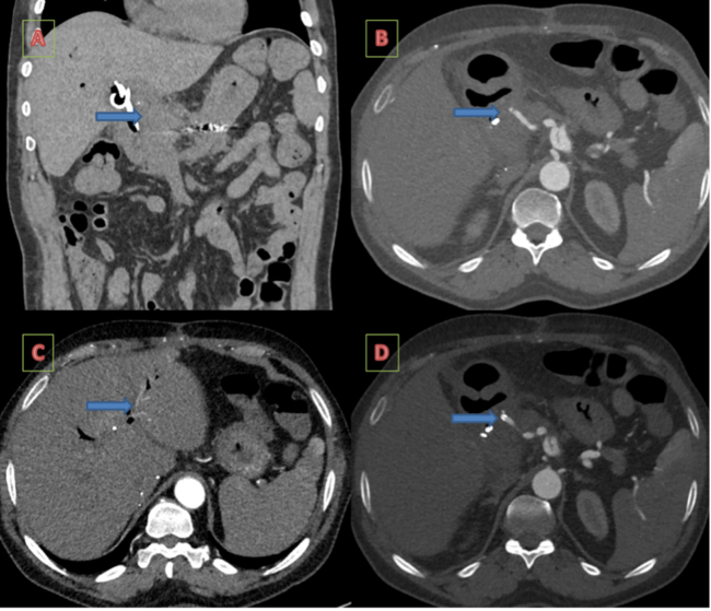


About Us
Vascular Disease Management™ (VDM), the official media partner of the International Symposium on Endovascular Therapy (ISET) and the Amputation Prevention Symposium (AMP), is a monthly peer-reviewed online journal that educates readers on the latest advancements in endovascular treatment strategies.
Learn more about ISET and AMP and register for the events here: