SEISMIQ: The Next-Generation Intravascular Lithotripsy System
Key Summary
- This review summarizes results from the RESTORE ATK (95 Rutherford 2-4 patients; superficial femoral artery/popliteal lesions) and RESTORE BTK (prospective, single-arm multicenter; 20 tibial patients) studies, which looked at the safety and efficacy of the SEISMIQ IVL System (Boston Scientific).
- In RESTORE ATK, 93.7% of lesions were severely calcified; residual stenosis was <50% in all patients and ≤30% in 86.3%, with 100% freedom from major adverse events at 30 days. RESTORE BTK reported no 30-day major adverse events and met its efficacy endpoint.
- The author views SEISMIQ as a promising option for severe peripheral calcification, citing selective energy delivery, up to 600 pulses per catheter, procedural flexibility, favorable early outcomes, and potential for future catheter-design innovations.
© 2026 HMP Global. All Rights Reserved.
Any views and opinions expressed are those of the author(s) and/or participants and do not necessarily reflect the views, policy, or position of Vascular Disease Management or HMP Global, their employees, and affiliates.
VASCULAR DISEASE MANAGEMENT. 2026;23(9):E127-E131
Abstract
Intravascular lithotripsy (IVL) has emerged as an effective strategy for treating heavily calcified peripheral arterial lesions. The SEISMIQ™ IVL System (Boston Scientific) introduces a novel laser-generated acoustic pressure-wave platform designed to fracture intimal and medial calcium. This unique design has 5 independently controlled emitter stations, allowing selective energy delivery and up to 600 pulses per catheter.
Clinical experience from the RESTORE ATK trial demonstrated procedural success in all 95 treated patients despite severe calcification in 93.7% of lesions. Primary outcome efficacy was met in all patients, with 100% freedom from major adverse events at 30 days and no reported perforation, distal embolization, abrupt closure, no reflow, or serious dissection. The RESTORE BTK trial similarly demonstrated favorable early safety and efficacy in tibial disease. Overall, laser-based IVL represents a promising advancement for calcium modification, offering procedural flexibility and favorable early clinical outcomes in challenging peripheral arterial disease.
Introduction
Intravascular lithotripsy (IVL) is a relatively recent novel technique for the treatment of severely calcific plaque lesions that utilizes acoustic shockwaves in a balloon-based delivery system. The technology was adopted from that used to treat renal calculi and merged into balloon-based angioplasty platforms, with the first to commercial market being electrical-current based (Shockwave, Johnson & Johnson). A substantial proportion of coronary and peripheral vascular lesions treated with transcatheter interventions contain moderate to severe calcific plaques, which are associated with lower procedural success rates, increased periprocedural adverse events, and unfavorable clinical outcomes compared to noncalcific plaques.1
Atherectomy, while helpful in certain instances and plaque morphologies, has failed to fully resolve the issue of severe calcific plaques, worsening patency and overall patient outcomes. IVL has been reported across multiple clinical studies in both peripheral and coronary circulations and has been shown to be safe and effective.2-5
The SEISMIQ™ IVL System (Boston Scientific) consists of a semicompliant balloon catheter integrated with 5 independently controlled lithotripsy emitter stations (10 total emitters) and a portable console. Unlike previous IVL technologies, the system delivers laser-generated pulsatile acoustic pressure waves to fracture intimal and medial calcium while minimizing injury to surrounding soft tissue. Balloon dilatation is then performed to open stenotic lesions.6
Efficacy and Trial Results
The Restore ATK trial6 enrolled patients with Rutherford category 2 to 4 disease with lesions located in the superficial femoral artery (SFA) or popliteal artery treated with the SEISMIQ IVL System. Target lesions had moderate to severe calcification, with more than 93.7% exhibiting severe calcification by adjudicated core lab review. Notably, this degree of calcification was greater than seen in the DISRUPT PAD II and DISRUPT PAD I trials (50% and 64.1%, respectively). The primary effectiveness endpoint was defined as procedural success, measured by an achievement of less than 50% residual diameter stenosis of the target lesion after treatment (with or without adjunctive percutaneous transluminal angioplasty [PTA] or stent placement). The primary safety endpoint was the composite incidence of new-onset major adverse events (MAEs) within 30 days, defined as death, clinically driven target lesion revascularization (CD-TLR), or major amputation of the target limb above the ankle.
A total of 95 patients with a mean age of 70.2 years were enrolled and treated, with 67.4% of patients identifying as male. Rutherford categories at baseline were predominantly category 3 (70.5%), with 23.2% classified as category 2 and 6.3% as category 4 (Table).
Table. The RESTORE ATK trial
| Intravascular lithotripsy (IVL) device | SEISMIQ™ IVL System (Boston Scientific) |
| N | 95 |
| Female (%) | 33 |
| Chronic total occlusions (%) | 22.1 |
| Lesion length (mm) | 96.0 ± 37.5 |
| Superficial femoral artery / popliteal artery (%) | 79 / 21 |
| Rutherford category 2 / 3 / 4 (%) | 23 / 71 / 6 |
| Severe calcification (core lab definition) (%) | 93.7 |
Residual stenosis was reduced to less than 50% in all 95 patients, which was also true when excluding adjunctive PTA therapy or stent placement. Of note, a more stringent threshold of 30% or less residual stenosis was achieved in 86.3% of patients. The primary safety endpoints were met with 100% freedom from MAEs at 30 days. Specifically, there were no cases of perforation, distal embolization, abrupt closure, no reflow, or dissections meeting serious adverse event criteria (n = 95).
The overall mean residual stenosis after IVL was 21.2%. Mean procedure duration was 62.3 minutes, and predilation occurred in 26.3% of cases to facilitate crossing target lesion or enhance imaging. Mean number of pulses delivered was 457.7 and 1.2 catheters per procedure.
The RESTORE BTK trial separately evaluated the safety and efficacy of the SEISMIQ IVL device in the below-the-knee setting.7 This was a prospective single-arm multicenter trial that enrolled 20 patients with Rutherford category 2 to 4 with de novo tibial lesions of 70% or greater stenosis and 150 mm or under in length. The primary safety endpoint was freedom from MAEs within 30 days after the index procedure. The primary efficacy endpoint was procedural success, defined as an acute reduction in target-lesion percentage diameter stenosis. At 30 days, no MAEs (including severe dissection, perforation, or unplanned major amputation of the target limb) and no CD-TLRs were reported, supporting the achievement of both the primary safety and efficacy endpoints in the trial.
Notable Differences in Technology
There are several notable differences in the technology that the physician operator should understand. The Shockwave device uses brief electrical discharges inside a saline-filled balloon catheter to create a rapid vapor plasma bubble. The collapse of this bubble generates localized sonic pressure waves that safely fracture hard calcium deposits in blocked arteries without harming soft vascular tissue.1-4 With the SEISMIQ device, the laser energy source is distributed through optical fibers converted to acoustic pressure waves by generating a plasma event as the energy hits each emitter's metallic target within the balloon. An initial plasma plume fractures the calcium, while a secondary cavitation bubble further expands the fractures. This difference in design leads to some notable differences for the physician end user.
First, the unit allows for delivery of 600 pulses per catheter, which is 200 more pulses per catheter compared with the Shockwave E8 (Johnson & Johnson).
Catheter lengths are 145 cm with 60 mm balloon lengths. The balloon catheter contains 5 emitter stations that are helically rotated along the length of the balloon catheter, and each station has 2 emitters that are positioned 180 degrees apart relative to the catheter. Each emitter station is offset by 72 degrees. Current balloon diameter offerings are 3.5 to 8 mm, all of which are 0.014-inch guidewire compatible and 6F sheath compatible. The console allows for 12 cycles with 50 pulses per 15-second cycle, and no lockout time between any subsequent cycle activations as opposed to the electrical current-based devices, which require a 10-second lockout period. The radiopaque emitter stations are easily seen by fluoroscopy, and the physician can “direct” the maximum pressure wave toward the most severe calcium deposits by orienting the calcium adjacent to the 2 parallel markers when seen at maximum separation. The unique laser-based pressure wave generation allows for emitter selectivity so the operator can choose to activate or deactivate any of the 5 emitter stations via the graphical console. This can allow for “saving” of certain emitter pulses when treatment is needed at multiple lesions but for shorter focal plaques.
Case Example
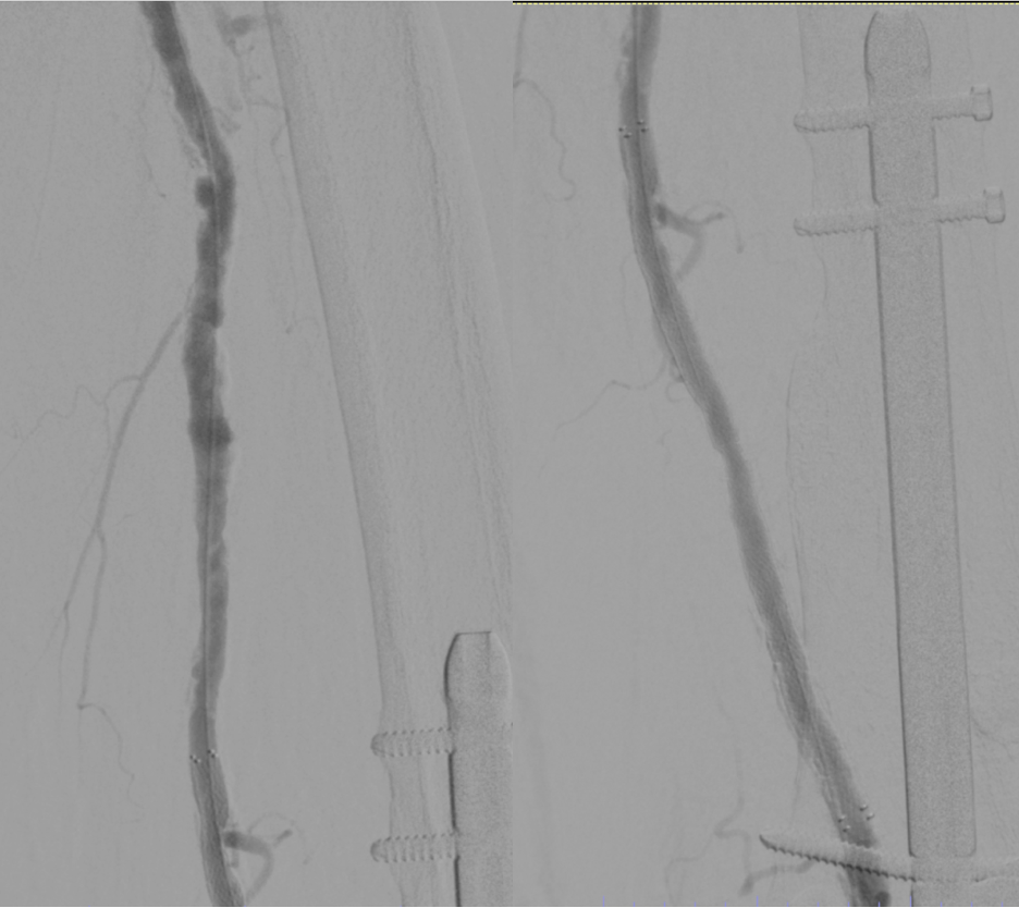
In this case, IVL was utilized for an SFA chronic total occlusion (CTO) in the setting of Rutherford category 5 chronic limb-threatening ischemia (CLTI). The patient is an 89-year-old woman with a history of coronary artery disease and hypertension, a 40 pack-year smoking history, and an abdominal aortic aneurysm (AAA) repaired in 2022 who presented to our clinic with a left medial ankle wound and significant rest pain relieved with dependence. Noninvasive testing by pulse volume record and eventually computed tomography angiography demonstrated a 12-cm long chronic occlusion of the distal SFA with severe intimal calcium in the proximal third (Figure 1).
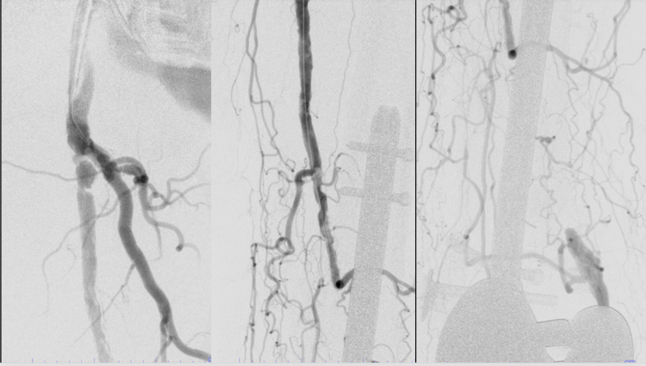
Access was obtained in antegrade fashion given the prior AAA repair and crossing attempted using a 0.014-inch weighted tip wire (Victory 14, Boston Scientific) and 0.018-inch Rubicon support catheter (Boston Scientific). Wire traversal could be obtained beyond the proximal cap but true luminal re-entry beyond to the native P1 popliteal artery could not, and device advancement was also unsuccessful due to severe calcium. Therefore, retrograde access was obtained via the posterior tibial artery, and a 0.014-inch wire was successfully advanced retrograde through the cap into the lumen above. Angioplasty was carried out from the pedal site (controlled antegrade retrograde tracking), allowing for successful antegrade wiring.
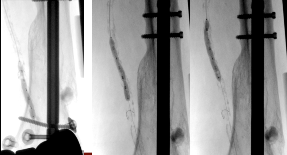
IVL was then carried out using a 6 x 60-mm SEISMIQ device and, using emitter selectivity, the resistant calcified proximal portion was effaced (Figure 2).
The balloon was then used via pedal access to treat a severe stenosis at the SFA origin adjacent to the sheath with successful effacement of the lesion (Figure 3).
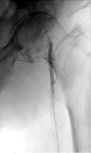
The SFA CTO was ultimately treated with a 6 x 150-mm Eluvia paclitaxel drug-eluting stent (Boston Scientific) with excellent results (Figure 4).
Conclusion
Laser-based IVL technology within the new SEISMIQ IVL System offers unique differences and potential advantages to operators over conventional electrical-based technologies. Data show extremely low rates of adverse events both in terms of vessel injury and embolization, as has been shown with electrical current-based IVL. The outcomes data are promising, with good lesion effacement in the face of severe calcific plaques. This novel means of laser-based cavitary pressure wave generation may portend to novel catheter designs such as longer balloon lengths, greater pressure wave generation, higher number of pulses per catheter, more emitters per balloon length, and longer catheter shaft lengths. Overall, the SEISMIQ IVL system offers exciting new treatment opportunities for difficult-to-treat severe peripheral calcification that has historically been a subset of the most difficult lesions to treat in patients with peripheral arterial disease and CLTI. n
Affiliations and Disclosures
From Atlantic Medical Imaging, LLC, Galloway, New Jersey.
The author reports no financial relationships or conflicts of interest regarding the content herein.
Manuscript accepted August 11, 2026.
Address for correspondence: Nicholas Petruzzi, MD, Atlantic Medical Imaging, 44 E. Jimmie Leeds Rd., Galloway, NJ 08205. Email: npetruzzi@atlanticmedicalimaging.com
References
1. Kereiakes DJ, Virmani R, Hokama JY, et al. Principles of intravascular lithotripsy for calcific plaque modification. J Am Coll Cardiol Interv. 2021;(12:1275-1292. doi:10.1016/j.jcin.2021.03.036
2. Ali ZA, Nef H, Escaned J, et al. Safety and effectiveness of coronary intravascular lithotripsy for treatment of severely calcified coronary stenoses: the Disrupt CAD II study. Circ Cardiovasc Interv. 2019;12(10):e008434. doi:10.1161/CIRCINTERVENTIONS.119.008434
3. Brodmann M, Holden A, Zeller T. Safety and feasibility of intravascular lithotripsy for treatment of below-the-knee arterial stenoses. J Endovasc Ther. 2018;25(4):499-503. doi:10.1177/1526602818783989
4. Brodmann M, Werner M, Holden A, et al. Primary outcomes and mechanism of action of intravascular lithotripsy in calcified, femoropopliteal lesions: results of Disrupt PAD II. Catheter Cardiovasc Interv. 2019;93(2):335-342. doi:10.1002/ccd.27943
5. Brodmann M, Werner M, Brinton TJ, et al. Safety and performance of lithoplasty for treatment of calcified peripheral artery lesions. J Am Coll Cardiol. 2017;70(7):908-910. doi:10.1016/j.jacc.2017.06.022
6. Brodmann M, Schlager O, Werner M, et al. Intravascular lithotripsy for peripheral artery calcification: 30-day and 6-month outcomes from the RESTORE ATK study. J Vasc Interv Radiol. 2026;37(6):108695. doi:10.1016/j.jvir.2026.108695
7. Zeller T. Evaluating the safety and efficacy of intravascular lithotripsy (IVL) technologies, including the RESTORE ATK and BTK clinical studies on peripheral artery calcification. Presented at: VIVA24, November 4, 2024; Las Vegas, NV.
Related Content