




Perforator-Based Thenar Flap for Reconstruction of a Traumatic Fingertip Wound Allowing Early Mobilization
© 2026 HMP Global. All Rights Reserved.
Any views and opinions expressed are those of the author(s) and/or participants and do not necessarily reflect the views, policy, or position of Wounds or HMP Global, their employees, and affiliates.
Abstract
Background. The conventional thenar flap is a reliable option for fingertip reconstruction; however, prolonged finger-to-palm immobilization may lead to postoperative joint stiffness. To address this limitation, the authors of the current case report developed a perforator-based modification of the distally based thenar flap that enables elevation of a longer flap and allows early finger mobilization. Case Report. A 22-year-old male presented with a painful volar pulp defect of the ring finger that persisted after healing by secondary intention. Reconstruction was performed using a distally based thenar flap incorporating a cutaneous perforator from the superficial palmar branch of the radial artery. Inclusion of the perforator enabled elevation of a longer flap, providing sufficient slack for tension-free inset and permitting limited finger motion while the flap remained attached. Early range of motion exercises were initiated on postoperative day 1, and the flap was divided 10 days after the initial surgery. Joint motion was preserved without contracture, and rapid functional recovery enabled an early return to work. Conclusion. This perforator-based modification allows elevation of a longer thenar flap with reliable vascularity. The additional flap length provides slack that permits early mobilization and may reduce the risk of postoperative joint stiffness compared with conventional thenar flaps.
Fingertip defect reconstruction requires durable, pliable, hairless, and well-padded tissue with excellent color and texture match. Among the available options, the thenar flap has long been considered a reliable method that satisfies these requirements.1,2 However, the conventional thenar flap is a random-pattern pedicled flap that requires prolonged finger-to-palm immobilization, which has been associated with postoperative metacarpophalangeal (MP) and proximal interphalangeal (PIP) joint contractures.1-3 In staged reconstruction of traumatic fingertip wounds, achieving durable coverage while minimizing immobilization-related functional impairment remains an important clinical consideration.
Orbay et al4 anatomically demonstrated that the palmar skin is supplied by multiple cutaneous perforators originating from the superficial palmar branch of the radial artery (SPBRA), thereby establishing the feasibility of designing palmar perforator flaps. Based on this concept, the authors of the current case report developed a perforator-based thenar flap that can be designed to achieve greater length than the conventional pedicled flap, thereby reducing direct constraint between the injured finger and the palm during fingertip reconstruction.
Postoperative immobilization is still required with this new technique. Nevertheless, the additional length of the flap provides a certain degree of slack, allowing limited finger motion and the initiation of gentle rehabilitation from postoperative day 1 while preventing complete joint restriction. This may help reduce the risk of postoperative joint stiffness while preserving the advantages of the thenar flap.
The current report describes this surgical technique, which uses a flap designed to provide sufficient slack for limited finger motion and immediate gentle rehabilitation, and presents its clinical feasibility in a representative case.
Case Report
A 22-year-old male sustained a volar pulp defect of the ring finger after his finger was caught in an industrial machine at work. The wound healed by secondary intention within 3 months. However, the patient continued to experience pain during pinching and pushing activities for more than 1 year, which prevented him from returning to work. Therefore, reconstruction using a thenar flap was performed to provide adequate soft-tissue padding. The patient provided written informed consent to publish details of his case.
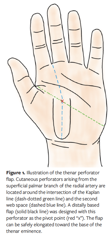
Orbay et al4 reported the presence of consistent cutaneous perforators originating from the SPBRA near the intersection of the Kaplan line and the second web space. Preoperative ultrasonography in the current case confirmed a perforator at this location, which was marked on the skin. A distally based perforator flap was designed with this vessel as the pivot point.
The flap was designed according to the defect size, with the perforator positioned at the base. The flap measured 20 mm in width and 55 mm in length, extending to the base of the thenar eminence (Figure 1).
The flap, including the thenar fascia, was elevated, with dissection proceeding from proximal to distal until the marked SPBRA perforator was reached (Figure 2). The vessels were contracted under tourniquet control and were difficult to visualize. Thus, meticulous dissection was performed around the perforator.
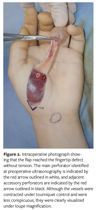
After refreshing the scar that had resulted from healing by secondary intention, the flap was inset using 5-0 nylon sutures. The flap was elevated at a greater length than in the conventional technique, which enabled suture to the fingertip without excessive tension, allowing the finger to be maintained in a more relaxed position and permitting a small degree of mobility while the flap remained attached. The donor site was closed to the greatest extent possible toward the base of the flap. The surgical duration was approximately 20 minutes.
Postoperatively, the finger was immobilized with a dorsal splint and covered with a bulky dressing. On postoperative day 1, flap perfusion was confirmed. Gentle flexion and extension exercises of the MP and PIP joints were initiated for approximately 30 minutes per day while the flap remained attached, ensuring that no excessive tension was applied to the flap. A certified hand therapist measured all ranges of motion. During this period, the ranges of motion with the pedicled flap in situ were as follows: MP joint, 90° flexion and 50° extension, and PIP joint, 100° flexion and 80° extension.
Ten days after the initial surgery, the flap was divided under local anesthesia. Neither skin maceration nor dermatitis related to prolonged joint flexion was observed. Two weeks after flap division, range of motion had recovered to 110° at the MP joint and 100° at the PIP joint, which were within the normal range. The patient was then able to return to work.
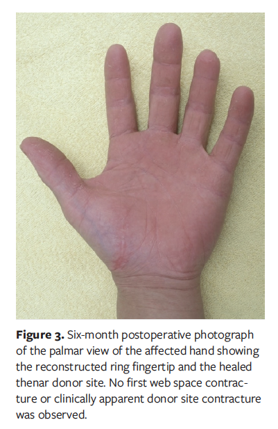
At the 6-month follow-up visit, the patient remained pain-free and had static 2-point discrimination of 7 mm over the reconstructed pulp. No hypertrophic donor site scarring or first web space contracture was observed (Figure 3).
Discussion
Unlike the conventional random-pattern pedicled thenar flap, the current technique incorporates a perforator from the SPBRA into a distally based pedicled flap, thereby providing a defined perforator-based vascular supply and allowing elevation of a longer flap. The contribution of the current modification lies not in the identification of the SPBRA perforator itself, which has been previously described, but in preserving the skin bridge while using the perforator-based vascular anatomy to extend the flap and provide sufficient slack for early controlled motion.
Omokawa et al5 anatomically demonstrated that perforators from the SPBRA communicate extensively with a longitudinal subdermal vascular network, providing the anatomical basis for safe longitudinal extension of thenar flaps. Consistent with this anatomical finding, Akita et al6 reported that thenar perforator flaps up to 65 mm in length could be raised, all of which survived completely. Accordingly, in the current case, the flap was extended to the base of the thenar eminence.
The major advantage of the current technique is that the additional length of the flap provides a degree of slack, which allows early mobilization. In contrast to conventional thenar flaps, in which the injured finger is directly fixed to the palm, complete joint immobilization can be avoided. Postoperative joint stiffness associated with prolonged finger-to-palm immobilization has long been a concern with conventional thenar flaps.3 Recent studies have shown that favorable functional outcomes can be achieved through refinements in flap design, appropriate positioning, and structured postoperative rehabilitation.1-3 However, these measures do not eliminate the inherent requirement for direct attachment of the injured finger to the palm until flap division, and joint motion during this period remains restricted. In the current technique, incorporation of the SPBRA perforator permitted elevation of a longer flap with sufficient slack, allowing controlled MP and PIP joint motion from postoperative day 1 while the flap remained attached. This structural difference, rather than postoperative rehabilitation alone, represents the principal functional advantage of the current modification over the conventional thenar flap.
The rapid recovery of joint motion after flap division further supports the potential functional benefit of this method. Barbato et al7 reported a mean time to return to work of 42 days (range, 30-120 days) among patients injured at work. More recently, Kang et al8 reported that patients treated with an extended thenar flap returned to work at a mean of 6.2 weeks. In the current case, the patient returned to work 24 days after the initial surgery and 14 days after flap division, suggesting relatively rapid functional recovery. Nevertheless, direct comparison between these reports should be interpreted cautiously because of differences in injury patterns, flap designs, rehabilitation protocols, occupational demands, and definitions of “return to work.” Moreover, because this is a single case, the early return to work cannot be attributed solely to the current modification.
The principal clinical implication of the current technique is that controlled rehabilitation can be initiated before flap division. Preservation of joint motion during the period of flap attachment may facilitate postoperative functional recovery and contribute to an earlier return to work. This technique may also be useful in patients for whom prolonged immobilization is considered undesirable, including older patients (ie, those older than 30 years), who have historically been considered at risk of joint stiffness.1,2 However, because the current patient was young, the potential benefit of this technique in preventing contracture in older patients remains to be established.
Akita et al6 reported a perforator-based thenar island flap supplied by the SPBRA, enabling single-stage fingertip reconstruction without temporary attachment of the injured finger to the palm. This represents a major advantage over conventional pedicled thenar flaps. However, complete islanding of the flap requires meticulous dissection around a small-caliber perforator and is technically more demanding than elevation of a skin-bridged pedicled flap. Recent literature has demonstrated the versatility and favorable outcomes of pedicled digital artery perforator flaps for fingertip reconstruction, while also documenting a wide variety of flap configurations that require precise perforator dissection.9 In contrast, the current technique preserves the skin bridge and does not require complete skeletonization of the perforator.
Surgical times cannot be directly compared across different studies. The current technique was completed within approximately 20 minutes, could be performed using surgical loupes alone, and did not require complete skeletonization of the perforator. Thus, it may represent a simple and practical reconstructive option in routine clinical practice.
The ability to maintain the finger in a less flexed position may represent another practical benefit of the current technique. In the current case, partial finger extension was possible while the flap remained attached, and neither skin maceration nor dermatitis was observed.
Overall, this perforator-based modification preserves the procedural simplicity associated with a skin bridge while permitting elevation of a longer flap and controlled joint motion before flap division. The current case demonstrates the technical feasibility of this approach; however, its proposed benefits in preventing stiffness and accelerating functional recovery require further evaluation.
Limitations
The current case report has limitations. The current distally based perforator flap is primarily suitable for volar fingertip defects and is not recommended for dorsal defects because of an unfavorable arc of rotation. In such cases, reconstruction using a proximally based conventional thenar flap may be more appropriate. In addition, this report describes a single young patient and does not include a control group treated with a conventional thenar flap. Therefore, the absence of joint contracture and the relatively early return to work cannot be conclusively attributed to the perforator-based modification or to early rehabilitation. The applicability of this technique to older patients and others at increased risk of postoperative joint stiffness, as well as the optimal flap length, degree of slack, rehabilitation protocol, and timing of flap division, should be evaluated in larger comparative studies.
Conclusion
The perforator-based thenar flap allows the elevation of a longer flap with a defined vascular supply by incorporating a perforator from the SPBRA. The additional flap length provides sufficient slack to permit controlled early mobilization while the flap remains attached and may reduce the risk of postoperative joint stiffness. This simple technique may represent a practical modification of the conventional thenar flap by reducing the functional burden associated with staged fingertip wound reconstruction.
Author and Public Information
Authors: Ko Nakao, MD1; Masafumi Yamaga, BSc2; Tetsu Domoto, MD1; Daiki Ito, MD1; and Mashu Tsujita, MD1
Affiliations: 1Department of Plastic and Reconstructive Surgery, Keio University School of Medicine, Tokyo, Japan; 2Department of Mechanical Engineering, Keio University, Yokohama, Japan
Acknowledgment: The authors would like to thank Enago for assisting in the English-language editing of the manuscript. A generative artificial intelligence tool (ChatGPT; OpenAI) was used solely to improve the English language of the manuscript; the authors reviewed all text and take full responsibility for the content of the manuscript.
Author Contributions: Ko Nakao: Conceptualization, surgical procedures, data collection, manuscript drafting, and corresponding author responsibilities. Masafumi Yamaga: Technical consultation, data interpretation, and manuscript revision. Tetsu Domoto: Surgical assistance and data collection. Daiki Ito: Postoperative management and data collection. Mashu Tsujita: Literature review and manuscript revision. All authors reviewed and approved the final manuscript.
Funding: This research did not receive any specific grant from funding agencies in the public, commercial, or not-for-profit sectors.
Disclosure: The authors disclose no financial or other conflicts of interest.
Data Availability: The data supporting the findings of this study are available from the corresponding author upon reasonable request.
Ethics Statement: Written informed consent to publish case details was obtained from the patient.
Correspondence: Ko Nakao, MD; Department of Plastic and Reconstructive Surgery, Keio University School of Medicine, 35 Shinanomachi, Shinjuku-ku, Tokyo 160-8582, Japan;
ko.comcomgrapes@gmail.com
Manuscript Accepted: August 14, 2026
References
1. Lemsanni M, Najeb Y, Chaouqui Y, Elkasseh M, Zoukal S. Fingertip injuries managed by a thenar flap: follow-up and long-term outcomes of 32 cases. Hand Surg Rehabil. 2021;40(4):484-490. doi:10.1016/j.hansur.2021.04.003
2. Huded V, Gautam G. Thenar flap: a workhorse flap for fingertip injuries. Cureus. 2025;17(8):e90785. doi:10.7759/cureus.90785
3. Li F, Tie JL, Kang YC, Tan JSW. Refinements in the design and division of the thenar flap to achieve better functional results. JPRAS Open. 2021;28:10-18. doi:10.1016/j.jpra.2021.01.010
4. Orbay JL, Rosen JG, Khouri RK, Indriago I. The glabrous palmar flap: the new free or reversed pedicled palmar fasciocutaneous flap for volar hand reconstruction. Tech Hand Up Extrem Surg. 2009;13(3):145-150. doi:10.1097/BTH.0b0133181ac9183
5. Omokawa S, Ryu J, Tang JB, Han J. Vascular and neural anatomy of the thenar area of the hand: its surgical applications. Plast Reconstr Surg. 1997;99(1):116-121. doi:10.1097/00006534-199701000-00018
6. Akita S, Kuroki T, Yoshimoto S, Rikihisa N, Satoh K. Reconstruction of a fingertip with a thenar perforator island flap. J Plast Surg Hand Surg. 2011;45(6):294-299. doi:10.3109/2000656X.2011.634549
7. Barbato BD, Guelmi K, Romano SJ, Mitz V, Lemerle JP. Thenar flap rehabilitated: a review of 20 cases. Ann Plast Surg. 1996;37(2):135-139. doi:10.1097/00000637-199608000-00003
8. Kang HJ, Sung SY, Whang JI, Hong JJ, Park HS, Kim JS. Extended thenar flap for two adjacent fingertip amputations. J Plast Reconstr Aesthet Surg. 2020;73(1):65-71. doi:10.1016/j.bjps.2019.07.002
9. Khan WU, Appukuttan A, Loh CYY. Homodigital pedicled digital artery perforator flaps for fingertip reconstruction: a review of flap options. JPRAS Open. 2022;34:199-218. doi:10.1016/j.jpra.2022.09.004