Is There a Chronic Wound-Patient Phenotype? A Retrospective Analysis of Real-World Overlap of Diabetic Foot and Venous Leg Ulcers
© 2026 HMP Global. All Rights Reserved.
Any views and opinions expressed are those of the author(s) and/or participants and do not necessarily reflect the views, policy, or position of Wounds or HMP Global, their employees, and affiliates.
Abstract
Background. The prevailing paradigm in wound care research assumes that diabetic foot ulcers (DFUs) and venous leg ulcers (VLUs) are distinct clinical entities. Objective. To evaluate and compare real-world patients and their wound characteristics in 2 large cohorts of patients with DFUs and VLUs. Methods. Aggregated data from a wound care–specific electronic health record documented from January 1, 2021, to December 31, 2022, were retrospectively analyzed. Comorbidities, medications, and concomitant wound types were compared. Results. Of 160 341 total wounds, there were 26 042 DFUs among 10 966 patients and 30 790 VLUs among 11 197 patients. Major comorbidities and medications were nearly identical between the cohorts. Most patients had obesity and hypertension, and 13.7% of patients with VLU and 16.1% of patients with DFU had an autoimmune disease. Among the approximately 70% of patients with multiple and/or concomitant wounds, 17.1% of patients with DFU had a VLU and 15.5% of patients with VLU had diabetic ulcers. Approximately 40% of patients in each cohort developed a new DFU or VLU over the 2-year period. Conclusion. Real-world patients with DFUs and VLUs exhibit overlapping clinical profiles and multiple wounds of different types, suggesting a shared chronic wound-patient phenotype. These findings challenge the established wound-specific disease paradigm, suggesting additive categories in the current approaches in wound care research.
Chronic wounds do not progress through the normal healing trajectory, often stalling in the inflammatory phase 4 weeks to 12 weeks after wound onset.1,2 Chronic wounds are an advanced complication of underlying disease and chronic conditions, including venous disease, arterial insufficiencies, diabetes, and unmanaged pressure and shear. A harmful cycle of prolonged inflammation that prevents wound closure is thought to result from ischemia-reperfusion injury that causes impaired, proinflammatory cellular activity and tissue hypoxia, lowering the natural immune defense and facilitating bacterial colonization and infection.2-7 The prevalence of chronic wounds is increasing at an alarming rate in the United States (US); among Medicare beneficiaries, prevalence rose from 14.5% in 2014 to 16.3% in 2019.8 Even more concerning is the growing burden of chronic wounds among patients younger than 65 years.8-10
Efforts to develop more effective wound treatments are accelerating, with the aim of reducing both patient suffering and the associated health care costs. However, most randomized controlled trials (RCTs) target a single, isolated wound of a specific type. RCTs typically enroll patients with relatively superficial wounds that are, on average, 3 to 5 times smaller than those encountered in routine clinical practice and assess healing by epithelialization over an unrealistically short time frame (eg, 12 weeks–16 weeks).11-13 Moreover, insufficient attention has been given to the underlying drivers of the chronic wound epidemic. Real-world patients are burdened by complex comorbidities that increase wound incidence and severity and impede wound healing but are largely excluded from RCTs.11-16 Real-world patients with chronic wounds have, on average, 8 serious comorbidities each, with some literature noting as many as 26 conditions per patient.11-14 Consequently, the patient journey in wound care often spans months or even years beyond the typical RCT time frame of 12 weeks to 16 weeks, with real-world patients seeing multiple providers at multiple sites of care.17,18 These patients’ multimorbidity may help explain why real-world healing rates at 12 weeks range from about 31% to 44%, despite the existence of efficacious advanced wound therapies.12
When efficacy, that is, the proof of principle of a treatment or intervention, is demonstrated only within a narrowly defined wound category in a few explanatory RCTs, little insight is gained into the real-world effectiveness (the beneficial effect of a treatment in real-world patients) of the treatment or intervention among complex patient populations.11-16,19 This noted paradox reflects a significant challenge in the field of wound management and arises from various factors, including adherence to treatment protocols, patient-specific variables, and the complexities of individual cases that are not fully addressed in clinical studies. The limitations of wound care research are further illustrated by the availability of wound-specific clinical practice guidelines developed primarily from studies of equally siloed wound types.20,21
To better understand the potential real-world benefit and generalizability of a treatment or intervention, real-world evidence (RWE) generated from real-world data (RWD) is used complementarily to RCT evidence. The US Food and Drug Administration (FDA) defines RWD as data routinely collected from electronic health records (EHRs), medical claims, registries, and other tools that inform on patient health status and care delivery and acknowledges that RWD may be essential to align clinical practice with the promising results of RCTs.13,16,22 Previously, the FDA required that identifiable patient RWD be submitted in marketing applications for medical devices. In December 2025, the FDA dropped this requirement, allowing for the increased use of RWE generated from deidentified patient databases and registries in its regulatory decision-making processes.23
The Wound Care Collaborative Community (WCCC), a 501(c)3 nonprofit recognized by the FDA, previously identified key barriers to wound care, including the lack of an understanding of the natural history of the underlying disease etiologies, the lack of innovative wound therapies, and the lack of timely access to treatments from which patients are likely to benefit.24 To overcome the limitations of RCTs and better understand the natural history of chronic wounds and the complete patient journey in wound care, the WCCC established an RWE group to promote the use of RWD and improve wound care research. This initiative aims to inform regulatory pathways for FDA approvals and guide payer coverage decisions to improve patient access and outcomes. An efficient and streamlined process of generating RWD is needed in wound care so that RWE is usable and applicable.17 Therefore, an RWD landscape analysis was initially performed in collaboration with B.R.I.D.G.E. To DATA (DGI, LLC) to identify the best RWD database for this project.25 The U.S. Wound Registry (USWR) was selected for the WCCC Natural History Project (NHP) because it is a national, wound-specific data repository generated from the direct transmission of structured EHR data, thus minimizing sample bias, and it is aggregated in a format readily available for analysis.13,16,25
Next, the WCCC RWE group used USWR data to understand the natural history of a broad spectrum of patients with diabetic foot ulcers (DFUs) and venous leg ulcers (VLUs) and to compare this real-world population with participants in wound RCTs. The WCCC RWE group chose to focus on the natural history of DFUs and VLUs, because DFUs are the most studied chronic wound type in wound care, whereas VLUs are the most common chronic ulceration.26,27 DFUs are ulcers located on the foot of patients with diabetes that develop in the setting of neuropathy or loss of protective sensation, peripheral vascular disease with ischemia, or both, and they can involve biomechanical abnormalities.28 VLUs are ulcerations that occur in the presence of venous hypertension.20
This first article of a 3-part retrospective analysis exploring the natural history of DFUs and VLUs analyzes 2 cohorts of patients with DFUs and VLUs, examining their comorbidities, concomitant medications, and wound characteristics to better understand their overall health status and wound burden. The subsequent articles in this series will explore the clinical course and progression of DFUs and VLUs (the “natural history” of real-world patients and their chronic wounds), providing a thought-provoking perspective on the complete patient journey and real-world outcomes.
Methods
Study EHR
In the USWR, research-grade data are captured by practitioners during routine clinical care using a highly structured, wound care–specific EHR (Intellicure; Intellicure, LLC).16 As the legal medical record, the EHR includes complete documentation of all patients and all wounds. Upon completion of each clinical encounter, the full structured record is automatically and securely transmitted to the USWR to ensure data integrity, completeness, and consistency. All patient data are aggregated, deidentified, and stored in a relational database.
The study EHR uses a structured, ontology-driven interface that facilitates clinical documentation at the point of care. The data collection and transfer processes, use of structured language, and interoperability features have been reported in detail previously.13,16,25 Briefly, to document a wound diagnosis, the EHR first prompts clinicians to choose the diagnostic pathway (in the current study, for a DFU or a VLU). This process uses Structured Query Language to accurately document associated underlying comorbidities, as well as wound location, depth, and severity. Within the relational database, these selections are systematically mapped to the most clinically precise International Classification of Diseases, Tenth Revision, Clinical Modification (ICD-10-CM) dual-code combinations,29 encompassing more than 30 000 clinical concepts, which guide the clinician in identifying the underlying disease (in the present study, diabetes or chronic venous insufficiency) based on the ulcer characteristics (eg, location and depth).
When venous disease was documented in the current study, this was done by clinical assessment (for example, a leg ulcer with hemosiderin and edema), and all VLUs were identified as Clinical-Etiology-Anatomy-Pathophysiology class 6, the most severe stage of venous disease. Clinicians were also required to classify DFUs using the Wagner grading system in the EHR, because Medicare coverage policy is linked to Wagner grade. The Wagner grade documented was cross-referenced to documentation of the deepest tissue type to identify DFUs with exposed bone and tendon, separate from the Wagner classification.
Data Collection
The retrospective analysis assessed data that had been prospectively collected over a defined period, specifically, from January 1, 2021, to December 31, 2022. This method allowed for a comprehensive review of information gathered during that time frame, providing insights into trends and outcomes relevant to the study objectives. During this time frame, 527 wound care providers contributed wound data at 149 participating clinics or facilities, including 95 outpatient clinics (83 hospital-based outpatient wound care centers and 12 office-based practices) in 29 US states. However, the registered clinicians could have documented EHR data at multiple sites per patient; thus, data were reported from 164 sites of care, including 62 residential facilities. In a handful of cases, data were collected from 7 inpatient facilities when a registered patient was hospitalized and their study clinician cross-referenced the hospitalization data to the USWR EHR; however, it is not common practice for clinicians to do so.
A Clinical Advisory Panel (CAP) of wound care experts from the WCCC provided study oversight. CAP members reviewed and provided advice on overall data processes and made recommendations to clarify research questions. For example, because the ICD-10-CM L97 code set includes designations for ulcers involving deep structures (eg, muscle and bone), some clinicians used these codes to document VLUs. To improve cohort specificity, the CAP excluded from analysis any VLUs with exposed deep structures or that were documented as being covered with eschar, findings which likely indicate arterial ulcers. Additionally, VLUs documented on the thigh were excluded, because this presentation is clinically unlikely.
The study dataset derived from all patients, their wounds, and their related visits during the study period, applying the following exclusions: patients with fewer than 3 total visits, nurse-only visits that did not involve an advanced practitioner, telehealth visits, and clinical problems that were not related to a wound (eg, lymphedema). Any patient with both DFU and VLU wound types was included in both cohorts, as long as they did not meet any exclusion criteria.
Medication data were obtained primarily from an electronic prescribing module (DrFirst) that was integrated into the EHR.
Data Analysis
Comorbidities were classified using the Elixhauser Comorbidity Index.30 To comprehensively analyze comorbidities that may not have been thoroughly recorded by practitioners, under the guidance of the CAP, imputations of comorbid conditions were made based on the following medications recorded in the EHRs: apixaban was imputed as atrial fibrillation; amlodipine, carvedilol, lisinopril, and/or losartan were imputed as hypertension; clopidogrel was imputed as peripheral arterial disease (PAD); and atorvastatin was imputed as hyperlipidemia. Obesity was imputed based on body mass index greater than or equal to 30. Thyroid disease was counted separately from autoimmune diseases, in accordance with the Elixhauser Comorbidity Index.30
The 30 most common medications ever documented for each patient cohort were summarized. A patient was considered to have received home health care when a home health agency was reported for some part of their time in service. Orders placed to a durable medical equipment company indicated that patients or their caregivers performed interval dressing changes between outpatient clinic visits.
Wound characteristics were compared between DFUs and VLUs and included wound age (patient-reported wound duration) and the area of the wound surface (length × width, hereafter referred to as wound area). For all patients, the number and types of DFUs and VLUs recorded in the EHR during the study period and their concomitant wounds (defined as wounds of any type simultaneously coexisting with the index DFU or VLU) were analyzed.
Categorical variables were summarized by counts and percentages. Descriptive statistics were used to summarize continuous variables. The natural orders of the burden of comorbidities, medications, and concomitant wound types were visualized and compared using treemaps created in Excel (Version 2608, Microsoft 365) for both datasets.
Results
A total of 160 341 wounds or ulcers were recorded in the USWR during the study period. The most common wound type was the generic chronic nonpressure ulcer (n = 35 743 [22.3%]), followed by VLUs (n = 34 236 [21.4%]) and DFUs (n = 26 042 [16.2%]); traumatic wounds (n = 23 207 [14.5%]) were nearly as prevalent as DFUs. Among the 34 236 VLUs initially identified, 3 444 (10.1%) were excluded from analysis for the following reasons: 2 768 (8.1%) for documented eschar, 935 (2.7%) for exposed deep tissue, and 67 (0.2%) for being located on the thigh. Finally, 2 VLUs from a pediatric patient were removed from the dataset because they did not meet the diagnostic criteria of VLU; clinical details specified that they were insect bites. The final dataset for analysis included 10 966 patients with 26 042 DFUs and 11 197 patients with 30 790 VLUs. However, there were 20 851 distinct patients with 56 832 distinct wounds, because there were 1 312 patients with multiple wounds of both etiologies who were included in both patient cohorts. These overlapping patients made up approximately 12% of each cohort and 6.3% of all patients analyzed.
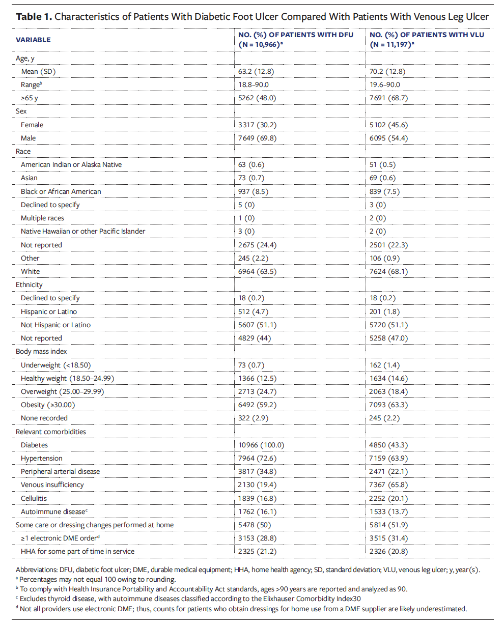
Table 1 summarizes the demographic and clinical characteristics of each patient cohort. All patients included in this anal ysis were adults (≥18 years). Patients with DFUs were generally younger than those with VLUs, with a mean (SD) age of 63.2 (12.8) years and 70.2 (12.8) years, respectively. Patients with DFU were predominantly male (69.8% [n = 7 649] vs 30.2% female [n = 3 317]), whereas there was a slight preponderance of males in the VLU cohort (54.4% [n = 6 095] vs 45.6% female [n = 5 102]). Race and ethnicity distributions were similar across both groups, including the proportion not reporting race or ethnicity. Body mass index data were available for nearly all patients and were similarly distributed across both cohorts, with approximately 61% of patients meeting criteria for obesity (Table 1).
Most patients in both cohorts had comorbidities documented: 10 264 patients (93.6%) in the DFU cohort had comorbidities (other than diabetes) documented, and 11 102 patients (99.2%) in the VLU cohort had any comorbidities documented. Only 1 patient in the DFU cohort (0%) and 26 patients in the VLU cohort (0.2%) had no comorbidities nor medications documented. Figure 1 displays treemaps of the 10 most common comorbidities among patients with DFUs and VLUs, using consistent color coding across panels to facilitate direct comparison. The overall burden and distribution of comorbidities were remarkably similar, with 8 shared conditions dominating both groups (80%). Differences aligned with expected clinical distinctions: Neuropathy and osteomyelitis were more prevalent in the DFU cohort, whereas congestive heart failure and lymphedema were more common in patients with VLU.
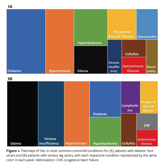
Table 1 provides additional evidence supporting the existence of a shared chronic wound-patient phenotype. Whereas 43.3% of all patients with VLU also had diabetes, 19.4% of patients with DFU had a documented diagnosis of venous insufficiency. Hypertension was highly prevalent in both cohorts, and PAD was also important in both cohorts. Of note, both patients with VLUs (13.79%) and those with DFUs (16.1%) had autoimmune disorders. These findings underscore the clinical complexity of chronic wound patients and challenge the traditional approach of managing DFUs and VLUs as entirely distinct disease entities.
Tables 2 and 3 list the 30 most commonly documented medications for patients with DFUs and VLUs, respectively. Notably, both patient populations shared 4 of the 5 most frequently prescribed medications. During the study period, at least 1 in 4 patients with DFUs and more than 1 in 5 patients with VLUs were prescribed antibiotics, while similar proportions were taking aspirin.
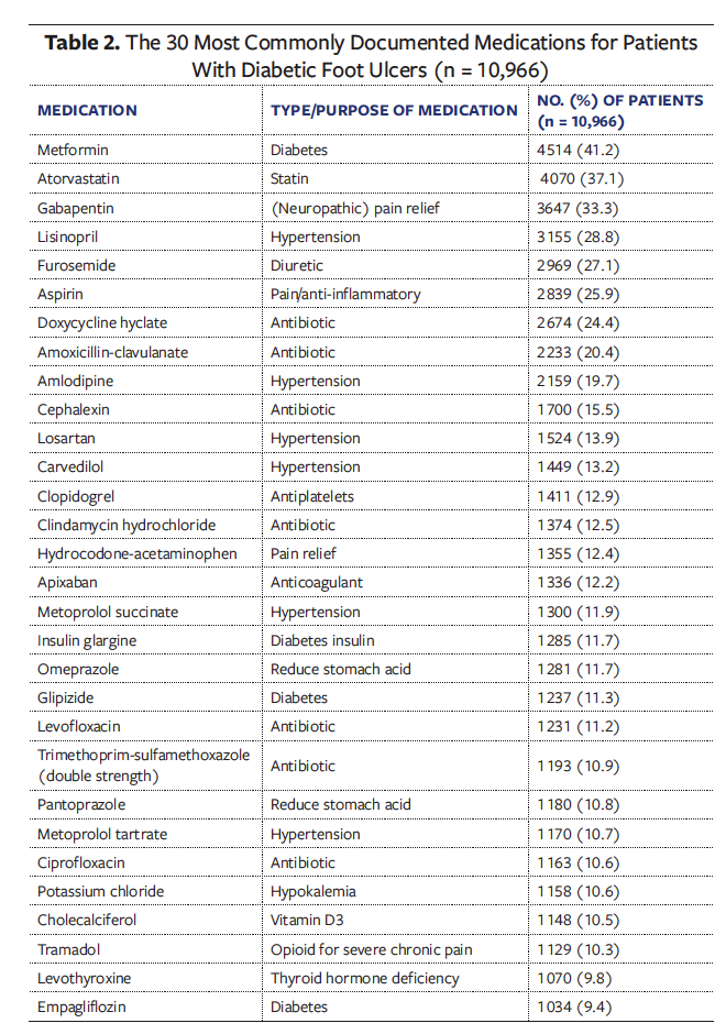
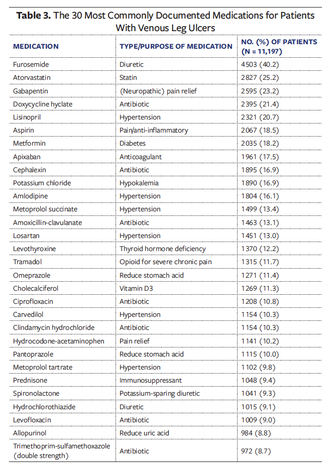
Figure 2 presents treemaps of the 10 most commonly prescribed medications for each patient cohort. Consistent with the overlapping comorbidity burden illustrated in Figure 1, the overall medication profiles were remarkably similar: 8 medications were shared across both cohorts (80%). These included agents used to manage diabetes, hypertension and edema, cardiovascular disease, pain, and infection. A key distinction was that potassium chloride—used to treat hypokalemia—was among the top 10 medications for patients with VLUs.
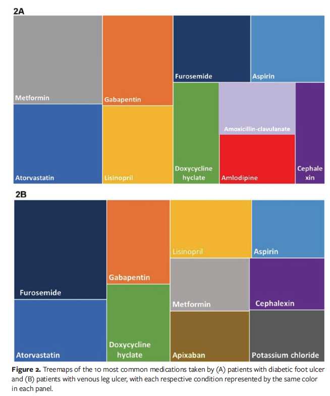
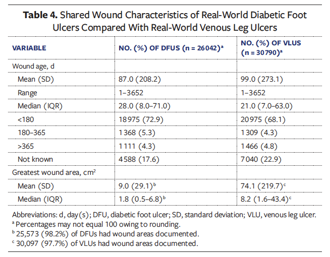
Table 4 compares key wound characteristics between DFUs and VLUs. Both wound types had a mean wound duration exceeding 84 days (12 weeks), with greater than 9% of wounds persisting for longer than 180 days, underscoring the chronic nature of these ulcers. On average, wound area was 8.2 times larger for VLUs than for DFUs.
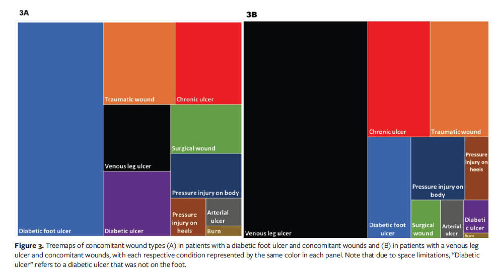
The percentage of patients with concomitant wounds was similar in both cohorts: 66.3% of patients with DFUs (n = 7 269) and 70.7% of patients with VLUs (n = 7 921). Table 5 shows the distribution of concomitant wounds that coexisted simultaneously with the index ulcer analyzed in each cohort. For patients with DFUs, the most common concomitant wound type was an additional DFU (69.7%). Similarly, patients with VLUs most frequently had another VLU (80.1%). Beyond this distinction, both groups demonstrated remarkably similar distributions of other concomitant wound types, including traumatic wounds, chronic ulcers, pressure injuries, and arterial ulcers, as illustrated in Figure 3 and detailed in Table 5.
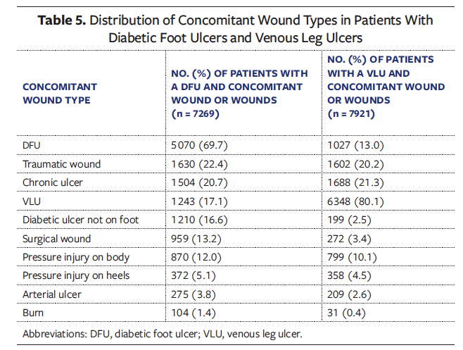
The treemaps of concomitant wounds for the 2 cohorts are nearly indistinguishable (Figure 3), further underscoring the shared chronic wound-patient phenotype. Patients with VLUs had a mean (SD) of 1.8 (1.2) concomitant VLUs (range, 1.0–11.9), and a mean of 2.5 (2.0) wounds of any type (range, 1.0–25.0). Patients with DFUs averaged 1.7 (1.1) concomitant DFUs (range, 1–12) and 2.7 (2.3) total concomitant wounds (range, 1–46). Notably, among patients with concomitant wounds, 17.1% of patients with DFUs had a concomitant VLU, and 15.5% of patients with VLUs had either a concomitant DFU or a nonfoot ulcer attributed to diabetes by the clinician (Table 5). Both groups were equally likely to develop new ulcerations during the course of treatment, with 41.2% of patients with DFU (n = 4 520) and 40.6% of patients with VLU (n = 4 551) developing new DFUs or VLUs while still in treatment for the original wound.
Discussion
Many findings in the present analysis support previously identified clinical associations. While racial and ethnic distributions were similar between groups, patients with DFUs were predominantly male, whereas a larger proportion of patients with VLUs were female, consistent with established associations between sex and wound type.9,29,30 Patients with VLUs were, on average, 7 years older than those with DFUs, and 68.7% were Medicare-eligible (≥65 years; Table 1). Only 48% of the DFU cohort was old enough for Medicare, which raises the concern that these complex problems are presenting at a younger age, which also has important implications to the workforce population that is covered under commercial payer plans.
Obesity and hypertension were the most prevalent comorbidities in both cohorts, while diabetes, PAD, other vascular disorders, autoimmune conditions, and wound infections contributed to the patient burden (Table 1, Figure 1). Although prior RWD studies have reported lower obesity rates in similar populations with DFUs and VLUs, hypertension remains the most consistently documented comorbidity, affecting the majority of patients.9,14,30-33
A particularly novel observation in the current analysis is the substantial prevalence of autoimmune disease, which has not been widely recognized in previous literature. Additionally, the high rates of diabetes and heart failure among patients with VLUs have important implications for both clinical trial design and clinical practice.
Medication history is rarely reported in wound care clinical trials, although when reported, diabetes treatment medications, antihypertensives, pain relievers, antibiotics, anticoagulants, and statins are commonly prescribed to clinical trial participants.9,34 In the present analysis, patients with DFUs and VLUs were similarly prescribed these medications. Antibiotics were among the most frequently used medications (Tables 2 and 3). While there were high rates of cellulitis observed in both cohorts in Table 1 and osteomyelitis was important among DFUs in Figure 1, debate continues regarding the possible overuse of antibiotics in hard-to-heal wounds. In a study of 350 chronic wounds treated by 20 clinicians, one-third of patients were prescribed antibiotics when they lacked clinical signs and symptoms of infection.35 Although the optimal use of antibiotics in these patients requires additional study, the prevalence of their use in the current analysis is important.
In the current study, the patterns of comorbidities, medication use, and concomitant wound types were strikingly similar between patients with DFUs and those with VLUs. As illustrated in Figures 1 through 3, the elements of each treemap align closely, forming a common chronic wound-patient phenotype that underscores the shared characteristics of these patient populations. The overlap in underlying medical conditions, medication profiles, and the frequent presence of multiple concomitant wound types challenges the prevailing notion that DFUs and VLUs should be categorized and treated as distinct clinical conditions. A chronic wound-patient phenotype is further suggested by the striking overlap of the various other wound types among the cohorts (Figure 3). A noteworthy finding is that approximately 11% (1 243/10 966) of patients with DFUs also had a VLU, and approximately 11% (1 226/11 197) of patients with VLUs had a DFU or other ulcer attributed to diabetes. Even more compelling, 6.3% of all patients in the current analysis ( 1312 of 20 851) were present in both cohorts, indicating that, in some cases, comparisons between DFUs and VLUs are literally being made between different wounds in the same patient. Future research should comparatively analyze the differences between patients with VLUs, patients with DFUs, and patients with both DFUs and VLUs. The current findings emphasize the limitations of wound-specific clinical practice guidelines; many patients with DFUs need edema management, and clinicians frequently overlook or underestimate lower extremity venous insufficiency and its implications for wound healing.27 Likewise, many patients with VLUs require diabetes management and protection from pressure.
The findings also raise another important question about wound care research: what’s in a wound name? Whether labeled a DFU or VLU, generic chronic ulcer, or pressure ulcer/injury, these lesions often occur in the same medically complex patients. This strongly suggests that the treatments used for a specific wound type are not directed at broader aspects of basic pathophysiology and underlying etiology. This insight carries profound implications for clinical trial design, regulatory frameworks, payer coverage policy, and the delivery of care to patients with chronic ulcers. These findings can perhaps also explain why results obtained using advanced wound treatments in the setting of RCTs do not usually yield similar results when the same advanced treatments are used in real-world settings. Complex patients with multimorbidity in the real world see multiple providers beyond the wound care setting, each of whom treats specific problems of the patient, including but not limited to primary care providers, rheumatologists, dermatologists, infectious diseases specialists, and potentially, vascular and plastic surgeons. Due to lack of wound education and highly siloed specialty guidelines,20,21 the standard of care, assessment and treatment of infection, and overall management of the underlying disease or diseases that caused the wound are fragmented. Consequently, the patient may experience a vicious cycle of poor outcomes. To holistically address the underlying factors that impair healing, wound care and wound care research must shift from a wound-centric paradigm to one that is patient-centric and addresses underlying etiologies.15,24,31
Wound care clinicians should approach patient care in accordance with the common wound care maxim, do not treat the hole in the patient but the patient as a whole. In the present study of RWD, the sum of all parts related to chronic DFUs and VLUs reveals a possible chronic wound-patient phenotype that merits further attention in wound care research.
Limitations
The registry used in the current study is subject to fewer patient or wound selection biases, because all wounds in all patients were documented in the EHR, which internally calculated the practitioner and facility charges, making this study unique in its ability to capture multiple concomitant wounds by type. While inpatient data typically are not included in the registry, it should be noted that these data would not help in understanding time to closure or final outcome, because inpatient stays are short and wound healing time frames are long. As has been noted in Medicare claims analysis, the majority of both cost and care for all wound types, including VLUs, happens in the outpatient setting.8
The primary limitation of the current study lies in the variability of clinical documentation inherent to all RWD analysis. Implausible wound measurements and occasional missing data may affect data quality and interpretation.16,36 Although wounds were thoroughly documented, comorbidities and medications were likely underreported by clinicians; many sites maintained 2 separate databases, which may not have fully overlapped, leading to potential gaps in the information about comorbidities. Among the 6.4% of patients in the DFU cohort and 0.8% in the VLU cohort without comorbidities documented, medications were imputed for their conditions, leaving only 1 patient in the DFU cohort and 26 in the VLU cohort without this information. Although clopidogrel was considered to be an imputation for PAD, this medication is also widely used for other conditions, such as coronary artery disease, cerebrovascular disease, recent myocardial infarction, and stroke prevention, or after certain cardiac interventions, such as stent placements. It is also worth noting that at the time of the present retrospective analysis, glucagon-like peptide-1 (GLP-1) receptor agonists were not in widespread use to manage diabetes, regulate blood sugar, and support weight loss.
Arterial assessment and other variables of quality of care were not analyzed in the current study. However, the majority of patients with DFUs (56.1% [n = 6 154]) and VLUs (57.8% [n = 6 473]) did not have a single arterial assessment documented during the study period, demonstrating variability of clinical documentation and introducing the challenges of providing comprehensive wound management strategies in real-world practice, which requires further investigation.
Because not all participating providers used the electronic durable medical equipment ordering system, counts for patients who required dressings for home use are also likely underestimated in Table 1. It is possible to link USWR data to claims via a tokenization process, although this process is often considered cost-prohibitive outside of pharmacologic studies. Doing so can provide a more complete, longitudinal picture of the patient journey.36
The USWR overcomes limitations inherent to RWD analysis because of the robustness of its data; the RWE generated by the USWR incorporates the essential elements of data interoperability needed for usable RWE and patient-centered research: standardized data elements, structures, and transfers.17 All patients and all DFUs and VLUs were included in the current analysis, thus eliminating the patient selection bias that plagues wound care research. The inclusion of nearly 21 000 patients with nearly 57 000 wounds collected prospectively from across the US means that the results are broadly generalizable to patients seen in clinical wound care settings.
The patients represented in the USWR were treated by practitioners working in settings that exclusively provide wound care, suggesting that this population likely reflects more severe and chronic wounds than those typically treated by primary care providers in general outpatient settings. The present study overcame a common limitation of most real-world datasets on chronic wounds—namely, the lack of specific ulcer code sets within the ICD-10-CM system—by ensuring accurate diagnosis coding at the point of care through a unique clinician interface.16 Improvements are needed in these code sets for diagnosing VLUs, DFUs, and arterial ulcers to ensure that wounds and ulcers are properly identified by specific codes, rather than by the underlying conditions associated with them. A related limitation that cannot be resolved with better diagnosis coding is the potential difficulty clinicians face in accurately identifying the wound type in the first place, given the overlapping clinical features and syndromes present in these patients. In the current study, highly specific diagnosis codes for DFUs and VLUs were assigned to what might have been clinically ambiguous wound types.
Finally, healing rates and outcomes were not explored in the current article, but they will be analyzed in depth for each cohort in the second and third articles in the NHP series.
Conclusion
The first analysis of the WCCC NHP challenges the conventional paradigm of wound care research in which DFUs and VLUs have been studied as distinct entities. Longitudinal data from real-world populations reveal substantial overlaps in patient comorbidities, medication profiles, and wound types. The possible existence of a chronic wound-patient phenotype may inform a paradigm shift to a more integrated, patient-centered approach to wound care research and treatment, which has significant implications for the optimization of real-world clinical care and the design of clinical trials. A new generation of clinical guidelines in wound care is urgently needed that integrate the foundational elements of DFU and VLU care and the overall management of the underlying disease (eg, hypertension, hyperlipidemia, and diabetes management) into a unified, comprehensive treatment strategy that includes off-loading; compression; and nutritional, arterial, and hemoglobin A1c assessment, and that addresses all wounds in a medically complex patient and not just those wounds classified into silos by artificial nomenclature that disregards mixed etiologies. The second and third articles in the NHP delve deeper into the patient clinical experience and journey of each DFU and VLU cohort and provide a complete analysis of outcomes.13,16
Author and Public Information
Authors: Vickie R. Driver, DPM, MS1; Lucian G. Vlad, MD2; Marissa J. Carter, PhD, MA3; Joseph Rolley, BS, MSIA4; David J. Margolis, MD, PhD5; Teresa L. Z. Jones, MD6; Lisa Gould, MD, PhD7; and Caroline E. Fife, MD8,9
Affiliations: 1Elson S. Floyd College of Medicine, Washington State University, Spokane, WA, USA; 2Wake Forest University School of Medicine, Winston-Salem, NC, USA; 3Strategic Solutions, Bozeman, MT, USA; 4JTR Business Consulting, LLC, Doylestown, PA, USA; 5Department of Dermatology, University of Pennsylvania Perelman School of Medicine, Philadelphia, PA, USA; 6National Institute of Diabetes and Digestive and Kidney Diseases, National Institutes of Health, Bethesda, MD, USA; 7Warren Alpert Medical School, Brown University, Providence, RI, USA; 8Baylor College of Medicine, Houston, TX, USA; 9Intellicure, LLC, The Woodlands, TX, USA
Acknowledgments: The authors would like to thank Kristen Eckert (independent consultant) for her assistance in data analysis, drafting, and editing the manuscript, and Ben LeBoutillier (Intellicure, LLC) for his assistance with data collection and extraction.
Contributions: All authors contributed equally to this work.
Funding: The Wound Care Collaborative Community funded this study.
Disclosures: V.R.D. is president, chair, and a volunteer member of the Wound Care Collaborative Community (WCCC). M.J.C is a volunteer member of and consultant for WCCC. C.E.F. is the chief medical officer of Intellicure, LLC, which received funding for this project. All remaining authors are volunteer members of WCCC and disclose no financial or other conflicts of interest.
Ethical Approval: The Woodlands IRB (The Woodlands, TX) reviewed the study protocol and determined that secondary analysis of deidentified data was considered exempt research.
Correspondence: Lucian G. Vlad, MD; Wake Forest University School of Medicine, 1 Medical Center Blvd, Winston-Salem, NC 27157: lucian.vlad@wfusm.edu
Manuscript Accepted: August 14, 2026
References
1. Kyaw BM, Järbrink K, Martinengo L, Car J, Harding K, Schmidtchen A. Need for improved definition of “chronic wounds” in clinical studies. Acta Derm Venereol. 2018;98(1):157-158. doi:10.2340/00015555-2786
2. Zhao R, Liang H, Clarke E, Jackson C, Xue M. Inflammation in chronic wounds. Int J Mol Sci. 2016;17(12):2085. doi:10.3390/ijms17122085
3. Mustoe T. Understanding chronic wounds: a unifying hypothesis on their pathogenesis and implications for therapy. Am J Surg. 2004;187(5A):65S-70S. doi:10.1016/S0002-9610(03)00306-4
4. Demidova-Rice TN, Hamblin MR, Herman IM. Acute and impaired wound healing: pathophysiology and current methods for drug delivery, part 1. Normal and chronic wounds: biology, causes, and approaches to care. Adv Skin Wound Care. 2012;25(7):304-314. doi:10.1097/01.ASW.0000416006.55218.d0
5. Stojadinovic A, Carlson JW, Schultz GS, Davis TA, Elster EA. Topical advances in wound care. Gynecol Oncol. 2008;111(2 Suppl):S70-S80. doi:10.1016/j.ygyno.2008.07.042
6. Mast BA, Schultz GS. Interactions of cytokines, growth factors, and proteases in acute and chronic wounds. Wound Repair Regen. 1996;4(4):411-420. doi:10.1046/j.1524-475X.1996.40404.x
7. Schreml S, Szeimies RM, Prantl L, Karrer S, Landthaler M, Babilas P. Oxygen in acute and chronic wound healing. Br J Dermatol. 2010;163(2):257-268. doi:10.1111/j.1365-2133.2010.09804.x
8. Carter MJ, DaVanzo J, Haught R, Nusgart M, Cartwright D, Fife CE. Chronic wound prevalence and the associated cost of treatment in Medicare beneficiaries: changes between 2014 and 2019. J Med Econ. 2023;26(1):894-901. doi:10.1080/13696998.2023.2232256
9. Gethin G, Vellinga A, Tawfick W, et al. The profile of patients with venous leg ulcers: a systematic review and global perspective. J Tissue Viability. 2021;30(1):78-88. doi:10.1016/j.jtv.2020.08.003
10. Mervis JS, Kirsner RS, Lev-Tov H. Protocol for a longitudinal cohort study: determination of risk factors for the development of first venous leg ulcer in people with chronic venous insufficiency, the VEINS (venous insufficiency in South Florida) cohort. BMJ Open. 2019;9(1):e023313. doi:10.1136/bmjopen-2018-023313
11. Carter MJ, Fife CE, Walker D, Thomson B. Estimating the applicability of wound care randomized controlled trials to general wound-care populations by estimating the percentage of individuals excluded from a typical wound-care population in such trials. Adv Skin Wound Care. 2009;22(7):316-324. doi:10.1097/01.ASW.0000305486.06358.e0
12. Fife CE, Eckert KA, Carter MJ. Publicly reported wound healing rates: the fantasy and the reality. Adv Wound Care (New Rochelle). 2018;7(3):77-94. doi:10.1089/wound.2017.0743
13. Driver VR, Vlad LG, Carter MJ, et al. Re-examining the real-world natural history of diabetic foot ulcers in a large, retrospective analysis reveals patients with multiple wounds and comorbidities. Wounds. 2026;38(10):262-277. doi:10.25270/wnds/26032.
14. Eckert KA, Fife CE, Carter MJ. The impact of underlying conditions on quality-of-life measurement among patients with chronic wounds, as measured by utility values: a review with an additional study. Adv Wound Care (New Rochelle). 2023;12(12):680-695. doi:10.1089/wound.2023.0098
15. Fortin M, Dionne J, Pinho G, Gignac J, Almirall J, Lapointe L. Randomized controlled trials: do they have external validity for patients with multiple comorbidities? Ann Fam Med. 2006;4(2):104-108. doi:10.1370/afm.516
16. Driver VR, Vlad LG, Carter MJ, et al. Multiple wounds and comorbidities drive the real-world natural history of venous leg ulcers in a retrospective analysis. Wounds. 2026;38(10):278-291. doi:10.25270/wnds/26033
17. Rodriguez P, Shields M, Locke T, Canter B. Building a Holistic View for Patient-Focused Evidence: A Policy Framework for High Quality Source Data Collection, Curation, and Linkage. Duke Margolis Institute for Health Policy; 2025. Accessed August 19, 2025. https://healthpolicy.duke.edu/sites/default/files/2025-07/Building_Holistic_View_Patient-Focused_Evidence.pdf
18. Ennis WJ, Fibeger E, Messner K, Meneses P. Wound healing outcomes: the impact of site of care and patient stratification. Wounds. 2007;19(11):286-293.
19. Porzsolt F, Wiedemann F, Phlippen M, et al. The terminology conflict on efficacy and effectiveness in healthcare. J Comp Eff Res. 2020;9(17):1171-1178. doi:10.2217/cer-2020-0149
20. O’Donnell TF Jr, Passman MA. Clinical practice guidelines of the Society for Vascular Surgery (SVS) and the American Venous Forum (AVF)--Management of venous leg ulcers. Introduction. J Vasc Surg. 2014;60(2 Suppl):1S-2S. doi:10.1016/j.jvs.2014.04.058
21. Hingorani A, LaMuraglia GM, Henke P, et al. The management of diabetic foot: a clinical practice guideline by the Society for Vascular Surgery in collaboration with the American Podiatric Medical Association and the Society for Vascular Medicine. J Vasc Surg. 2016;63(2 Suppl):3S-21S. doi:10.1016/j.jvs.2015.10.003
22. Center for Drug Evaluation and Research, Center for Biologics Evaluation and Research. Data standards for drug and biological product submissions containing real-world data. U.S. Food and Drug Administration; December 2023. Accessed August 19, 2025. https://www.fda.gov/regulatory-information/search-fda-guidance-documents/data-standards-drug-and-biological-product-submissions-containing-real-world-data
23. U.S. Food and Drug Administration. Use of real-world evidence to support regulatory decision-making for medical devices: guidance for industry and Food and Drug Administration staff. December 18, 2025. Accessed January 16, 2026. https://www.fda.gov/media/190201/download
24. Driver VR, Walthall H, Oropallo A, et al. Collaboration encourages innovation: setting new standards in wound care with the Wound Care Collaborative Community expert panel consensus recommendations. Wounds. 2024;36(12):410-418. doi:10.25270/wnds/24186
25. DGI, LLC. B.R.I.D.G.E. TO DATA. Accessed September 4, 2026. https://www.bridgetodata.org/
26. Falanga V, Isseroff RR, Soulika AM, et al. Chronic wounds. Nat Rev Dis Primers. 2022;8(1):50. doi:10.1038/s41572-022-00377-3
27. Raffetto JD, Ligi D, Maniscalco R, Khalil RA, Mannello F. Why venous leg ulcers have difficulty healing: overview on pathophysiology, clinical consequences, and treatment. J Clin Med. 2020;10(1):29. doi:10.3390/jcm10010029
28. Lavery LA, Suludere MA, Attinger CE, et al. WHS (Wound Healing Society) guidelines update: diabetic foot ulcer treatment guidelines. Wound Repair Regen. 2024;32(1):34-46. doi:10.1111/wrr.13133
29. World Health Organization. International Statistical Classification of Diseases and Related Health Problems. 10th rev. World Health Organization; 2016. Accessed September 14, 2026. who.int
30. Elixhauser A, Steiner C, Harris DR, Coffey RM. Comorbidity measures for use with administrative data. Med Care. 1998;36(1):8-27. doi:10.1097/00005650-199801000-00004
31. Garavello A, Fransvea P, Gilardi S, Fiamma P. Venous ulcers: look at the patient, not at the ulcer! Risk factors and comorbid conditions in venous ulcers of lower limbs. Minerva Cardiol Angiol. 2023;71(5):575-581. doi:10.23736/S2724-5683.23.06130-6
32. Eckert AJ, Zimny S, Altmeier M, et al. Factors associated with diabetic foot ulcers and lower limb amputations in type 1 and type 2 diabetes supported by real-world data from the German/Austrian DPV registry. J Diabetes. 2024;16(2):e13531. doi:10.1111/1753-0407.13531
33. Meloni M, Izzo V, Giurato L, Lázaro-Martínez JL, Uccioli L. Prevalence, clinical aspects and outcomes in a large cohort of persons with diabetic foot disease: comparison between neuropathic and ischemic ulcers. J Clin Med. 2020;9(6):1780. doi:10.3390/jcm9061780
34. Jockenhöfer F, Gollnick H, Herberger K, et al. Aetiology, comorbidities and cofactors of chronic leg ulcers: retrospective evaluation of 1 000 patients from 10 specialised dermatological wound care centers in Germany. Int Wound J. 2016;13(5):821-828. doi:10.1111/iwj.12387
35. Raspovic KM, Wukich DK, Naiman DQ, et al. Effectiveness of viable cryopreserved placental membranes for management of diabetic foot ulcers in a real world setting. Wound Repair Regen. 2018;26(2):213-220. doi:10.1111/wrr.12635
36. Serena TE, Gould L, Ousey K, Kirsner RS. Reliance on clinical signs and symptoms assessment leads to misuse of antimicrobials: post hoc analysis of 350 chronic wounds. Adv Wound Care (New Rochelle). 2022;11(12):639-649. doi:10.1089/wound.2021.0146
37. Mahlapuu M, Sidorowicz A, Mikosinski J, et al. Evaluation of LL-37 in healing of hard-to-heal venous leg ulcers: a multicentric prospective randomized placebo-controlled clinical trial. Wound Repair Regen. 2021;29(6):938-950. doi:10.1111/wrr.12977